MRI for MS: How Magnetic Resonance Imaging Diagnoses and Monitors Multiple Sclerosis
MRI for MS explained: how scans detect lesions, recommended protocols, contrast use, and what brain and spine images reveal about multiple sclerosis...
MRI for MS is the single most important imaging tool clinicians use to diagnose, stage, and follow multiple sclerosis throughout a patient's life. Multiple sclerosis is a chronic autoimmune disease in which the immune system attacks the myelin sheath surrounding nerve fibers in the brain, spinal cord, and optic nerves. Because demyelinating plaques are too small and too subtle for CT or ultrasound to resolve reliably, magnetic resonance imaging fills a gap no other modality can match, offering exquisite soft-tissue contrast that exposes lesions long before they cause permanent disability.
The diagnostic power of MRI in multiple sclerosis stems from its ability to highlight differences in water content and tissue chemistry. Inflamed, demyelinated white matter holds more free water than healthy tissue, so it lights up on T2-weighted and FLAIR sequences as bright, ovoid foci. Radiologists then look at the size, shape, distribution, and orientation of these lesions to decide whether they fit the McDonald criteria, the internationally accepted standard for diagnosing relapsing-remitting and primary progressive forms of the disease.
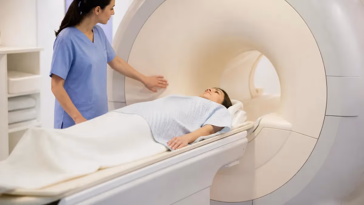
For patients new to neurology, the first MRI is often a frightening moment. Many people are referred after a single episode of optic neuritis, numbness, or unexplained vertigo called a clinically isolated syndrome. The scan determines whether that episode is a one-off neurological hiccup or the opening chapter of MS. Knowing what to expect — including how long the scan lasts, whether contrast will be used, and how the radiologist reads the images — makes the process less intimidating and helps patients participate in their own care.
Beyond the initial diagnosis, MRI plays an indispensable role in monitoring treatment. Disease-modifying therapies are evaluated not only by clinical relapses but also by the appearance of new T2 lesions or enhancing lesions on follow-up scans. A medication that controls symptoms but fails to suppress radiographic activity is often considered a partial response, prompting a switch in therapy. This concept of "NEDA" — no evidence of disease activity — has become a treatment goal directly driven by MRI findings.
Modern MS imaging protocols are highly standardized thanks to consensus guidelines from MAGNIMS in Europe and CMSC in North America. These protocols specify slice thickness, sequence selection, contrast administration, and even patient positioning to ensure that scans done in different facilities can be compared longitudinally. For a deeper look at common patterns radiologists describe, see our overview of common MRI findings across the brain and spine.
This guide walks through every aspect of MRI for multiple sclerosis: the physics that makes lesion detection possible, the specific sequences used, contrast considerations, brain versus spinal cord imaging, interpretation challenges, and the questions patients ask most. Whether you are a newly diagnosed patient, a technologist preparing for the registry, or a medical student rotating through neuroradiology, understanding how MRI illuminates this disease equips you to read reports with confidence and ask sharper questions.
The stakes are high — accurate, timely imaging directly influences whether patients receive treatment early enough to prevent irreversible neurological damage. Fortunately, the technology has improved dramatically over the past two decades, and the clinical workflow built around it has become one of the most refined in all of radiology.
MRI for MS by the Numbers
Recommended MS MRI Protocols
Includes 3D T2-FLAIR, axial T2, 3D T1 pre and post-gadolinium, and diffusion-weighted imaging. Slice thickness 3 mm or less. This protocol forms the cornerstone of every initial MS workup and serves as the comparison reference for all future scans.
Sagittal T2, STIR, and T1 post-contrast through the cervical and upper thoracic cord. Axial T2 at suspicious levels. Critical because nearly 90% of MS patients have cord lesions, and finding even one can satisfy dissemination-in-space criteria.
Coronal STIR and fat-suppressed T1 post-gadolinium of the orbits with thin slices. Used when patients present with visual symptoms suggestive of optic neuritis, often the first manifestation of multiple sclerosis in young adults.
Streamlined version focused on 3D FLAIR and T1 post-contrast to detect new or enhancing lesions. Skips redundant sequences when prior imaging is available, reducing scan time to roughly 25 minutes while preserving diagnostic yield.
Modified sequences accounting for smaller anatomy and faster cardiac motion. Often performed without gadolinium when possible due to long-term concerns about contrast retention in young patients. Sedation may be required for children under seven.
The pathophysiology of multiple sclerosis explains why MRI is so uniquely suited to its detection. When immune cells breach the blood-brain barrier and attack myelin, they trigger an inflammatory cascade that disrupts the tight junctions of capillaries, increases local water content, and damages oligodendrocytes — the cells that produce myelin. Each of these changes alters the magnetic relaxation properties of tissue water, which is precisely what MRI measures. T2 prolongation makes lesions appear bright, while T1 shortening from gadolinium leakage reveals active inflammation.
Lesion distribution follows characteristic patterns that radiologists learn to recognize on sight. The classic locations include the periventricular white matter (touching the lateral ventricles), the juxtacortical and cortical regions, the infratentorial structures like the brainstem and cerebellum, and the spinal cord. MS plaques are typically ovoid, oriented perpendicular to the ventricular surface — a configuration known as Dawson's fingers — because they track along the deep medullary veins where inflammation begins.
The McDonald criteria, updated most recently in 2017, formalize how these imaging findings support diagnosis. A patient must demonstrate dissemination in space (lesions in at least two of four typical MS locations) and dissemination in time (either a new lesion on follow-up imaging or simultaneous enhancing and non-enhancing lesions on a single scan). These criteria allow neurologists to diagnose MS earlier and more accurately than relying on clinical relapses alone, sometimes after just one symptomatic episode.
Differential diagnosis remains a constant concern. Many conditions mimic MS on MRI, including small vessel ischemic disease, migraine-related white matter changes, neuromyelitis optica spectrum disorder, MOG antibody disease, vasculitis, Susac syndrome, and progressive multifocal leukoencephalopathy. Each of these has subtle distinguishing features, and the radiologist's job is to consider the clinical context, lesion morphology, and enhancement patterns to suggest the most likely diagnosis or recommend additional workup.
Patients often wonder what makes MS lesions look different from the small white spots common in older adults. Vascular lesions tend to be rounded, located in subcortical white matter, and lack a periventricular distribution. MS lesions, by contrast, hug the ventricles, extend into the corpus callosum (especially the undersurface where the callososeptal interface lives), and frequently appear in the spinal cord — a location vascular disease almost never affects in isolation. Understanding the value of MRI with and without contrast helps clarify when enhancement is necessary.
Field strength matters more for MS imaging than for many other indications. A 3.0 Tesla magnet doubles the signal-to-noise ratio compared to 1.5 Tesla and detects roughly 20-30% more small lesions, particularly in the cortex and juxtacortical regions. Some research centers use 7 Tesla scanners to visualize the central vein sign — a tiny vessel running through the middle of a plaque that is highly specific for MS and helps distinguish it from mimickers.
The clinical value of all this imaging detail extends beyond diagnosis. Lesion burden correlates loosely with disability, atrophy measurements predict long-term outcomes, and the presence of contrast-enhancing lesions identifies patients in a window of acute inflammation when corticosteroids or escalation of disease-modifying therapy may meaningfully change the disease trajectory.
Key MRI Sequences Used for MS
Fluid-attenuated inversion recovery is the workhorse sequence for MS brain imaging. It suppresses signal from cerebrospinal fluid while leaving demyelinated tissue bright, making periventricular plaques pop against the dark ventricles. Without FLAIR, lesions touching the ventricular margins would blend into the surrounding CSF on conventional T2 images, which is why FLAIR has become non-negotiable in every modern MS protocol.
Three-dimensional FLAIR sequences acquired with isotropic voxels allow the radiologist to reformat images in any plane and detect lesions that might be missed on traditional 2D acquisitions. This is particularly valuable for small cortical and juxtacortical lesions, which are now recognized as important markers of disease severity and cognitive involvement. The trade-off is a longer acquisition time, typically five to seven minutes per 3D FLAIR.
Strengths and Limitations of MRI for MS
- +Exquisite sensitivity for demyelinating lesions even before symptoms appear
- +No ionizing radiation, allowing safe repeated scans over decades of follow-up
- +Detects both active inflammation and chronic damage in a single examination
- +Standardized protocols permit reliable comparison between institutions
- +Identifies disease activity that may be silent on neurological examination
- +Guides treatment escalation decisions through NEDA assessment
- +Offers excellent visualization of the spinal cord, optic nerves, and brainstem
- −Cannot distinguish active from chronic lesions without contrast administration
- −Gadolinium retention concerns limit repeated contrast use, especially in pediatrics
- −Long scan times can be difficult for patients with claustrophobia or tremor
- −Lesion burden correlates only modestly with clinical disability levels
- −Mimickers like small vessel disease and NMO can produce overlapping appearances
- −Cortical lesions remain underdetected even at 3T field strengths
Preparing for Your MRI for MS
- ✓Remove all metal objects including jewelry, hairpins, hearing aids, and removable dental work
- ✓Inform the technologist of any implanted devices, pacemakers, or aneurysm clips
- ✓Complete a screening form listing all prior surgeries and metallic implants
- ✓Disclose any history of kidney disease before gadolinium contrast administration
- ✓Arrange for someone to accompany you if anxiety or sedation is anticipated
- ✓Wear comfortable clothing without metal zippers, snaps, or underwire
- ✓Eat and drink normally unless your physician gives specific instructions
- ✓Bring a list of current medications including any recent steroid pulses
- ✓Mention pregnancy or breastfeeding status — protocols may be modified
- ✓Bring prior outside scans on CD so the radiologist can perform direct comparison
The 3 mm Rule
A new MS lesion must measure at least 3 mm in its longest axis and appear in two consecutive slices to count toward dissemination criteria. Tiny punctate foci under 3 mm are often dismissed as nonspecific. This threshold helps prevent overdiagnosis and ensures that follow-up scans reliably identify true disease activity rather than imaging artifacts or normal variation between exams.
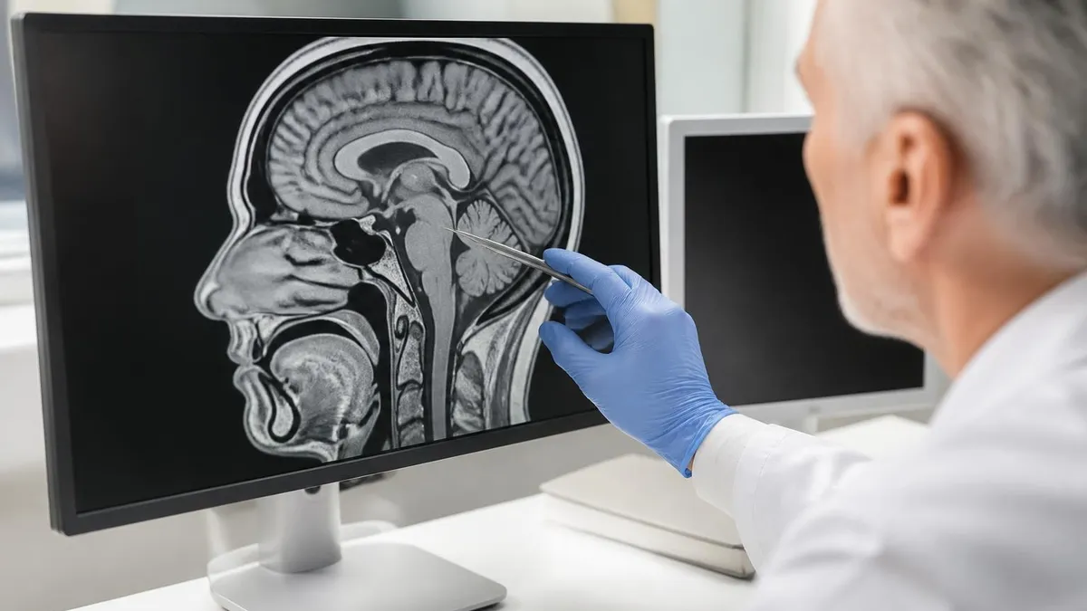
Reading an MS MRI report can feel like decoding a foreign language. Radiologists use precise terminology to convey lesion location, count, morphology, and activity status — every word carries clinical weight. The report typically begins with a description of overall brain volume, noting any atrophy in the corpus callosum, thalamus, or cortex. Volume loss often precedes clinical symptoms by years, making it a sensitive marker of neurodegeneration that complements the more obvious lesion counts.
The lesion inventory comes next. The radiologist describes the number, size, and distribution of T2 hyperintense lesions across the four McDonald regions: periventricular, juxtacortical/cortical, infratentorial, and spinal cord. They will note whether any lesions show contrast enhancement, indicating active inflammation, and whether any appear as "black holes" on T1 — hypointense areas representing more severe tissue destruction and axonal loss with worse prognostic implications than typical T2 plaques.
Comparison with prior studies is the heart of follow-up reporting. The radiologist will explicitly state whether new T2 lesions, new enhancing lesions, or enlarging lesions are present compared to the previous examination. This direct comparison drives the NEDA-3 framework, which considers a patient stable only if they have no clinical relapses, no MRI activity, and no disability progression. Achieving NEDA on a given disease-modifying therapy is a strong indicator of treatment success.
Pseudotumoral or tumefactive lesions warrant special attention. These are large MS plaques, often greater than 2 cm, that can mimic brain tumors or abscesses. They typically show incomplete ring enhancement (open toward the cortex), have less mass effect than expected for their size, and may show characteristic peripheral diffusion restriction. Misdiagnosis can lead to unnecessary biopsies, so recognizing the imaging pattern is essential, especially in younger patients without other systemic illness.
The optic nerve and spinal cord findings deserve dedicated paragraphs in any thorough MS report. Optic nerve enhancement on coronal post-contrast images confirms acute optic neuritis. Spinal cord lesions in MS are typically less than two vertebral segments long and occupy less than half the cord's cross-section — features that help distinguish them from the longitudinally extensive lesions seen in neuromyelitis optica, where lesions often span three or more vertebral bodies.
Brain atrophy quantification is moving from research into clinical practice. Automated software measures whole-brain volume, gray matter, and specific structures like the thalamus, then compares these to age-matched norms. An accelerated rate of atrophy — typically more than 0.4% per year — signals ongoing neurodegeneration even when no new focal lesions appear. This concept of "silent progression" is reshaping how clinicians think about disease activity in apparently stable patients.
Finally, the impression summarizes the radiologist's overall assessment, often using standardized phrases like "findings consistent with multiple sclerosis with active disease" or "stable disease without evidence of new inflammatory activity." These phrases feed directly into clinical decision-making, so reading them carefully and asking the neurologist to clarify any uncertainty is always worthwhile.
Although gadolinium-based contrast agents are generally safe, trace amounts can deposit in brain tissue with repeated administrations. Patients with severe kidney disease face a small risk of nephrogenic systemic fibrosis. Always disclose pregnancy, breastfeeding, kidney problems, or prior contrast reactions before your scan, and ask whether contrast is essential or could be deferred for routine follow-up imaging.
Long-term monitoring of multiple sclerosis with MRI has evolved into a structured science. The standard approach calls for a baseline scan at diagnosis, a follow-up six months after starting a new disease-modifying therapy to establish a new reference point, and then annual or biannual scans thereafter depending on clinical stability. Patients with highly active disease, those switching medications, or those entering pregnancy planning often require more frequent imaging to catch breakthrough activity early.
Choosing where to have your scans done makes a real difference in image quality and consistency. Whenever possible, follow-up scans should occur on the same scanner using identical protocols and slice positioning. Subtle differences in field strength, sequence parameters, or patient positioning can introduce false-positive or false-negative findings on comparison. Patients moving cities or switching insurance plans should request that their prior images be transferred so the new radiologist has the proper reference. Tools to find a local MRI scan can help maintain continuity of care.
The concept of NEDA — no evidence of disease activity — has reshaped MS treatment goals. NEDA-3 requires three negatives: no clinical relapses, no new or enlarging T2 lesions and no enhancing lesions on MRI, and no disability progression on the Expanded Disability Status Scale. NEDA-4 adds a fourth criterion: no accelerated brain volume loss. Achieving NEDA-4 on a given therapy is increasingly viewed as the modern benchmark of treatment success, though it remains difficult to attain over many years.
Pediatric MS presents unique imaging challenges. Children typically have higher lesion burden at presentation than adults but recover better between relapses. Their developing brains complicate the interpretation of atrophy measurements, and clinicians must weigh gadolinium use carefully given concerns about long-term retention. Most pediatric protocols minimize contrast and rely heavily on FLAIR and DWI to characterize lesions and rule out alternative diagnoses like ADEM or MOG antibody disease.
Pregnancy raises another set of considerations. MRI without contrast is considered safe throughout pregnancy and is often performed when clinically necessary to assess disease activity, especially postpartum when relapse risk peaks. Gadolinium is generally avoided during pregnancy because it crosses the placenta and accumulates in amniotic fluid, with unclear long-term effects on the fetus. Many MS specialists schedule a postpartum scan three to six months after delivery to assess disease control once contrast can be safely administered again.
Beyond the standard clinical protocol, several emerging techniques are reshaping the field. The central vein sign, visualized on susceptibility-weighted imaging at 3T or higher, is highly specific for MS and helps distinguish it from mimickers. Paramagnetic rim lesions identify chronic active plaques with smoldering inflammation that may predict more aggressive disease courses. Spinal cord atrophy measurements, particularly of the cervical cord, correlate with disability better than brain atrophy in many patients.
The future of MS imaging is increasingly quantitative and increasingly automated. Artificial intelligence tools now automate lesion counting, atrophy measurement, and even prediction of future disease trajectory based on baseline imaging features. While these tools are not yet a replacement for human radiologists, they assist in catching subtle changes that the eye might miss and standardize measurements across institutions. For patients, this means more precise treatment decisions and earlier intervention when disease activity flares.
Practical preparation for your MS MRI begins days before the appointment. Stay well hydrated in the 24 hours before your scan, especially if gadolinium contrast will be used — adequate hydration speeds clearance through the kidneys and reduces the small risk of contrast-related complications. Avoid heavy caffeine if anxiety is a concern, and if you have severe claustrophobia, ask your neurologist for a short-acting anti-anxiety medication well in advance rather than the morning of your appointment.
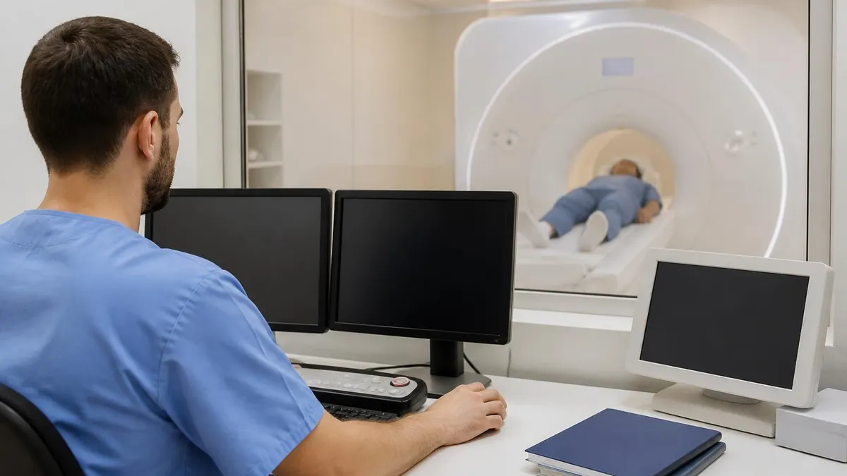
On scan day, arrive 30 minutes early to complete safety screening forms. The technologist will ask about implants, surgeries, possible metal fragments in the eyes, and pregnancy status. Wear loose-fitting clothing without metallic components, leave jewelry and valuables at home or with a companion, and bring your insurance card and physician referral. Most facilities will provide a hospital gown if your clothing has zippers or metal snaps that could cause artifact.
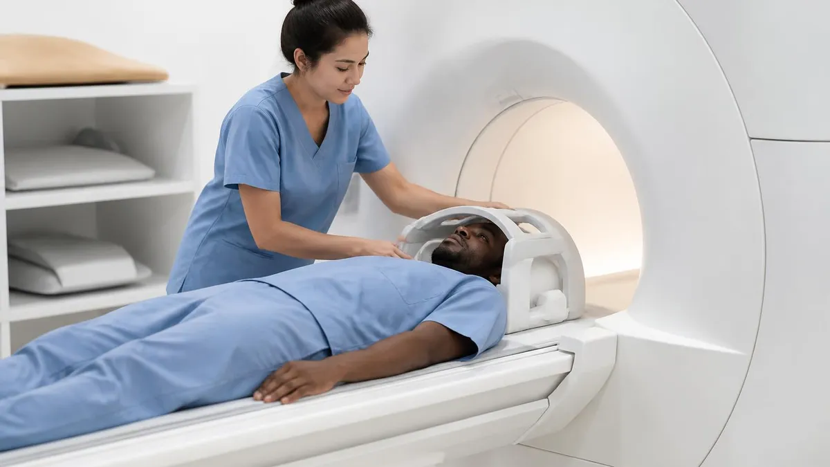
During the scan itself, you will lie supine on a padded table that slides into the bore of the magnet. The technologist will provide ear protection because the gradient coils generate loud clanging and knocking sounds. A small mirror system or video goggles may be offered so you can watch a movie or look out toward your support person. A squeeze ball or call button stays in your hand throughout the scan in case you need to communicate or stop the exam.
Lying still is the single most important contribution you make to image quality. Even small head movements blur the images and may require sequences to be repeated, extending the total exam time. Take slow, steady breaths, focus on a single point on the bore ceiling, and try to relax your jaw and shoulders. The exam typically lasts 45 to 60 minutes for a complete brain and cervical spine MS protocol, though follow-up scans without contrast can be completed in 25 to 30 minutes.
If contrast is part of the order, an IV will be placed in your arm before the scan begins. About two thirds of the way through the exam, the technologist will inject gadolinium, then acquire the post-contrast sequences. You may feel a brief cool sensation along your arm or a metallic taste in your mouth, but these sensations pass within seconds. Allergic reactions to modern macrocyclic gadolinium agents are extremely rare, occurring in fewer than one in ten thousand administrations.
After the scan, you can resume normal activities immediately. Drink extra water for the next 24 hours if contrast was used. The radiologist typically interprets the images within 24 to 72 hours, depending on the facility and whether comparison studies are available. Your neurologist will review the report, often with you in person, and decide whether any changes to your treatment plan are warranted. Bring a list of questions to that appointment — understanding your imaging is a critical part of partnering in your own care.
Finally, keep copies of your imaging. Request that the facility provide either a CD or, increasingly, secure cloud-based access to your DICOM files. Storing your own copy ensures that if you move, switch insurance plans, or need a second opinion, the actual images travel with you rather than just the report. Many patients build a personal imaging archive that proves invaluable when reviewing the trajectory of their disease over a lifetime.
MRI Questions and Answers
About the Author
Medical Laboratory Scientist & Clinical Certification Expert
Johns Hopkins UniversityDr. Sandra Kim holds a PhD in Clinical Laboratory Science from Johns Hopkins University and is certified as a Medical Technologist (MT) and Medical Laboratory Scientist (MLS) through ASCP. With 16 years of clinical laboratory experience spanning hematology, microbiology, and molecular diagnostics, she prepares candidates for ASCP board exams, MLT, MLS, and specialist certification tests.