AI MRI Analysis Free: How Artificial Intelligence Is Transforming MRI Interpretation in 2026 June
🧠 AI MRI analysis free tools explained — how AI reads scans, top platforms, accuracy stats, and what it means for patients and radiologists.
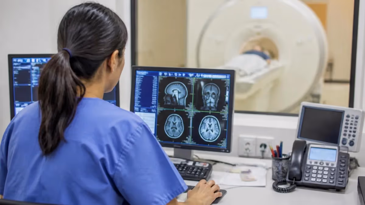
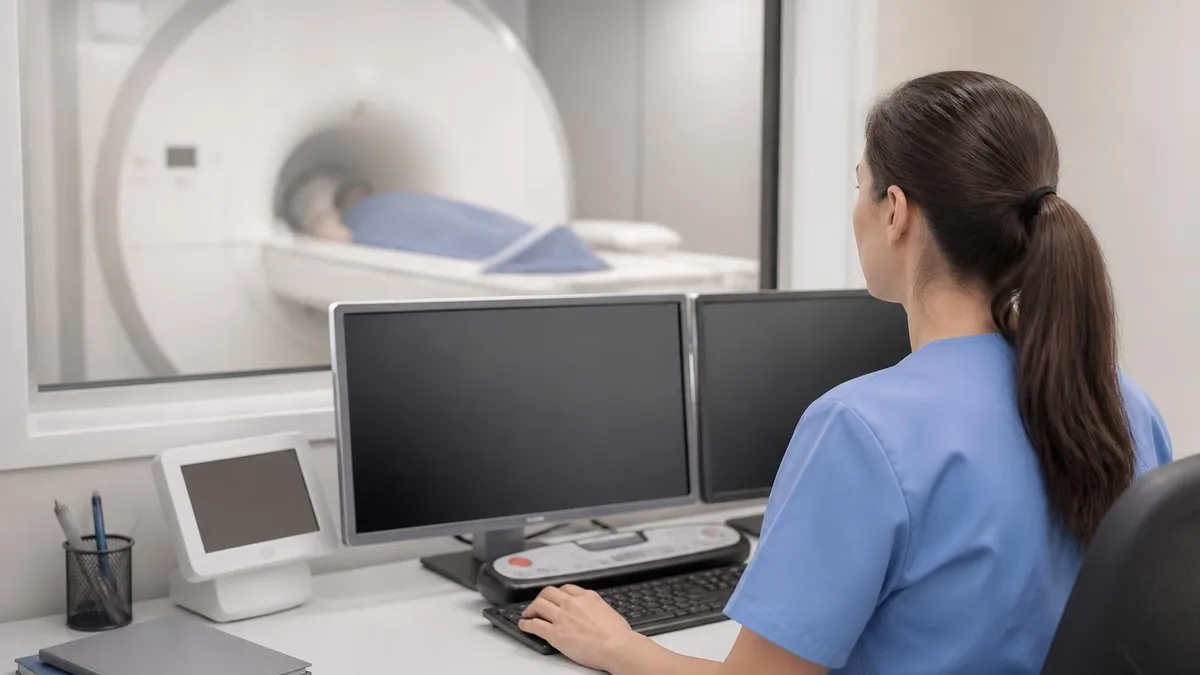
AI mri analysis free tools have moved from research curiosity to clinical reality faster than almost any other technology in diagnostic imaging. Just five years ago, the idea that a software algorithm could scan a brain MRI in under two minutes and flag a potential tumor with radiologist-level accuracy seemed like science fiction.
Today, hospitals across the United States are deploying these systems every day, and patients are increasingly asking their providers whether AI played a role in reading their images. Understanding what these tools do, how accurate they are, and which free options exist can help both patients and aspiring MRI technologists make better-informed decisions.
At its core, AI-driven MRI analysis uses deep learning models — specifically convolutional neural networks trained on millions of labeled scans — to detect patterns in image data that correlate with specific diagnoses. These models learn to recognize subtle signal differences in T1-weighted, T2-weighted, FLAIR, and diffusion-weighted sequences, building an internal representation of what healthy tissue looks like versus pathological tissue. When a new scan arrives, the model compares incoming pixel-level data against those learned patterns and assigns probability scores to different findings, from white-matter lesions to meniscal tears to spinal cord compression.
The speed advantage alone is staggering. A trained radiologist requires roughly 20 to 30 minutes to thoroughly read a complex brain MRI. Leading AI platforms like Aidoc, Viz.ai, and Enlitic can process the same study in under 90 seconds and surface critical findings — such as intracranial hemorrhage or pulmonary embolism — to the top of a radiologist's worklist immediately. This triage function means that life-threatening conditions get seen faster, reducing the time from image acquisition to treatment decision in emergency departments where every minute genuinely matters.
For MRI students and technologists preparing for board exams, understanding AI analysis is increasingly non-negotiable. The ARRT and ARMRIT credentialing bodies have both expanded their content outlines to include questions about computer-aided detection, AI workflow integration, and the ethical responsibilities of technologists working alongside automated systems. Knowing how AI tools process diffusion-weighted sequences, for instance, connects directly to exam content you will encounter. For a deeper look at one of the sequences AI analyzes most frequently, explore our guide on ai mri analysis and diffusion-weighted imaging techniques.
Free AI MRI analysis tools are increasingly available for educational and research purposes. Platforms such as MD.ai, ITK-SNAP with AI plugins, and cloud-based research environments from institutions like the National Institutes of Health allow students and researchers to upload anonymized DICOM files and receive automated segmentation or detection outputs at no cost. These tools are not cleared for clinical diagnosis without radiologist oversight, but they provide invaluable hands-on experience with the technology shaping the future of the field. Several academic medical centers have also released open-source models on GitHub that anyone can run locally with a compatible GPU.
The regulatory landscape governing AI MRI tools in the United States is more nuanced than many practitioners realize. The FDA has created a specific pathway — the De Novo classification and 510(k) clearance process — for AI-based Software as a Medical Device (SaMD). As of early 2026, more than 750 AI-enabled medical imaging devices have received FDA clearance, with the majority covering radiology applications including MRI.
Cleared tools carry specific indications, meaning an AI cleared to detect intracranial hemorrhage cannot be marketed as detecting stroke ischemia unless separately validated and cleared for that indication. This matters because it defines exactly where human radiologist oversight remains legally and clinically required.
Patient experience with AI-analyzed MRI is also evolving. Informed consent discussions increasingly touch on whether automated systems will process images, how that data is stored, and what happens when AI and radiologist conclusions diverge. Some large health systems have adopted policies requiring explicit disclosure, while others treat AI-assisted reading as a standard-of-care tool no different from a PACS workstation enhancement. For patients receiving MRI scans today, asking your imaging center about their AI tools is entirely reasonable and increasingly common as public awareness grows.
AI MRI Analysis by the Numbers
How AI Analyzes an MRI Scan: Step by Step
Image Acquisition & DICOM Transfer
Preprocessing & Normalization
Feature Extraction via Neural Network
Lesion Detection & Segmentation
Worklist Prioritization & Alert
Radiologist Review & Final Report
Free and low-cost AI MRI analysis platforms have multiplied rapidly since 2022, driven by open-source model releases, academic partnerships, and cloud computing cost reductions. For MRI students, technologists, and researchers without access to enterprise clinical AI systems, these tools provide a genuine window into how automated image interpretation works in practice. The most accessible category is open-source segmentation software, where tools like FreeSurfer, FSL, and 3D Slicer offer automated brain structure segmentation that has been refined over decades of NIH-funded research. These are not marketing-grade AI — they are the same pipelines used in peer-reviewed neuroimaging studies worldwide.
MD.ai is another platform worth knowing about. While their enterprise tier is commercial, they offer a free researcher account that allows you to upload anonymized DICOM studies and apply pre-trained AI models for tasks including lung nodule detection, brain lesion labeling, and knee cartilage segmentation. The interface is intuitive, and the platform is widely used in medical AI research collaborations. Graduate students and residents at affiliated institutions often use MD.ai to annotate datasets for model training, giving them hands-on experience with the annotation workflows that underpin commercial AI development.
Google's Medical Imaging Suite and Amazon Web Services HealthLake both offer limited free-tier access to medical imaging AI APIs. These cloud platforms provide pre-trained models for tasks like pathology detection and imaging data management. For developers building healthcare applications or researchers testing AI pipelines, the free usage tiers are enough to run meaningful experiments. Both platforms also provide documentation and sample code in Python, making them accessible to anyone with basic programming skills and a dataset of anonymized MRI files to work with.
Open-source deep learning models hosted on GitHub and Hugging Face have become another major resource. Research groups at institutions like Stanford, Johns Hopkins, and Massachusetts General Hospital regularly release trained model weights alongside their published papers, allowing anyone to download and run inference on their own hardware. Models covering brain tumor segmentation (based on the BraTS challenge dataset), multiple sclerosis lesion detection, and lumbar spine grading are all freely available. Running these models requires a Python environment, PyTorch or TensorFlow, and ideally a GPU — but cloud notebooks like Google Colab can handle inference for smaller datasets at no cost.
For patients curious about AI analysis of their own MRI scans, the landscape is more limited. A small number of direct-to-consumer platforms exist that claim to provide AI-generated insights from uploaded scans, but these are not FDA-cleared for diagnostic purposes and should never substitute for radiologist interpretation. That said, tools like Notal Vision's home OCT monitoring and similar consumer-grade imaging AI point toward a future where patients may have more direct access to AI-assisted image review. Any such output should always be discussed with your treating physician before drawing conclusions about your health.
Educational AI tools for MRI board exam preparation represent a different category of free resource. Several question banks and study platforms now incorporate AI-generated case images and interactive anatomy tools that help learners connect scan appearances to pathophysiology. These do not analyze real patient scans, but they do train pattern recognition skills in a way that traditional textbooks cannot. Practicing these skills alongside your formal exam preparation reinforces both the technical MRI physics concepts and the clinical interpretation skills that appear on the ARRT registry examination and related credentialing tests.
Integration between free AI tools and standard medical imaging viewers is improving steadily. OHIF Viewer, an open-source web-based DICOM viewer, now supports plugin architectures that allow AI inference engines to overlay results directly on the viewer interface. Researchers can connect open-source models to OHIF and create a workflow that closely mimics what radiologists see in commercial enterprise systems. This hands-on experience with real DICOM data and AI overlays is increasingly valuable for anyone entering the field, as AI-integrated PACS environments are becoming the norm rather than the exception in U.S. radiology departments of all sizes.
AI MRI Analysis: Accuracy, Limitations, and Clinical Comparisons
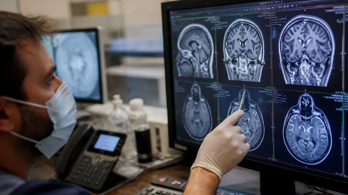
Published validation studies consistently show that top-tier AI MRI platforms achieve sensitivity rates between 90 and 96 percent for high-contrast findings like intracranial hemorrhage and large brain metastases. Specificity rates, however, are more variable — typically ranging from 85 to 93 percent depending on the training dataset and the imaging center's scanner protocols. The most rigorous validations use multi-site datasets drawn from diverse patient populations, because AI models trained on data from a single academic center often underperform when deployed at community hospitals with different scanners, field strengths, and patient demographics.
Importantly, AI performance is not static. Models trained on 1.5T data may perform differently on 3T acquisitions without dedicated retraining. Vendors are required to disclose the training conditions in their FDA submission, so clinicians can assess whether a cleared tool was validated on equipment similar to their own. Ongoing real-world performance monitoring — tracking AI output against final radiologist reports over time — is considered best practice and is increasingly required by institutional credentialing committees evaluating AI tool deployments.
AI MRI Analysis: Benefits and Drawbacks
- +Dramatically faster triage: critical findings flagged in under 90 seconds, reducing time to treatment
- +Consistent performance unaffected by fatigue, shift length, or time of day
- +Quantitative measurements like tumor volume are more reproducible than manual radiologist estimates
- +Worklist prioritization ensures the sickest patients are seen first, improving emergency care outcomes
- +Free open-source tools make AI MRI experience accessible to students and researchers globally
- +Continuous learning models improve over time as they process more validated clinical data
- −AI models can fail silently on rare pathologies or heavily motion-degraded images without obvious warning
- −Training dataset bias may cause underperformance in diverse patient populations not well-represented in training data
- −FDA-cleared AI tools have narrow indications — one cleared tool cannot legally substitute for another
- −Integration with existing PACS and EMR systems often requires significant IT infrastructure investment
- −Liability and accountability remain unclear when AI output contributes to a diagnostic error
- −Over-reliance on AI alerts can create automation bias, where radiologists accept AI findings without adequate scrutiny
Checklist: How to Evaluate an AI MRI Analysis Platform
- ✓Confirm FDA 510(k) or De Novo clearance and verify the specific cleared indication matches your intended use case.
- ✓Request the clinical validation study and check that the training and test datasets reflect your patient population.
- ✓Ask for performance data stratified by patient age, sex, race, and scanner field strength (1.5T vs. 3T).
- ✓Evaluate DICOM routing and PACS integration: confirm the AI can receive studies automatically without manual upload.
- ✓Check average processing time from image acquisition to AI output delivery for your expected study volume.
- ✓Verify the platform supports configurable sensitivity and specificity thresholds so your radiologists can tune alert behavior.
- ✓Review the vendor's post-market surveillance plan and how model updates are validated and deployed.
- ✓Confirm that the platform provides explainable AI output — color overlays or attention maps that radiologists can review.
- ✓Assess data privacy and security: confirm HIPAA compliance, BAA availability, and data retention policies.
- ✓Pilot the tool on a retrospective dataset of known cases from your institution before committing to full deployment.
AI Does Not Replace the Radiologist — It Changes What the Radiologist Does
Studies from major academic medical centers consistently show that AI-assisted radiology workflows reduce critical finding turnaround times by 40 to 60 percent without replacing radiologist oversight. The radiologist's role shifts from volume scanning to complex interpretation, communication, and quality assurance — skills that AI cannot replicate. MRI technologists who understand AI workflows will be more valuable team members as these tools become universal across U.S. imaging departments.
AI integration into clinical MRI workflows in the United States has accelerated dramatically since 2023, driven by reimbursement clarity, FDA clearance momentum, and growing evidence from prospective clinical trials. Major health systems including Mayo Clinic, Cleveland Clinic, and Mass General Brigham have published outcome data showing measurable improvements in time-to-diagnosis for stroke and intracranial hemorrhage when AI triage tools are deployed. These studies have helped convert skeptical radiologists and hospital administrators, accelerating purchasing decisions that previously stalled in committee for years.
Workflow integration is where many AI deployments succeed or fail in practice. The most successful implementations share a common feature: seamless, automated DICOM routing that requires no additional steps from the technologist or radiologist. When AI requires manual image upload or separate login to a secondary portal, adoption drops precipitously. The best enterprise platforms integrate directly into the existing PACS and radiology information system, delivering AI annotations as an overlay within the viewer the radiologist already uses every day. This invisible integration is what separates successful deployments from expensive tools that gather dust.
Radiologist acceptance of AI tools has evolved considerably. Early resistance centered on concerns about AI replacing jobs, but the dominant view in 2026 is more nuanced. Most practicing radiologists now see AI as a productivity and safety tool — one that catches the findings they might miss at the end of a long shift, not one that threatens their role. Subspecialty radiologists, particularly neuroradiologists and musculoskeletal radiologists, are increasingly using AI-generated measurements as a starting point for their reports, editing AI output rather than generating measurements from scratch, saving several minutes per complex study.
The technologist's role in AI-assisted workflows is often underappreciated. MRI technologists are responsible for image quality, and image quality directly determines AI performance. A technologist who understands how motion artifact, improper coil placement, or incorrect sequence parameters degrade AI output can proactively prevent failures before they reach the radiologist. This technical awareness is becoming a differentiator in the job market — technologists who can troubleshoot AI-assisted systems command higher salaries and more senior roles in large imaging centers and academic medical departments.
Quantitative AI applications are transforming serial MRI monitoring for chronic disease. In multiple sclerosis care, AI platforms can automatically compare the current brain MRI to all prior studies, counting new or enlarging T2 lesions and calculating total lesion volume change with millimeter precision. This longitudinal tracking, which historically required a neuroradiologist to manually compare multiple studies side by side, can now be done automatically with results appended directly to the radiology report. The clinical impact is significant: neurologists receive more precise, reproducible treatment response data, enabling earlier adjustments to disease-modifying therapy regimens.
Oncology is another domain where AI MRI analysis is creating tangible clinical value. Response Assessment in Neuro-Oncology (RANO) criteria require precise, reproducible tumor measurement on serial brain MRIs for patients receiving chemotherapy or immunotherapy. Manual measurement is time-consuming and varies between readers. AI segmentation tools trained on gadolinium-enhancing tumor regions can produce RANO-compliant measurements in seconds, with inter-reader variability far below that of manual methods. Several major cancer centers have incorporated AI-generated tumor measurements into their standard oncology workflow, and payer recognition of AI-facilitated oncology imaging is growing alongside clinical adoption.
The future of AI in MRI clinical workflows will likely involve foundation models — large, multi-task AI systems trained across multiple imaging modalities and anatomical regions simultaneously. Early research suggests that these foundation models, analogous to large language models in text AI, may generalize better to rare pathologies and diverse patient populations than narrow single-task models trained on specific datasets.
Several groups are building MRI foundation models using data from millions of studies, and initial results published in high-impact journals like Radiology and Nature Medicine suggest that the performance ceiling for AI MRI analysis is considerably higher than current deployed systems have achieved.
No FDA-cleared AI MRI analysis tool is approved to generate a final diagnostic report without radiologist review. Using AI output as a standalone diagnosis — without a licensed radiologist signing the final report — violates FDA clearance conditions and may constitute unauthorized practice of medicine. Patients should always receive a final report generated by a board-certified radiologist, regardless of which AI tools the imaging center uses to assist in interpretation.
For MRI technologists and students preparing for certification examinations, understanding AI analysis has become a genuine exam content area rather than optional enrichment. The ARRT's MRI examination content outline includes computer-aided detection, quality assurance of AI-assisted tools, and the technologist's responsibility in ensuring image quality that supports reliable automated analysis. Exam writers have recognized that today's new technologists will spend their entire careers working alongside AI systems, making foundational literacy in how these systems work an essential professional competency rather than advanced specialized knowledge.
Physics preparation remains foundational even in an AI era. The AI models that analyze MRI are ultimately interpreting signal intensities, contrast behavior, and spatial relationships that are governed by the same physics principles covered on the ARRT examination.
Understanding why T1 and T2 relaxation times differ between tissue types, how gadolinium-based contrast agents alter signal characteristics, and how diffusion restriction manifests on DWI sequences gives you genuine insight into what AI is detecting. Technologists who understand the physics beneath the AI output are better positioned to identify when a scan's technical quality may be undermining the AI's reliability, and to escalate appropriately.
Anatomy and pathology remain the clinical core of MRI competency. AI tools excel at detecting findings when they match patterns from their training data, but interpreting the clinical significance of those findings still requires deep anatomical knowledge.
When an AI flags an abnormal signal in the posterior limb of the internal capsule, understanding why that location matters for corticospinal tract function — and therefore for motor prognosis after stroke — requires human anatomical and physiological reasoning. Practice tests that focus on anatomy-pathology correlations build exactly this kind of knowledge. Use our resources on ai mri analysis alongside pathology practice to connect imaging findings to clinical context.
Registry preparation strategy should integrate AI awareness throughout rather than treating it as a separate topic. When you study white matter disease, consider how AI detects lesion load. When you study contrast enhancement patterns, consider what the AI is measuring when it segments an enhancing tumor. This integrated approach reinforces both the traditional content areas and the AI context simultaneously, making your study time more efficient. Many candidates who struggle with the registry examination are studying individual facts in isolation rather than building connected clinical reasoning schemas that mirror how the real imaging workflow operates.
Clinical experience alongside AI tools is increasingly available even to students and new graduates. Many large imaging centers now offer clinical rotations specifically designed to introduce learners to AI-assisted workflows, with structured observation of how AI annotations are reviewed, confirmed, or overridden by attending radiologists.
Seeking out these rotations — or asking your clinical site whether they use AI tools and requesting exposure to that workflow — can significantly differentiate your residency or job applications. Program directors and hiring managers have begun explicitly asking candidates about AI tool experience, and candidates who can speak fluently about their practical exposure stand out.
Continuing education in AI is growing rapidly across the MRI profession. The American Society of Radiologic Technologists and the Society for MRI Education both offer webinar series, online modules, and conference workshops specifically covering AI in clinical MRI practice. Many of these are available free to student members or at reduced cost through institutional affiliations. Building a habit of engaging with this continuing education early in your career ensures that you stay current as the technology evolves, rather than scrambling to catch up when your department deploys a new AI system and expects fluency from day one.
The bottom line for anyone in the MRI field is this: AI is not replacing the MRI technologist or the radiologist in any near-term realistic scenario, but it is fundamentally changing what excellence in those roles looks like. The most valuable practitioners in 2026 and beyond will be those who combine deep physics and anatomy knowledge, strong technical skills for image quality assurance, and working fluency with AI-assisted imaging systems. The combination of those three competencies — not any one of them alone — is what defines the next generation of MRI professional.
Practical preparation for a career that increasingly involves AI MRI analysis starts with building the right foundational habits now, regardless of where you are in your training. The most important habit is developing consistent, systematic image review routines — because AI tools are built to augment systematic review, not to substitute for it. Radiologists and technologists who review images in a structured, region-by-region sequence catch more findings and make better use of AI alerts than those who scan images impressionistically. This discipline develops through practice, and it is exactly the skill that structured practice testing reinforces.
Invest time in understanding DICOM — the file format that underlies all medical imaging including MRI. DICOM tags contain metadata about acquisition parameters, patient demographics, and scanner settings that AI systems use to contextualize their analysis. A technologist who understands DICOM header information can troubleshoot mislabeled studies, incorrect sequence parameters, and other data quality issues that degrade AI performance. Free DICOM viewers like RadiAnt, Horos, and OsiriX allow you to open and inspect DICOM files on your own computer, exploring the metadata that AI systems rely on alongside the image data they analyze.
Networking with radiologists who work in AI-integrated environments can accelerate your learning considerably. Many academic medical centers host grand rounds, departmental presentations, and public webinars where radiologists present their experience with AI tools, including candid discussions of where the tools have helped and where they have fallen short.
Following academic radiologists on professional social media platforms like LinkedIn and X (formerly Twitter) exposes you to the ongoing clinical and research conversation about AI MRI, including links to new papers, conference presentations, and opinion pieces that reflect the current state of the field at a level of detail that textbooks cannot provide.
Consider building a personal learning project around free AI MRI tools. Download FreeSurfer or FSL, obtain a publicly available neuroimaging dataset like OpenNeuro or the Human Connectome Project, and run automated brain segmentation on a few subjects. Document what the tool outputs, how long it takes, and where the segmentations look anatomically plausible versus where they fail. This kind of hands-on experimentation develops practical intuition about AI MRI analysis that no amount of reading can substitute for, and it produces a tangible portfolio item you can describe in job interviews and residency applications.
Quality assurance skills are more important in AI-integrated MRI departments than ever before. AI performance monitoring requires comparing AI outputs against radiologist ground truth on an ongoing basis, tracking metrics like positive predictive value, sensitivity, and specificity over time.
When AI performance degrades — which can happen if scanner software updates change image characteristics, if patient population demographics shift, or if a new pathology appears that differs from training data — someone needs to detect the drift and escalate to the vendor or clinical leadership. Technologists and quality assurance specialists with the statistical literacy to understand these metrics are in growing demand across large imaging departments.
Finally, keep patient communication at the center of your professional identity regardless of how technology evolves. AI systems analyze pixels. They do not explain findings to anxious patients, adjust their communication style for health literacy, or recognize when a patient needs an extra moment of reassurance before entering the magnet bore. The human dimensions of MRI practice — patient-centered communication, procedural safety monitoring, and empathetic care — are permanent differentiators that no AI tool will replace. Technologists who combine technical AI fluency with genuine patient-centered care will define the highest standard of MRI practice for the foreseeable future.
Whether you are studying for your ARRT registry examination, entering a clinical rotation, or exploring free AI MRI tools for the first time, the resources available today are more comprehensive and accessible than they have ever been. Take advantage of the open-source tools, academic datasets, and online communities that exist to help you build real competency. The combination of rigorous exam preparation and practical AI awareness creates a foundation for a long, rewarding career at the intersection of medicine, technology, and patient care.
MRI Questions and Answers
DWI MRI: Diffusion-Weighted Imaging Explained (2026 Guide)
Side Effects of Brain MRI: Understanding Risks, Safety, and What Every Patient Should Know
MRI Lumbar Spine Without Contrast: Complete Guide to the Scan, What It Shows, and How to Prepare
MRI History: From Nuclear Resonance to Modern Scanners
MRI of Cervical Spine: Complete Guide to Imaging, Anatomy, Pathology, and Patient Preparation
About the Author
Medical Laboratory Scientist & Clinical Certification Expert
Johns Hopkins UniversityDr. Sandra Kim holds a PhD in Clinical Laboratory Science from Johns Hopkins University and is certified as a Medical Technologist (MT) and Medical Laboratory Scientist (MLS) through ASCP. With 16 years of clinical laboratory experience spanning hematology, microbiology, and molecular diagnostics, she prepares candidates for ASCP board exams, MLT, MLS, and specialist certification tests.
Join the Discussion
Connect with other students preparing for this exam. Share tips, ask questions, and get advice from people who have been there.
View discussion (4 replies)