MRI Contrast Dye: Gadolinium, Safety & When Used
MRI contrast dye explained: gadolinium types, side effects, allergic reactions, NSF risk, and when contrast is truly needed for your scan.
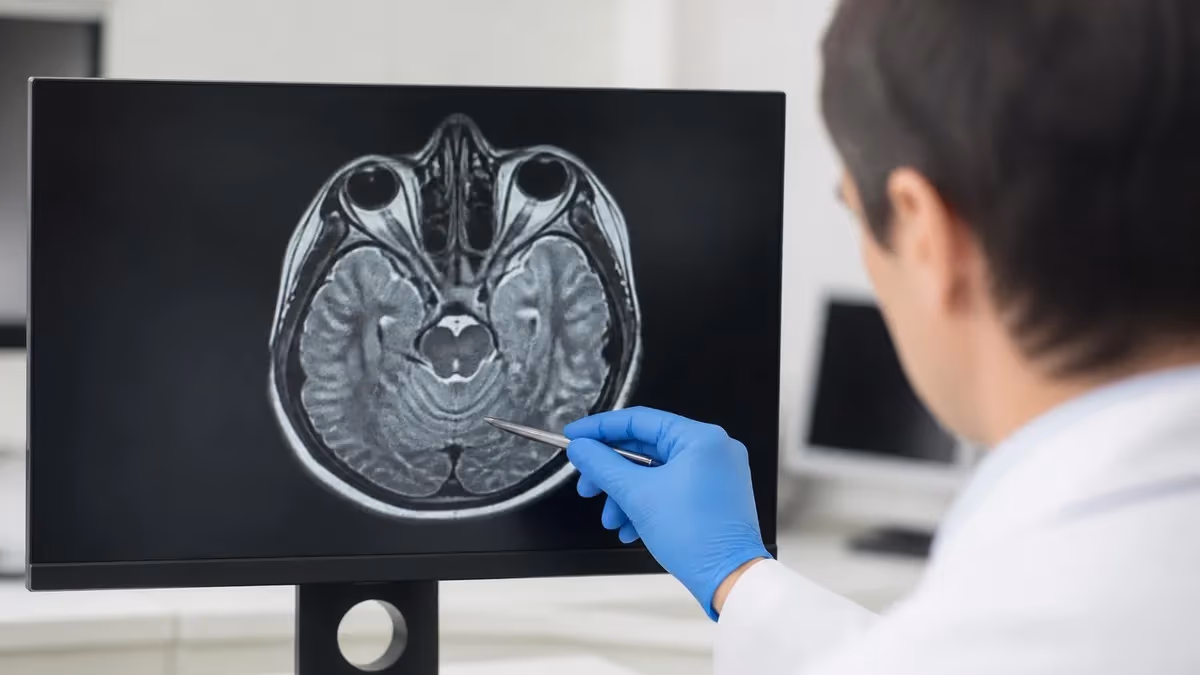
When your doctor orders an MRI, the order slip often includes two words that raise eyebrows: with contrast. That phrase means you will receive an MRI contrast dye, almost always a gadolinium-based contrast agent (GBCA), through an IV line shortly before or during the scan. The dye does not contain iodine and it is nothing like the contrast used in CT scans.
Instead, it is a metal ion wrapped inside a protective molecular cage that travels through your bloodstream and changes how nearby tissues appear on the magnetic resonance images. The result is a sharper, more informative scan that gives your radiologist far more confidence in the final report.
So what is MRI contrast dye, really? It is a clear, water-like liquid containing a paramagnetic metal called gadolinium. Paramagnetic simply means the metal disturbs the magnetic field in a controlled way, which shortens the relaxation time of nearby water molecules. When those water molecules behave differently, the MRI scanner produces a brighter signal in tissues where the dye accumulates, such as tumors with leaky blood vessels, areas of active inflammation, or organs with high blood flow. That contrast between enhancing and non-enhancing tissue is what makes the dye so diagnostically powerful.
Roughly half of all MRI exams use contrast, and for good reason. Gadolinium dramatically improves the visibility of tumors, infections, blood vessels, inflammation, and small lesions that might otherwise blend into surrounding tissue. Still, plenty of patients ask the same questions before getting injected. Is it safe?
What if I have kidney problems? Can I refuse contrast dye for an MRI? What does an allergic reaction to MRI dye actually look like? This guide answers all of that in plain English, walks you through the most common GBCAs, explains who should be cautious, and shows you when an MRI without contrast is just as useful as one with the dye.
Before we go deeper, it helps to anchor a few definitions. The term contrast dye MRI refers to any magnetic resonance scan performed after IV gadolinium has been administered. The phrase injection dye for MRI describes the act of giving that agent. MRI no contrast simply means the same machine and the same body region were scanned without dye. None of these phrases describes a different machine or a different type of MRI. The hardware is identical; the only variable is whether gadolinium is circulating in your bloodstream during the imaging sequence.
The numbers above tell a reassuring story. Severe reactions to gadolinium are extraordinarily rare, the agent has been used billions of times since the late 1980s, and the most feared complication, nephrogenic systemic fibrosis (NSF), almost never occurs in patients with healthy kidneys. That does not mean MRI contrast is risk-free, but it does mean the benefit-to-risk ratio is very favorable for most people. Knowing the basics helps you walk into the imaging suite informed rather than anxious, and it makes the consent conversation with the technologist much more productive.
Patients sometimes confuse the IV dye in an MRI with the iodine-based contrast used in CT or with the chalky barium drink used in some X-ray studies. They are three completely different substances with three completely different safety profiles. The MRI inject dye you receive is gadolinium-based, contains no iodine, and almost never causes the warmth or flushing sensation people remember from a CT scan. Most patients describe the MRI dye as feeling like nothing at all, although a small minority notice a brief cool sensation as the saline flush runs through the IV.
Below we unpack the four pillars every patient should understand: when contrast is actually indicated, what types of GBCAs you might receive, what side effects to watch for, and what alternatives exist if you cannot have gadolinium for any reason. Each piece of this story matters, because the right answer for one patient is rarely identical to the right answer for another.
A 40-year-old marathon runner with a knee injury, a 70-year-old with stage 4 chronic kidney disease, and a 28-year-old being worked up for new-onset seizures all need different conversations about MRI dye, even if they end up in the same scanner the same afternoon.
Gadolinium in plain English
Pure gadolinium is a heavy metal and toxic on its own. To make it safe for medical use, chemists bind it tightly inside an organic molecule called a chelate. The resulting compound, a gadolinium-based contrast agent, passes through your body and is filtered out by your kidneys within about 24 hours. The chelate keeps the metal locked up so it cannot interact with your tissues, which is why GBCAs have such a strong safety record when kidney function is normal.
Not every MRI needs contrast. A simple knee MRI for a torn meniscus, a lumbar spine scan for a herniated disc, or a brain scan for a routine headache often produces excellent images without any injection. Contrast is reserved for situations where the radiologist needs to highlight blood flow, separate scar tissue from active disease, or pick out a tiny lesion that would otherwise be invisible. Think of contrast as a spotlight: when the scan is already well-lit by anatomical detail, the spotlight is unnecessary, but when the target is hidden in shadow, it can change the entire diagnosis.
Doctors who order MRIs follow guidelines from the American College of Radiology, the European Society of Urogenital Radiology, and specialty societies that publish appropriateness criteria. These documents spell out which clinical scenarios warrant contrast and which do not. Your referring physician usually checks the appropriate protocol before sending the order, but the radiologist supervising the scan has the final say and can add or remove contrast based on what the early images show. The four structure cards below summarize the four big questions patients ask most often about that decision.
The Four Pillars of MRI Contrast
Tumor detection and staging, brain or spine infections, MS plaque activity, vascular imaging (MRA), suspected metastases, post-surgical follow-up, and inflammatory bowel disease assessment all benefit from gadolinium enhancement.
Two structural classes exist: macrocyclic agents (Dotarem, Gadavist, ProHance) and linear agents (Magnevist, Multihance, Omniscan, Eovist). Macrocyclic agents are now preferred because they hold gadolinium more tightly.
Most people feel nothing. Mild reactions include nausea, headache, or a cool sensation at the IV site. True allergic reactions occur in less than 1 in 10,000 patients and range from hives to, very rarely, anaphylaxis.
Non-contrast MRA, diffusion-weighted imaging, ultrasound with microbubbles, CT with iodinated contrast, and PET imaging can all substitute depending on the clinical question. Your radiologist picks the safest equivalent.
Once you know contrast is on the menu, the next question is which gadolinium agent you will receive. Different hospitals stock different brands based on cost, contracts, and the type of imaging they specialize in. A liver-focused center may favor Eovist for its biliary excretion, while a multiple sclerosis clinic typically reaches for Dotarem or Gadavist because of their stability and brain enhancement profile. As a patient, you usually do not get to choose the agent, but you can ask which one will be used and request a macrocyclic option if you are concerned about gadolinium retention.
The tabs below break down four practical topics: the common agents you might see on your paperwork, the indications that justify contrast, your rights if you want to refuse it, and the kidney-related concern called NSF that drives most of the screening questions you will be asked. Each tab is written to stand alone, so feel free to jump straight to the topic that worries you most.
Gadolinium Agents and Patient Choice
Magnevist (gadopentetate dimeglumine) was the first FDA-approved GBCA in 1988 and is a linear ionic agent now used less often because of stability concerns. Multihance (gadobenate dimeglumine) is a linear ionic agent with stronger liver uptake, making it useful for hepatic imaging. Dotarem (gadoterate meglumine) is a macrocyclic ionic agent considered one of the most stable options and is preferred for brain and spine work, particularly in patients with reduced kidney function. Other names you may see include Gadavist, ProHance, Omniscan, and Eovist.

Before the contrast goes in, the technologist will run through a short safety checklist with you. Answering accurately is the single most important thing you can do to keep the exam safe. Even small details, like a kidney infection two years ago or a mild hives reaction during a previous scan, can change which agent is used or whether contrast is given at all.
Technologists are trained to ask the same questions in slightly different ways to make sure nothing slips through, so do not be surprised if you hear about allergies three times. It is a feature, not a glitch.
If your kidney function has not been checked in the last six months and you are over 60, diabetic, or have a history of kidney disease, the imaging center will likely run a quick blood test on arrival. Many facilities now use point-of-care creatinine analyzers that return a result within five minutes, so this rarely delays the scan. The test screens for the very specific scenario where reduced kidney clearance plus an older linear GBCA could trigger NSF.
Always disclose: pregnancy or breastfeeding, current dialysis treatment, severe kidney disease, prior contrast reactions of any kind, asthma, multiple drug allergies, sickle cell disease, and any recent contrast injections within the last 24 hours. If you are unsure about your kidney function, ask whether a point-of-care creatinine test is available. Five extra minutes of screening prevents the vast majority of avoidable complications.
Pre-scan preparation is straightforward but the small details matter. Use the checklist below to walk into the appointment ready. Most facilities ask you to fast for four hours before contrast to reduce nausea, although clear fluids are usually fine. Hydration before and after the scan helps your kidneys clear the gadolinium quickly, and a glass of water every hour for the rest of the day is a simple post-scan habit your kidneys will appreciate.
If you have ever had an allergic reaction to MRI dye, your radiologist may prescribe a short prep with oral steroids and an antihistamine starting 12 hours before the scan. This protocol blunts the immune response and lets most previously reactive patients tolerate a second exposure without trouble. Bring the prescription bottle with you so the technologist can confirm the dose was taken on schedule.
Diabetics taking metformin once had to stop the medication around contrast scans, but that rule applied to iodinated CT contrast, not gadolinium. You do not need to pause metformin for an MRI with gadolinium. Likewise, breastfeeding mothers can continue nursing after a gadolinium injection; the amount of dye that crosses into breast milk is vanishingly small and the FDA considers it safe. Pregnancy is a different matter because gadolinium does cross the placenta. Most guidelines reserve contrast MRI in pregnancy for cases where the diagnostic benefit clearly outweighs theoretical fetal risk.
Pre-MRI Contrast Patient Checklist
- ✓Confirm your most recent kidney function (eGFR or creatinine) test result
- ✓List every prior contrast reaction, even mild ones such as hives or nausea
- ✓Disclose pregnancy, possible pregnancy, or current breastfeeding status
- ✓Bring a list of all current medications, especially diuretics and NSAIDs
- ✓Stop eating four hours before, but keep drinking water unless told otherwise
- ✓Remove all metal, jewelry, hearing aids, and any transdermal medication patches
- ✓Ask whether the scan can be performed without contrast if you have concerns

So is contrast worth it for your specific situation? That depends entirely on what your doctor is trying to find. The comparison table below lists the most common MRI indications and shows where contrast adds real diagnostic value versus where a plain scan does the job just as well. Use it as a starting point for a conversation with your radiologist or referring physician, not as a substitute for medical advice. Imaging protocols are individualized, and your medical history may shift the calculus.
Patients commonly worry about the side effects of MRI without contrast versus with contrast, but the side effect profile of a non-contrast scan is almost nonexistent. The trade-off is purely diagnostic. A non-contrast MRI cannot reliably distinguish active MS plaques from old ones, characterize small liver lesions, or detect tiny pituitary tumors.
If those are not relevant to your case, skipping contrast costs you nothing and spares you the IV stick. The strongest cases for contrast are oncology follow-up, neurology workups involving demyelinating disease, infections of the brain or spine, and vascular imaging that needs to map blood flow rather than just anatomy.
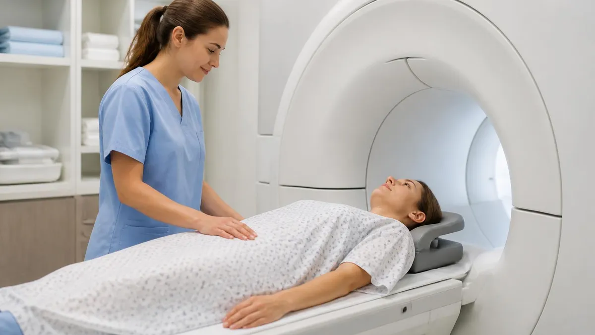
Many people also ask whether the contrast injection itself hurts. The honest answer is that the IV placement is the only uncomfortable part, and it feels like any other blood draw. Once the catheter is in, the gadolinium itself is painless. A few patients notice a brief cool wave as the saline flush follows the dye, and an even smaller subset report a strange taste in the mouth that lasts about a minute. None of these sensations indicate a problem; they are normal and pass quickly.
Reactions to MRI contrast, while rare, do happen. Mild reactions appear in roughly 1 to 2 percent of injections and usually involve nausea, a metallic taste, dizziness, or mild itching that resolves on its own within minutes. Moderate reactions, such as widespread hives or wheezing, occur in fewer than 1 in 10,000 patients.
Severe anaphylactic reactions are extraordinarily rare at about 1 in 30 million injections, but the imaging suite is always stocked with epinephrine, oxygen, and trained staff who can respond within seconds. If you have ever reacted to gadolinium, your radiologist may pre-medicate you with steroids and antihistamines or switch to a different chemical class entirely. Modern protocols make repeated MRI exams feasible even for patients with a documented reaction history.
Long-term gadolinium retention has also drawn attention in recent years. Trace amounts of gadolinium can deposit in the brain, bones, and skin after repeated GBCA exposure, particularly with older linear agents. To date no clinically harmful effects have been definitively linked to retention in patients with normal kidneys, but the FDA now requires a Medication Guide and recommends using the lowest dose needed. If you anticipate many scans over your lifetime, ask your team to use a macrocyclic agent whenever possible. Examples include Dotarem, Gadavist, and ProHance.
Some patients develop a condition called gadolinium deposition disease, a controversial diagnosis characterized by persistent symptoms such as bone pain, brain fog, skin thickening, and burning sensations after a contrast MRI. The medical community is still studying whether this represents a true clinical syndrome distinct from background complaints, but the FDA has acknowledged the reports. If you have unusual symptoms after a contrast scan, document them and discuss with both your referring doctor and the imaging center. Honest feedback helps the field refine guidelines over time.
Patients consistently ask the same handful of questions in the days before a contrast-enhanced MRI. The answers below cover the most common concerns, from how the injection feels to whether you can drive home afterward. If your specific question is not addressed, call the imaging department ahead of time. Technologists field these calls every day and would rather answer a question on the phone than reschedule a scan because of a last-minute concern in the changing room.
One last reminder: the goal of a contrast MRI is to answer a clinical question your doctor cannot answer any other way. That answer might confirm a benign finding and save you from invasive testing, or it might catch a treatable condition early. The few minutes spent with an IV in your arm are usually a very small price for the information the scan delivers.
Walk in informed, ask questions, and trust the screening process the team puts you through. If you want to test how much you have learned, the practice quizzes linked above are a quick way to lock in the key facts about MRI contrast dye, gadolinium safety, and modern imaging protocols.
Imaging technology continues to evolve. Newer non-contrast MR angiography sequences can now visualize arteries and veins without any injection, and ultra-high-field 3T and 7T scanners produce images sharp enough that some indications no longer require contrast at all. AI-driven post-processing also extracts more information from each scan, which may further reduce the need for dye over time. Until those tools fully mature, gadolinium remains the workhorse for enhanced MRI, and understanding what it is, why it is used, and when to question it puts you firmly in the driver's seat for your own care.
MRI Questions and Answers
About the Author
Medical Laboratory Scientist & Clinical Certification Expert
Johns Hopkins UniversityDr. Sandra Kim holds a PhD in Clinical Laboratory Science from Johns Hopkins University and is certified as a Medical Technologist (MT) and Medical Laboratory Scientist (MLS) through ASCP. With 16 years of clinical laboratory experience spanning hematology, microbiology, and molecular diagnostics, she prepares candidates for ASCP board exams, MLT, MLS, and specialist certification tests.