Pituitary Macroadenoma MRI: Complete Guide to Imaging, Protocols, and Interpretation
Complete guide to pituitary macroadenoma MRI — 📗 protocols, sequences, dynamic contrast imaging, and interpretation tips for radiology professionals.
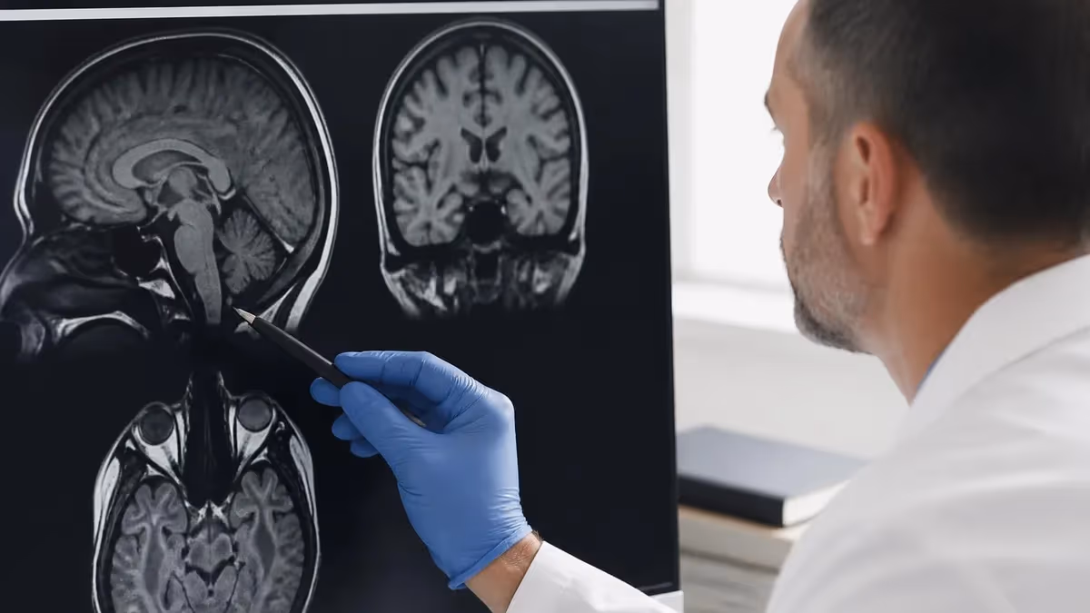
A pituitary macroadenoma mri is the gold-standard imaging study for evaluating tumors of the sella turcica that measure 10 millimeters or larger. These benign neoplasms arise from the anterior pituitary gland mri cpt code and can produce dramatic visual, endocrine, and neurological symptoms when they expand beyond the bony confines of the sella. MRI provides unmatched soft-tissue contrast for differentiating tumor from normal pituitary parenchyma, optic chiasm, cavernous sinus, and surrounding cerebrospinal fluid spaces, making it essential for surgical planning.
Unlike microadenomas, which require sophisticated dynamic contrast mri techniques to detect, macroadenomas are typically visible on standard T1-weighted and T2-weighted sequences. However, the imaging protocol must still be meticulous to characterize tumor extension into the cavernous sinus, compression of the optic apparatus, invasion of the sphenoid sinus, and the presence of intratumoral hemorrhage or cystic change. Each of these findings directly influences surgical approach, expected outcomes, and the likelihood of gross total resection.
Radiologists and MRI technologists working in neuroimaging encounter pituitary cases frequently. Endocrinologists order pituitary protocols when patients present with hyperprolactinemia, acromegaly, Cushing disease, or visual field defects suggesting chiasmal compression. Emergency departments order them when a patient presents with sudden severe headache, ophthalmoplegia, or visual loss suggesting pituitary apoplexy. Understanding the imaging protocol and interpretive nuances ensures these patients receive timely, accurate diagnoses.
The modern pituitary mri cpt protocol has evolved considerably. While 1.5T systems remain widely used, 3T scanners have become the preferred platform because of their superior signal-to-noise ratio, which permits thinner slices and higher in-plane resolution. Dedicated pituitary protocols typically include small field-of-view coronal and sagittal T1 sequences before and after gadolinium administration, T2-weighted imaging, and increasingly, three-dimensional volumetric sequences that allow multiplanar reconstruction.
Macroadenomas demonstrate characteristic imaging features that experienced readers recognize immediately. The classic snowman or figure-of-eight appearance results from waist-like constriction at the diaphragma sellae as the tumor expands superiorly into the suprasellar cistern. Cystic change, hemorrhage, and calcification can complicate the appearance. Differentiating macroadenoma from other sellar and parasellar lesions — including craniopharyngioma, meningioma, Rathke cleft cyst, and metastasis — relies on careful evaluation of signal characteristics, enhancement pattern, and anatomic relationships.
This comprehensive guide walks through everything radiology professionals need to know about pituitary macroadenoma MRI, from sequence selection and slice planning to identifying cavernous sinus invasion and recognizing apoplexy. Whether you are preparing for the MRI registry exam, building a neuro protocol library, or simply seeking to interpret pituitary studies more confidently, the material below covers the technical and clinical foundations. For background on imaging terminology used throughout, see the MRI medical abbreviation reference guide.
By the end of this article you will understand the optimal pulse sequences, the role of dynamic contrast imaging, the Knosp grading system for cavernous sinus invasion, the differential diagnosis of sellar masses, and the post-operative imaging considerations that follow transsphenoidal surgery. These concepts are tested heavily on advanced MRI registry examinations and form the foundation of neuro-MRI competency.
Pituitary Macroadenoma MRI by the Numbers
Essential Sequences in the Pituitary MRI Protocol
Small field-of-view coronal T1 with 2-3 mm slices through the sella establishes baseline signal of the gland, identifies hemorrhage, and characterizes the posterior pituitary bright spot before contrast administration.
High-resolution coronal T2 imaging demonstrates cystic components, edema in adjacent brain parenchyma, and the relationship of tumor to the optic chiasm, which appears as a low-signal horizontal band above the sella.
Rapid serial coronal T1 acquisitions during gadolinium injection capture the differential enhancement kinetics between normal pituitary tissue and adenomatous tissue, which enhances more slowly than the surrounding gland.
Sagittal post-contrast imaging shows superior tumor extension into the suprasellar cistern, infundibular deviation, and inferior extension into the sphenoid sinus, essential information for transsphenoidal surgical planning.
Isotropic 3D T1 SPGR or MP-RAGE sequences permit thin-slice multiplanar reformatting, useful for assessing complex tumor geometry, neuronavigation planning, and post-operative follow-up comparisons across time points.
Understanding the anatomy surrounding the pituitary macroadenoma is fundamental to producing diagnostic images and to interpreting them accurately. The pituitary gland sits within the sella turcica, a bony depression in the sphenoid bone shaped roughly like a Turkish saddle. The diaphragma sellae, a fold of dura mater, forms the roof of the sella and is pierced centrally by the pituitary stalk, also called the infundibulum, which connects the gland to the hypothalamus above.
Immediately above the sella lies the suprasellar cistern, a cerebrospinal fluid space bordered by the optic chiasm, the third ventricle, and the circle of Willis. The optic chiasm itself sits roughly 5 to 10 millimeters above the diaphragma sellae in most patients, though anatomic variants known as pre-fixed and post-fixed chiasms shift this relationship and have implications for the pattern of visual field defect produced by tumor compression.
Laterally, the cavernous sinuses flank the sella on each side. These dural venous spaces house the internal carotid artery in its cavernous segment, along with cranial nerves III, IV, V1, V2, and VI. The medial wall of the cavernous sinus is thin and often the first structure breached by an aggressively invading macroadenoma. Tumor extending into the cavernous sinus can encase the carotid artery and compress the cranial nerves, producing ophthalmoplegia, facial numbness, or vision loss.
Inferiorly, the sella sits directly above the sphenoid sinus, separated only by the thin bony floor. This anatomic relationship is the foundation of the transsphenoidal surgical approach, which allows neurosurgeons to access pituitary tumors through the nasal cavity and sphenoid sinus without entering the cranial vault. Pneumatization patterns of the sphenoid sinus — sellar, presellar, or conchal — affect the surgical corridor and should be noted on the radiology report.
The normal pituitary gland itself has two functionally and embryologically distinct lobes. The anterior lobe, or adenohypophysis, derives from oral ectoderm and accounts for roughly 80 percent of glandular volume. It produces growth hormone, prolactin, ACTH, TSH, LH, and FSH. The posterior lobe, or neurohypophysis, derives from neural ectoderm and stores oxytocin and vasopressin synthesized in the hypothalamus. On T1-weighted imaging, the posterior lobe typically demonstrates a characteristic bright signal known as the posterior pituitary bright spot.
Gland size and shape vary with age, sex, and physiological state. The normal adult pituitary measures up to about 8 millimeters in height in women and 6 to 7 millimeters in men, though it can reach 10 to 12 millimeters during pregnancy and lactation due to physiologic hyperplasia. Recognizing these normal variations prevents over-diagnosis of small adenomas and helps differentiate physiologic enlargement from pathology. For historical context on how MRI evolved into this anatomical tool, the history of MRI provides useful background.
The infundibulum normally lies in the midline, though minor deviation is common. Significant lateral deviation of the stalk away from the midline raises concern for a small ipsilateral adenoma or other space-occupying lesion. Width of the stalk should taper smoothly from the hypothalamus to its insertion on the posterior aspect of the gland; thickening suggests pathology such as hypophysitis, sarcoidosis, Langerhans cell histiocytosis, or germinoma.
MRI Practice Test Questions
Prepare for the MRI - Magnetic Resonance Imaging exam with our free practice test modules. Each quiz covers key topics to help you pass on your first try.
MRI Knowledge
MRI Exam Questions covering Knowledge. Master MRI Test concepts for certification prep.
MRI Physics
Free MRI Practice Test featuring Physics. Improve your MRI Exam score with mock test prep.
MRI Anatomy and Pathology
MRI Test Prep for MRI Anatomy and Pathology. Practice MRI Quiz questions and boost your score.
MRI Anatomy and Positioning
MRI Questions and Answers on MRI Anatomy and Positioning. Free MRI practice for exam readiness.
MRI Contrast Agents
Free MRI Quiz on MRI Contrast Agents. MRI Exam prep questions with detailed explanations.
MRI Patient Care and Positioning
MRI Practice Questions for MRI Patient Care and Positioning. Build confidence for your MRI certification exam.
Signal Characteristics of Pituitary Macroadenomas
On non-contrast T1-weighted imaging, the typical pituitary macroadenoma is isointense to slightly hypointense compared with the normal pituitary gland and gray matter. This subtle signal difference can make small adenomas difficult to detect without contrast, but macroadenomas are usually conspicuous because of their size and the architectural distortion they produce in the sellar region.
When intratumoral hemorrhage occurs, T1 signal varies depending on the age of blood products. Acute and hyperacute hemorrhage appears isointense or slightly hyperintense, whereas subacute hemorrhage containing methemoglobin produces strikingly bright T1 signal. Recognizing T1 hyperintensity is critical because it suggests apoplexy, prior hemorrhage, or, less commonly, high protein content within a cystic component of the tumor.
Should You Use 3T or 1.5T for Pituitary Imaging?
- +Higher signal-to-noise ratio enables thinner slices and higher resolution
- +Better detection of small adenomas and subtle invasion of the cavernous sinus
- +Improved depiction of the optic chiasm and surrounding cranial nerves
- +Shorter scan times for equivalent image quality reduce motion artifact
- +Superior dynamic contrast temporal resolution captures tumor enhancement kinetics
- +3D volumetric sequences produce isotropic voxels ideal for multiplanar reformatting
- +Better visualization of microadenomas in Cushing disease workups
- −More susceptible to susceptibility artifacts near the sphenoid sinus air spaces
- −Increased chemical shift artifact at the bone-marrow and fat-tissue interfaces
- −Higher specific absorption rate may limit some sequence parameters
- −Implant safety considerations may be more restrictive at higher field strength
- −Equipment and maintenance costs significantly higher than 1.5T systems
- −Acoustic noise levels typically louder at 3T scanners
- −Patient claustrophobia concerns sometimes worsened in higher field bore designs
Technologist Checklist for Pituitary Macroadenoma MRI
- ✓Confirm contraindications to gadolinium contrast and review renal function labs
- ✓Verify patient safety screening for implants and prior surgical hardware
- ✓Position the patient supine with head in a dedicated head coil and immobilize gently
- ✓Acquire localizer images in three planes to identify the sella turcica accurately
- ✓Set small field of view of 16-20 cm centered on the sella for high resolution
- ✓Use 2-3 mm slice thickness with no gap on coronal and sagittal T1 sequences
- ✓Run pre-contrast coronal T1 and T2 sequences through the entire sellar region
- ✓Prepare power injector with gadolinium dose and 20 mL saline flush
- ✓Begin dynamic contrast sequence at the start of injection for optimal timing
- ✓Acquire post-contrast coronal and sagittal T1 with the same slice prescription as pre-contrast
- ✓Review all sequences for motion, coverage, and adequate contrast before releasing patient
- ✓Document any incidental findings and forward complete series to the reading radiologist
Dynamic Contrast Timing Is Everything
The window for optimal tumor-to-gland contrast on dynamic imaging is narrow — typically the first 30 to 60 seconds after gadolinium reaches the pituitary. Beginning the dynamic series too late causes the tumor and gland to equilibrate, eliminating the contrast difference that makes adenomas visible. Always start the dynamic acquisition at the moment of injection.
The Knosp grading system is the most widely used framework for assessing cavernous sinus invasion by pituitary macroadenomas on coronal MRI. Developed by Engelbert Knosp and colleagues in 1993, the system uses the position of the tumor relative to imaginary tangent lines drawn through the intracavernous and supraclinoid segments of the internal carotid artery on the coronal plane. The grade directly influences surgical strategy, expected resection rates, and prognosis.
Grade 0 indicates tumor that remains medial to the medial tangent of the carotid artery and shows no extension toward the cavernous sinus. Grade 1 tumor extends past the medial tangent but does not cross the intercarotid line, the imaginary line connecting the centers of the intracavernous and supraclinoid carotid segments. Grade 2 tumor extends past the intercarotid line but does not cross the lateral tangent of the carotid artery.
Grade 3 and grade 4 represent true cavernous sinus invasion. Grade 3 tumor extends past the lateral tangent of the carotid, and Knosp grade 3 is further subdivided into 3A for invasion of the superior cavernous compartment and 3B for invasion of the inferior compartment. Grade 4 tumor completely encases the intracavernous carotid artery on all sides. Grades 3B and 4 are associated with the highest rates of subtotal resection and the lowest rates of biochemical remission for functioning adenomas.
Beyond Knosp grading, several other imaging features influence prognosis and surgical planning. Tumor consistency, which can sometimes be inferred from T2 signal intensity, affects ease of resection. Soft, suckable tumors are easier to remove than firm fibrous tumors that require sharp dissection. Densely granulated growth-hormone secretors that appear hypointense on T2 tend to be firmer and harder to resect completely through a transsphenoidal approach.
The relationship of the tumor to the optic chiasm dictates urgency of intervention and predicts visual recovery after surgery. Chiasmal compression with kinking or significant T2 hyperintensity in the chiasm itself suggests longer-standing or more severe compression and may correlate with incomplete visual recovery. Patients with progressive visual field loss, particularly bitemporal hemianopia, generally require timely surgical decompression regardless of biochemical status.
Suprasellar extension is measured as the distance from the diaphragma sellae to the most superior aspect of the tumor on midline sagittal imaging. Tumors with greater than 10 millimeters of suprasellar extension or those producing the classic snowman appearance with waist-like constriction at the diaphragma are more challenging to remove completely through standard transsphenoidal approaches and may require extended endoscopic or transcranial routes.
Inferior extension into the sphenoid sinus, lateral extension into the cavernous sinus, and posterior extension into the clivus or prepontine cistern should all be carefully documented. Giant adenomas, defined as those greater than 4 centimeters, often have multidirectional extension and represent particular surgical challenges. The radiology report should include measurements in all three planes, descriptive assessment of all involved compartments, and explicit Knosp grading when cavernous sinus involvement is present or suspected.
Pituitary apoplexy is a neurosurgical emergency characterized by acute hemorrhage or infarction within a pre-existing adenoma. Patients present with sudden severe headache, visual loss, ophthalmoplegia, and altered mental status. On MRI, look for T1 hyperintensity from methemoglobin, fluid-fluid levels indicating layering blood products, and a hemorrhagic mass within the sella. Rapid recognition is critical because surgical decompression and corticosteroid replacement can be life-saving.
The differential diagnosis of a sellar or parasellar mass on MRI extends well beyond pituitary macroadenoma, and every neuroradiologist must consider alternative diagnoses before settling on adenoma as the working interpretation. The age of the patient, the presence of calcification, the pattern of enhancement, and the relationship of the lesion to surrounding structures all guide the differential. Misdiagnosis can lead to inappropriate surgery, missed neoplasms, and adverse outcomes.
Craniopharyngioma is the most important alternative diagnosis in pediatric and adolescent patients, though adamantinomatous and papillary subtypes have bimodal age distributions extending into adulthood. Classic imaging features include calcification visible on CT, a multicystic component with the cystic fluid showing variable T1 and T2 signal due to high protein and cholesterol content, and an enhancement pattern dominated by solid nodular components. The presence of calcification essentially excludes pituitary adenoma.
Rathke cleft cyst is a benign developmental lesion arising from remnants of Rathke pouch. These cysts typically sit between the anterior and posterior lobes of the pituitary, demonstrate variable T1 and T2 signal depending on protein content, and characteristically lack internal enhancement, though they may show a thin rim of enhancement from compressed normal pituitary. An intracystic nodule with low T2 signal is highly specific for Rathke cleft cyst.
Meningioma of the tuberculum sellae, planum sphenoidale, or diaphragma sellae can mimic a macroadenoma with suprasellar extension. Meningiomas typically demonstrate intense homogeneous enhancement, a dural tail of enhancement extending along adjacent dura, and a broad base of attachment to the bony skull base. The pituitary gland is usually separable from the mass on careful imaging, distinguishing meningioma from a sellar primary lesion.
Other considerations include hypophysitis, an inflammatory condition of the pituitary that produces stalk thickening and gland enlargement, often in postpartum women or patients on checkpoint inhibitor immunotherapy. Metastasis to the pituitary occurs most often from breast and lung primaries and may preferentially involve the posterior pituitary, producing diabetes insipidus. Lymphoma, germinoma, and chordoma round out the differential for sellar masses with atypical features.
Post-operative imaging after transsphenoidal surgery presents its own interpretive challenges. Early post-operative scans typically demonstrate packing material in the sphenoid sinus and surgical bed, which can mimic residual tumor on T1 imaging. Fat packing appears bright on T1, while gel foam and other absorbable materials enhance variably. Comparison with pre-operative imaging and knowledge of the surgical approach are essential for accurate interpretation. For broader context on when other modalities might supplement or replace MRI in specific clinical scenarios, the article on MRI alternatives offers useful comparison.
Long-term follow-up imaging assesses for tumor recurrence, regrowth of residual tumor, and treatment response in patients receiving medical therapy or radiation. Stable scans at 3 months, 1 year, and annually thereafter are typical for completely resected non-functioning adenomas, while functioning tumors require correlation with biochemical markers. New or growing enhancement within the surgical bed warrants prompt attention, and dedicated comparison with prior studies side by side prevents missed recurrences.
Practical tips for producing diagnostic pituitary macroadenoma MRI studies begin with patient preparation and positioning. Explain the procedure thoroughly to reduce anxiety, since claustrophobia and motion are common reasons for non-diagnostic studies. Position the head in a dedicated head coil with secure but comfortable immobilization, and place the patient in a slightly flexed neck position when possible to align the sella perpendicular to the scanner z-axis. This alignment simplifies coronal slice prescription.
Slice prescription is the single most important technical decision for pituitary imaging. Coronal slices should be prescribed perpendicular to the floor of the sella on a sagittal localizer, with the prescription block extending from the anterior aspect of the optic chiasm posteriorly through the dorsum sellae. Sagittal slices should cover from one cavernous sinus to the other, with overlap acceptable to ensure full coverage. Use the smallest field of view that captures the entire sella with a small margin, typically 16 to 20 centimeters.
For dynamic contrast imaging, temporal resolution matters more than spatial resolution within reason. Aim for individual phase durations of 10 to 15 seconds, with a total of 5 to 7 phases covering approximately 60 to 90 seconds after injection. The dynamic series should begin precisely at the start of contrast injection, not at the end, to capture the early phase when tumor-to-gland contrast is greatest. Many scanners offer bolus tracking or fluoroscopic triggering for optimal timing.
Contrast administration follows standard protocols with weight-based dosing at 0.1 millimoles per kilogram of body weight for most macrocyclic agents. Power injection at 2 milliliters per second followed by a 20 milliliter saline flush provides reproducible bolus geometry. Always confirm intravenous access is patent and review contraindications including pregnancy, severe renal impairment with GFR less than 30, and known contrast allergy. Premedication protocols exist for patients with prior mild reactions.
Image review and quality control should occur before releasing the patient. Check that the sella is fully covered in both coronal and sagittal planes, that there is no significant motion blurring, and that contrast enhancement is well visualized. If any sequence is non-diagnostic, repeat it while the patient is still on the table rather than recalling them later. Communicate any concerning incidental findings, such as pituitary apoplexy, immediately to the radiologist or referring physician.
For radiology residents and MRI technologists preparing for board examinations, pituitary imaging is a high-yield topic that appears regularly on the ARRT MRI registry, the American Board of Radiology core examination, and various subspecialty examinations. Focus your study on protocol design, sequence selection, signal characteristics on each pulse sequence, the Knosp grading system, and the differential diagnosis of sellar masses. Hands-on review of cases at the workstation cements concepts faster than textbook reading alone.
Finally, remember that imaging is only one piece of the pituitary tumor evaluation. Correlation with clinical presentation, hormonal panels, visual field testing, and surgical findings completes the picture. Functioning adenomas may be small but biochemically significant, while non-functioning macroadenomas can grow to impressive size before producing mass effect symptoms. A complete radiology report integrates imaging findings with clinical context whenever possible to maximize value for the referring physician and patient.
MRI Questions and Answers

MRI Medical Abbreviation: What MRI Stands For and Why It Matters

The History of MRI: From Discovery to Modern Medicine

Knee MRI Images: A Complete Guide to Reading, Understanding, and Interpreting Knee Scans

Noise of MRI Machine: Why MRI Scanners Are So Loud and What to Expect
MRI Alternatives: When CT, Ultrasound, X-Ray, and PET Make More Sense Than an MRI
About the Author
Medical Laboratory Scientist & Clinical Certification Expert
Johns Hopkins UniversityDr. Sandra Kim holds a PhD in Clinical Laboratory Science from Johns Hopkins University and is certified as a Medical Technologist (MT) and Medical Laboratory Scientist (MLS) through ASCP. With 16 years of clinical laboratory experience spanning hematology, microbiology, and molecular diagnostics, she prepares candidates for ASCP board exams, MLT, MLS, and specialist certification tests.
Join the Discussion
Connect with other students preparing for this exam. Share tips, ask questions, and get advice from people who have been there.
View discussion (6 replies)