Hand MRI: What It Shows, How It Works, and What to Expect 2026 July
Hand MRI explained: what it diagnoses, how the scan works, prep tips, contrast use, costs, and how to read your results. Complete patient guide. 🟢
A hand MRI is one of the most detailed imaging tests available for evaluating the soft tissues, bones, tendons, ligaments, and nerves of the hand and wrist. Unlike X-rays, which primarily show bones, or ultrasound, which has limited depth penetration, magnetic resonance imaging produces high-resolution cross-sectional images that reveal subtle pathology invisible on other modalities. For patients with unexplained hand pain, swelling, weakness, or numbness, a hand MRI often becomes the diagnostic test that finally provides clarity after weeks or months of uncertainty.
Physicians order hand MRIs for a wide range of conditions, including suspected scaphoid fractures, triangular fibrocartilage complex (TFCC) tears, ligament injuries, tendon ruptures, ganglion cysts, inflammatory arthritis, infections, and tumors. The hand contains 27 bones, more than 30 muscles, and an intricate network of tendons and nerves packed into a small anatomical space. MRI is uniquely suited to evaluate this complex region because it can simultaneously visualize bone marrow edema, cartilage thinning, and soft tissue inflammation in a single examination.
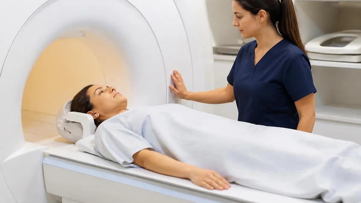
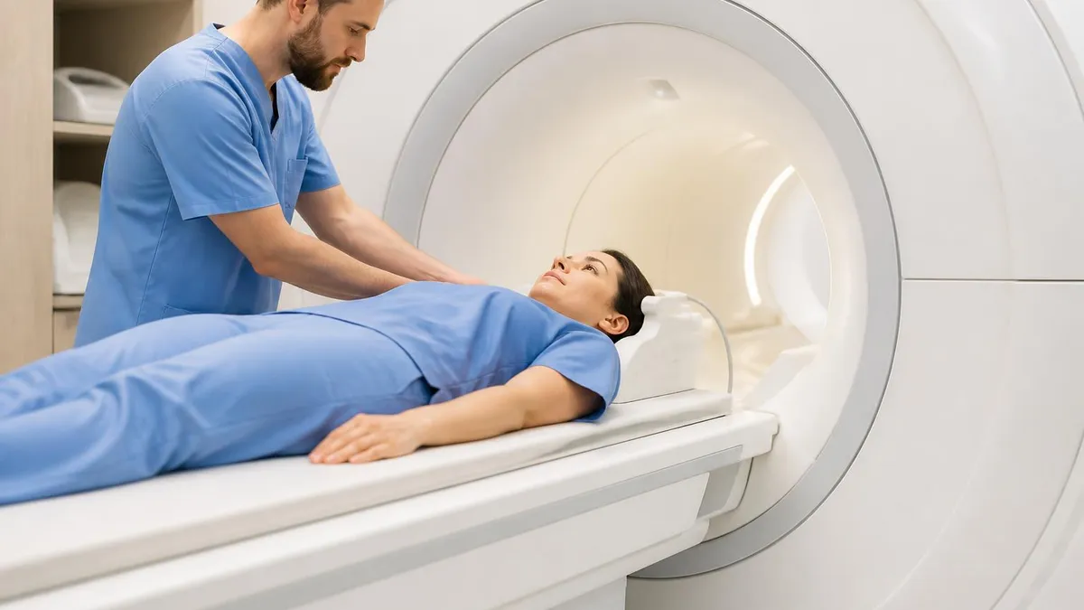
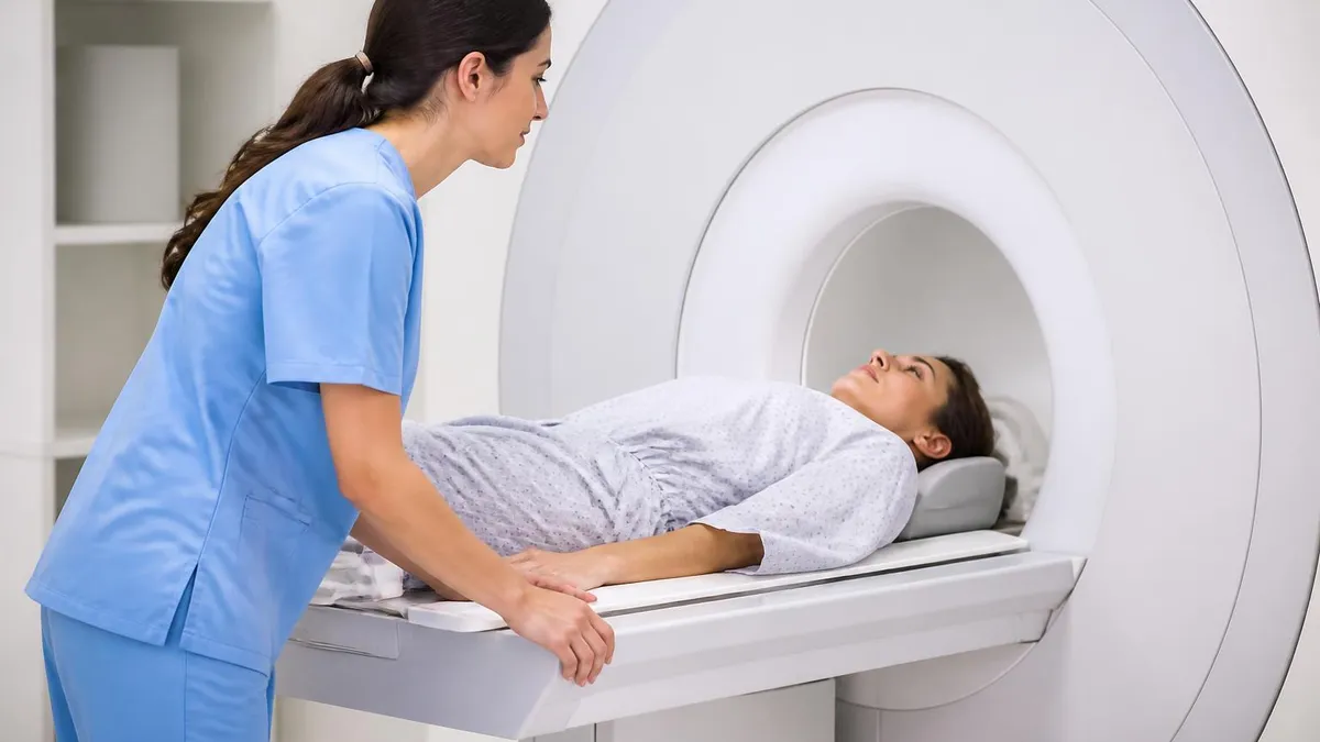
The exam typically takes 30 to 45 minutes and requires the patient to keep the hand still inside a dedicated hand or wrist coil. The coil is a specialized antenna placed around the hand to improve image quality and resolution. Some patients undergo the scan with the arm extended overhead in a "superman" position, while others lie on their back with the hand at their side, depending on scanner type and protocol. For more on contrast use, see our guide on mri with and without contrast cpt.
Hand MRI scans can be performed with or without intravenous gadolinium contrast. Non-contrast studies are common for routine pain evaluation, ligament tears, and fractures. Contrast-enhanced studies are reserved for suspected infections, tumors, inflammatory arthritis, or post-surgical evaluation where vascular enhancement patterns help distinguish active disease from scar tissue. Your radiologist or referring physician will decide which protocol is most appropriate based on the clinical question being asked.
Field strength matters significantly in hand imaging. Most modern hand MRIs are performed on 1.5 Tesla or 3 Tesla scanners, with 3T offering superior resolution for fine cartilage and ligament detail. Some specialized centers use dedicated extremity scanners with smaller bore openings designed specifically for hands and feet, which improve patient comfort while still producing diagnostic-quality images. The choice of scanner often depends on the specific pathology being investigated.
Insurance coverage for hand MRI usually requires prior authorization in the United States. Most insurers want documentation that simpler imaging tests, such as X-rays, have been performed first and that conservative treatments like splinting or physical therapy have been attempted. Out-of-pocket costs range widely depending on whether the scan is done at a hospital, imaging center, or specialized orthopedic clinic, and whether contrast is used during the examination.
This guide walks through everything you need to know about hand MRI, including what conditions it diagnoses, how to prepare, what happens during the scan, what your radiology report means, and how results guide treatment. Whether you are a patient awaiting your first scan or a healthcare professional refreshing your knowledge of indications and protocols, you will find detailed, evidence-based information drawn from current radiology and orthopedic practice guidelines used across the United States.
Hand MRI by the Numbers
Common Indications for Hand MRI
Scaphoid and other carpal bone fractures often hide on X-ray for days. MRI detects bone marrow edema within hours of injury, making it the gold standard when clinical suspicion is high but radiographs are negative.
Tears of the scapholunate ligament, lunotriquetral ligament, and triangular fibrocartilage complex cause chronic wrist pain. MRI arthrography improves detection sensitivity, especially for partial tears and central perforations.
Evaluation of flexor and extensor tendons for tenosynovitis, ruptures, and entrapment conditions like de Quervain tenosynovitis. MRI shows fluid in tendon sheaths and the integrity of the tendon substance itself.
Early rheumatoid arthritis causes synovitis and erosions detectable on MRI long before X-rays show changes. Contrast-enhanced MRI is increasingly used to monitor disease activity and treatment response in inflammatory conditions.
Ganglion cysts, giant cell tumors of tendon sheath, glomus tumors, and rare soft tissue sarcomas all have characteristic MRI appearances. Contrast helps distinguish solid tumors from fluid-filled cysts.
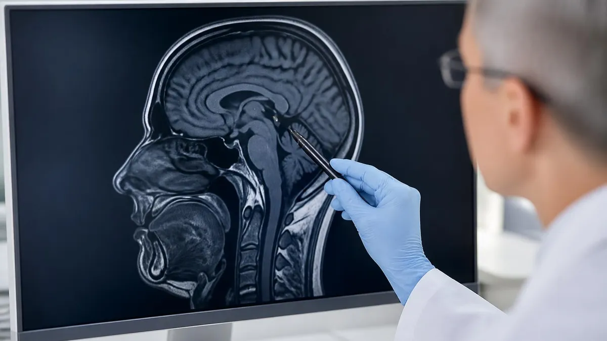
Hand MRI uses powerful magnetic fields and radio waves to generate images, without any ionizing radiation. When you enter the scanner, hydrogen protons in your tissues align with the magnet's field. Brief radiofrequency pulses tip these protons out of alignment, and as they relax back to equilibrium, they emit signals that the receiver coil captures. Computer algorithms then convert these signals into the detailed gray-scale images radiologists interpret. The physics behind this process is identical whether you are scanning a brain, a knee, or a fingertip.
For hand imaging specifically, technologists position the affected hand inside a dedicated wrist or hand coil that fits snugly around the anatomy. Coil design has a major impact on image quality because closer proximity between the antenna and the tissue produces higher signal-to-noise ratio. Newer multi-channel coils with 8, 16, or even 32 individual elements can capture data in parallel, dramatically reducing scan times while maintaining or improving spatial resolution compared to older single-channel designs from a decade ago.
Standard hand MRI protocols include several pulse sequences, each highlighting different tissue properties. T1-weighted images show anatomic detail with bright fat and dark fluid, ideal for evaluating bone marrow and cortical bone. T2-weighted and fluid-sensitive sequences like STIR or fat-saturated T2 make fluid bright, which helps detect edema, inflammation, and tears. Proton density sequences provide a balance between anatomy and pathology detection, while gradient echo sequences are particularly useful for cartilage evaluation in small joints. Curious about the broader history? Read about the history of MRI.
Slice thickness for hand MRI is typically 2 to 3 millimeters, which is much thinner than slices used for larger body parts like the abdomen or pelvis. Thin slices are essential because the structures being evaluated, such as individual finger pulleys or the scapholunate ligament, may only be a few millimeters wide. Image acquisition occurs in three planes, axial, coronal, and sagittal, with each plane providing complementary information about anatomy and any pathology present.
When intravenous gadolinium contrast is administered, it shortens T1 relaxation time in tissues with high vascularity, making them appear bright on post-contrast T1-weighted images. Inflammation, infection, and tumor tissue typically enhance because they have increased blood flow or disrupted capillary membranes. Comparing pre-contrast and post-contrast images allows radiologists to identify subtle abnormalities and differentiate active disease from chronic scar tissue, which generally does not enhance significantly.
MR arthrography is a specialized technique where a radiologist injects a dilute gadolinium solution directly into the wrist joint before the scan. This distends the joint capsule and allows the contrast to flow through any ligament or cartilage tears, dramatically improving sensitivity for intrinsic ligament injuries and TFCC perforations. The procedure adds about 30 minutes to your total appointment time and carries a small risk of infection or allergic reaction, but the diagnostic yield is substantially higher than standard MRI for certain conditions.
Image quality depends on the patient remaining still throughout acquisition. Even small movements can cause motion artifact that obscures findings or requires sequences to be repeated. Technologists position patients carefully, often with foam pads and tape, to minimize involuntary movement. For pediatric patients or those with claustrophobia, sedation or open-bore scanners may be considered, though dedicated extremity scanners often eliminate claustrophobia concerns since only the hand enters the magnet bore.
MRI Practice Test Questions
Prepare for the MRI - Magnetic Resonance Imaging exam with our free practice test modules. Each quiz covers key topics to help you pass on your first try.
MRI Knowledge
MRI Exam Questions covering Knowledge. Master MRI Test concepts for certification prep.
MRI Physics
Free MRI Practice Test featuring Physics. Improve your MRI Exam score with mock test prep.
MRI Anatomy and Pathology
MRI Test Prep for MRI Anatomy and Pathology. Practice MRI Quiz questions and boost your score.
MRI Anatomy and Positioning
MRI Questions and Answers on MRI Anatomy and Positioning. Free MRI practice for exam readiness.
MRI Contrast Agents
Free MRI Quiz on MRI Contrast Agents. MRI Exam prep questions with detailed explanations.
MRI Patient Care and Positioning
MRI Practice Questions for MRI Patient Care and Positioning. Build confidence for your MRI certification exam.
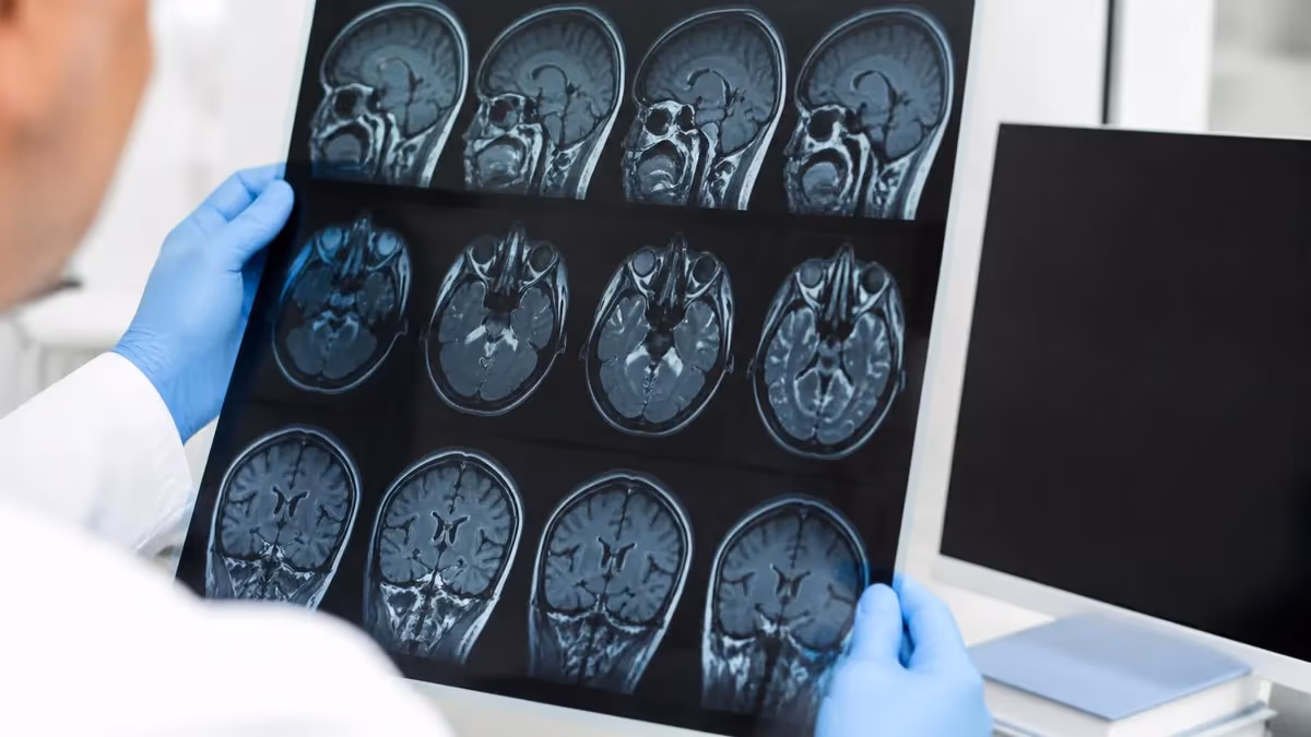
Anatomy Visualized on Hand MRI
Hand MRI clearly visualizes all 27 bones, including the 8 carpal bones, 5 metacarpals, and 14 phalanges. Cortical bone appears dark on most sequences, while marrow has a bright fatty signal on T1-weighted images. Fluid-sensitive sequences reveal marrow edema, which is the earliest sign of stress reactions, occult fractures, osteomyelitis, and avascular necrosis of the lunate or scaphoid.
Joint surfaces show articular cartilage as a thin intermediate-signal band between bones. Erosions, osteophytes, subchondral cysts, and effusions all have characteristic appearances. The radiocarpal, midcarpal, distal radioulnar, carpometacarpal, metacarpophalangeal, and interphalangeal joints can each be evaluated for arthritis, fluid, synovial proliferation, and bony abnormalities in a single comprehensive examination.

Hand MRI vs Other Imaging Tests
- +No ionizing radiation, safe for repeat imaging and pregnant patients with caution
- +Superior soft tissue contrast compared to CT, X-ray, or ultrasound
- +Detects occult fractures and bone marrow edema within hours of injury
- +Visualizes tendons, ligaments, nerves, and cartilage simultaneously
- +Can be performed with or without contrast based on clinical need
- +Multiplanar imaging shows pathology from axial, coronal, and sagittal views
- +Gold standard for TFCC tears, scaphoid fractures, and inflammatory arthritis
- −Higher cost than X-ray, CT, or ultrasound, often $500 to $3,500
- −Requires prior insurance authorization in most US plans
- −Contraindicated for patients with certain pacemakers or metallic implants
- −Claustrophobia can limit tolerance for closed-bore scanners
- −Scan time of 30-45 minutes requires the patient to remain still
- −Limited availability in some rural areas compared to X-ray
- −Image quality degrades with metallic hardware from prior surgery
How to Prepare for Your Hand MRI
- ✓Complete the MRI safety screening form, listing all implants and prior surgeries
- ✓Remove all jewelry, watches, rings, and metal hair accessories before the scan
- ✓Wear comfortable clothing without metal zippers, buttons, or embroidery
- ✓Bring your physician's order and prior imaging studies on disc if available
- ✓Inform the technologist about any tattoos, especially older ones with metallic ink
- ✓Notify staff if you are pregnant, breastfeeding, or have kidney disease
- ✓Arrive 15-30 minutes early to complete intake and IV placement if needed
- ✓Avoid heavy meals just before the scan if contrast may cause nausea
- ✓Take prescribed anti-anxiety medication only as directed by your physician
- ✓Bring a list of current medications and known allergies to gadolinium
- ✓Empty your bladder before the scan since you cannot leave the room
- ✓Ask about hearing protection options since the scanner is very loud
Tell your technologist about any metal in your body
Even small metallic fragments from prior eye injuries, surgical clips, cochlear implants, or older pacemakers can be dangerous in the MRI scanner. Always disclose your complete medical history, including occupational metal exposure such as welding or machining, so staff can determine whether additional X-ray screening is needed before scanning.
Understanding your hand MRI report can feel overwhelming because radiology language is dense and technical. Most reports follow a standardized structure: clinical history, technique, comparison, findings, and impression. The findings section systematically describes each anatomic structure, while the impression summarizes the clinically relevant abnormalities. The impression is usually the most important paragraph for patients to focus on, though discussing the full report with your ordering physician is always recommended.
Common findings on hand MRI include bone marrow edema, which appears as bright signal on fluid-sensitive sequences and dark signal on T1-weighted images. Edema is nonspecific and can result from trauma, stress reactions, inflammation, infection, or transient bone marrow lesion syndromes. The pattern, location, and associated findings help narrow the differential diagnosis. For example, edema in the scaphoid waist after a fall is highly suggestive of an occult fracture even without a visible cortical line.
Ligament injuries are described as partial or complete tears, with grading based on the extent of fiber disruption. A complete scapholunate ligament tear often correlates with widening of the scapholunate interval on radiographs and may lead to dorsal intercalated segment instability if untreated. Partial tears are managed conservatively in many cases, while complete tears in active patients often require surgical repair or reconstruction to prevent long-term carpal collapse and arthritis.
Tendon abnormalities range from mild tenosynovitis, with fluid in the tendon sheath, to complete rupture with retraction. The extensor pollicis longus is particularly vulnerable to rupture after distal radius fractures or in patients with rheumatoid arthritis. Reports often mention the location of the tear, the length of the gap between torn ends, and the presence of associated muscle atrophy, which all influence surgical planning. Compare findings to common patterns in our review of common MRI findings.
Cyst formation is extremely common in the hand and wrist. Dorsal wrist ganglion cysts arise from the scapholunate ligament, while volar ganglions often originate from the radioscaphoid or scaphotrapezial joint. Mucous cysts at the distal interphalangeal joints are associated with osteoarthritis. MRI confirms cyst characteristics, identifies their stalk of origin, and rules out solid tumors that can mimic cysts on physical examination but require completely different management.
Inflammatory arthritis findings include synovitis, bone marrow edema, erosions, and tenosynovitis. Modern MRI scoring systems like RAMRIS (Rheumatoid Arthritis MRI Score) quantify disease activity and damage, helping rheumatologists adjust treatment intensity. The presence of erosions on MRI before they appear on radiographs is one of the strongest predictors of progressive joint damage and influences early aggressive treatment decisions with disease-modifying antirheumatic drugs or biologic therapies.
Incidental findings, such as small ganglion cysts, mild degenerative changes, or asymptomatic accessory muscles, are extremely common and usually do not require treatment. Your radiologist will distinguish between clinically significant abnormalities and incidental findings of no consequence. If you have questions about anything in your report, write them down and bring them to your follow-up appointment so your physician can address each concern in context with your symptoms and examination findings.
Patients with severe kidney disease (eGFR less than 30) face an increased risk of nephrogenic systemic fibrosis from certain gadolinium agents. Always disclose kidney problems, dialysis, prior contrast reactions, and pregnancy status. Modern macrocyclic agents have substantially lower risk profiles than older linear agents.
The cost of a hand MRI in the United States varies enormously depending on geography, facility type, and insurance status. Hospital-based imaging centers typically charge $1,500 to $3,500, while freestanding outpatient imaging centers and orthopedic clinics often charge $400 to $1,200 for the same examination. Adding contrast typically increases the cost by $200 to $500. Patients without insurance can often negotiate substantial discounts by paying cash at the time of service, sometimes reducing costs by 40 to 60 percent.
Insurance coverage requires meeting medical necessity criteria, which usually means documentation of persistent symptoms, failed conservative treatment, and abnormal physical examination findings. Most insurers require prior authorization, which can take 24 to 72 hours to process. Some plans require trying X-ray, physical therapy, or splinting for a defined period before approving advanced imaging. Working closely with your ordering physician's office staff often helps navigate these requirements smoothly. For help finding a local facility, see MRI scan near me.
Medicare typically covers hand MRI when ordered for an established diagnostic indication, with the patient responsible for 20 percent of the Medicare-approved amount after meeting the Part B deductible. Medicare Advantage plans may have different cost-sharing structures, including copayments ranging from $50 to $300 per advanced imaging study. Always verify coverage with your specific plan before scheduling to avoid surprise bills, particularly for facility fees that hospital-based centers charge in addition to professional fees.
Scheduling timelines depend on whether your scan is routine or urgent. Routine outpatient MRIs typically book one to four weeks out at busy centers, though some clinics offer next-day or same-week appointments. Emergency department patients with acute trauma usually receive MRI within hours when clinically indicated. Workers' compensation cases often require specific authorization from the insurer or case manager, which can add days to the scheduling process depending on the state and jurisdiction.
What to bring on the day of your scan includes your insurance card, photo identification, physician's order, list of current medications, and any prior imaging on disc. Wear comfortable clothing without metal, and plan to arrive 15 to 30 minutes early for paperwork. The technologist will explain the procedure, position you in the scanner, and check on you throughout the examination using an intercom. Most centers provide ear plugs or headphones with music to reduce the noise from gradient coils.
After the scan, you can immediately resume normal activities unless you received contrast and were specifically instructed otherwise. Results are typically available within 24 to 48 hours, though urgent or emergency department scans are usually read within an hour. Your ordering physician will discuss the findings and recommend next steps, which might include conservative management, specialist referral to a hand surgeon or rheumatologist, or in some cases additional imaging or procedures based on what was found.
Quality varies among facilities, so it is worth asking about the scanner type, coil specifications, and whether a fellowship-trained musculoskeletal radiologist will interpret your study. ACR (American College of Radiology) accreditation indicates that a facility meets rigorous standards for equipment, personnel, and image quality. If your first MRI is suboptimal due to motion or metal artifact, requesting repeat imaging at a higher-quality center can prevent missed diagnoses and unnecessary repeat tests months later.
Practical preparation for your hand MRI begins the day before your appointment. Get a good night's sleep so you can lie still comfortably during the 30 to 45 minute examination. Eat a light meal a few hours beforehand, especially if contrast will be administered, since some patients experience mild nausea after gadolinium injection. Stay well hydrated, which makes IV placement easier and helps your kidneys clear contrast efficiently after the scan is completed.
If you experience claustrophobia, talk to your physician about options before your appointment. Some patients benefit from a low dose of oral anxiolytic medication taken 30 to 60 minutes before the scan, though this requires a driver since the medication impairs reflexes for several hours. Other strategies include closing your eyes throughout the scan, focusing on deep breathing, or asking about a dedicated extremity scanner where only your hand enters the magnet bore while you sit in a chair.
Positioning matters significantly for image quality. Many hand MRIs are performed in the "superman" position, where you lie prone with the affected arm extended overhead. This position can be uncomfortable for older patients or those with shoulder problems. Newer extremity scanners allow scanning with the hand at your side, which is more comfortable but may not be available at all centers. Ask about positioning options when you schedule, particularly if you have shoulder pain or limited range of motion.
Communicate any symptoms or specific concerns to the technologist and radiologist before the scan begins. Pointing to the exact location of your pain helps the radiologist focus on that area during interpretation. If you can reproduce your symptoms with certain hand positions, mention this to the technologist so they can position you accordingly. Sometimes additional sequences in specific positions reveal pathology that would be missed with standard protocols, particularly for dynamic conditions like impingement or instability.
After the scan, request a copy of your images on disc or via patient portal. Bringing these to your follow-up appointment allows your physician to review the actual images, not just the written report. If you are referred to a specialist, having your own copy avoids delays from records requests. Many practices now provide secure online access to imaging studies, which makes it easy to share results with multiple providers or seek second opinions when complex findings warrant additional expert input.
Follow-up imaging may be recommended depending on initial findings. Bone marrow edema from a stress reaction typically resolves over 6 to 12 weeks with appropriate rest and activity modification. Inflammatory conditions are often monitored with follow-up MRI at 3 to 6 month intervals to assess treatment response. Post-surgical imaging is sometimes performed to evaluate healing or complications. Your physician will explain whether and when follow-up imaging is appropriate based on your specific diagnosis and treatment plan.
Finally, do not panic if your report mentions findings that sound serious. Many MRI findings have low clinical significance, and even concerning-sounding diagnoses often have excellent treatment outcomes when caught early. The whole purpose of getting a hand MRI is to identify what is causing your symptoms so that targeted, evidence-based treatment can be initiated. Trust the process, ask questions, follow up with your physician, and remember that high-quality imaging combined with expert interpretation gives you the best possible foundation for recovery.
MRI Questions and Answers


About the Author

Medical Laboratory Scientist & Clinical Certification Expert
Johns Hopkins UniversityDr. Sandra Kim holds a PhD in Clinical Laboratory Science from Johns Hopkins University and is certified as a Medical Technologist (MT) and Medical Laboratory Scientist (MLS) through ASCP. With 16 years of clinical laboratory experience spanning hematology, microbiology, and molecular diagnostics, she prepares candidates for ASCP board exams, MLT, MLS, and specialist certification tests.
Join the Discussion
Connect with other students preparing for this exam. Share tips, ask questions, and get advice from people who have been there.
View discussion (6 replies)