What Does an EEG Do? Everything You Need to Know About the EEG Test 2026 July
❓ Learn what does an EEG do, how the EEG test works, what it diagnoses, how long it takes, costs, and what to expect before and after.

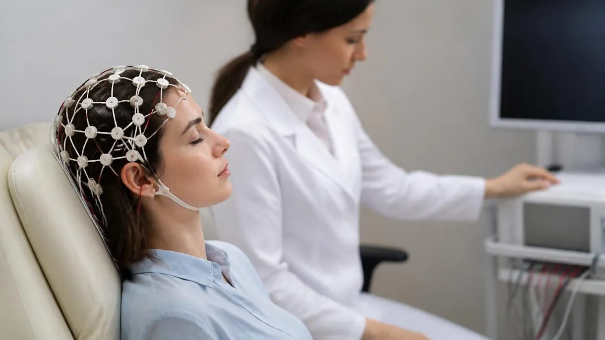
An EEG test — short for electroencephalogram — is one of the most important diagnostic tools in modern neurology, and understanding what does an EEG do can help patients and caregivers feel far more confident walking into a clinic. At its core, an EEG records the continuous electrical activity generated by millions of neurons firing in your brain.
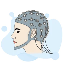
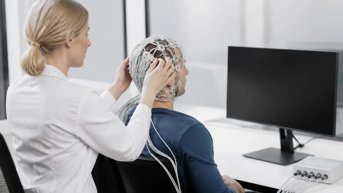
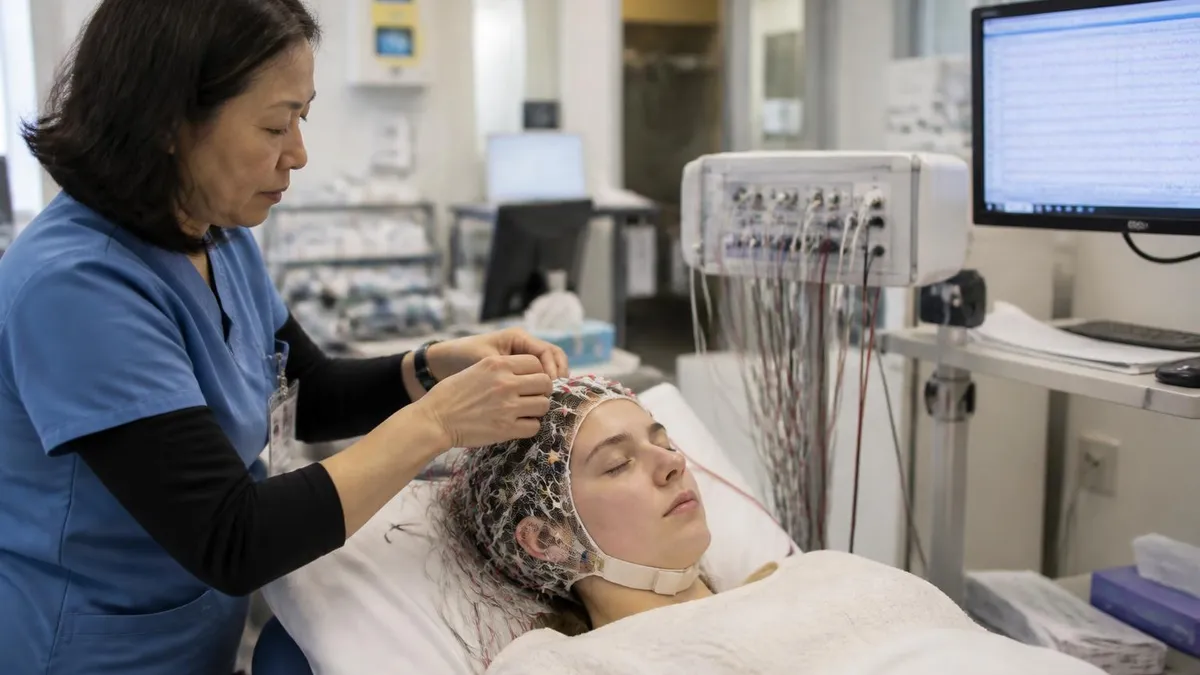
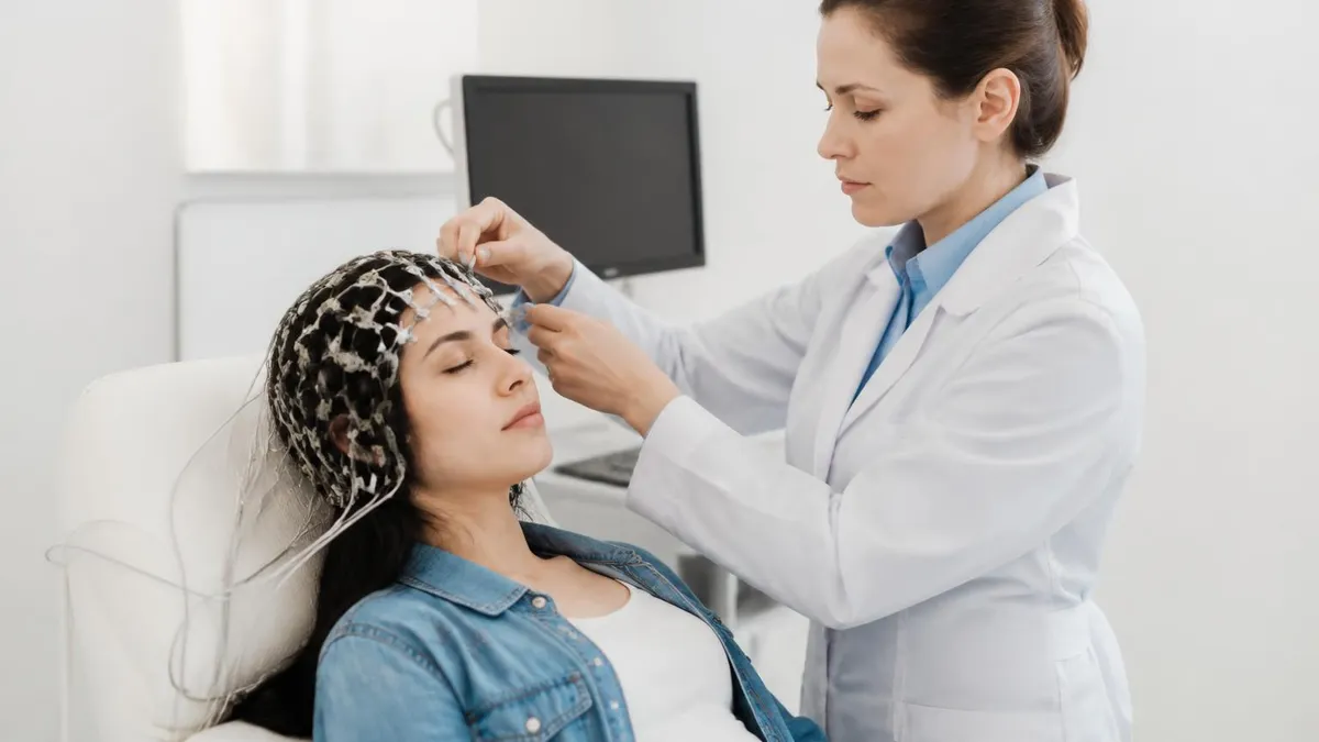
Small metal sensors called electrodes are placed on your scalp according to a standardized map, and they detect these tiny voltage fluctuations in real time, transmitting them to a computer that draws wavy lines representing your brain's activity. If you've ever wondered whether this test is similar to a heart monitor, our guide on what is an eeg test compared to an ECG breaks down those differences clearly.
The history of the EEG stretches back to 1924, when German psychiatrist Hans Berger made the first human recording, identifying what he called the alpha rhythm — the gentle 8–12 Hz oscillation that appears when you close your eyes and relax. Since then, the technology has evolved from cumbersome analog machines into fully digital systems capable of recording hundreds of channels simultaneously, streaming data wirelessly, and even performing automated spike detection using machine-learning algorithms. Despite all these advances, the fundamental principle remains unchanged: neurons communicate electrically, and the EEG is our window into that communication.
Doctors order an EEG medical test for a wide range of clinical questions. Most commonly, a neurologist will request one when a patient has experienced unexplained seizures, recurrent blackouts, or episodes of abnormal movement.
The test is also ordered to evaluate sleep disorders such as narcolepsy, assess the depth of anesthesia or sedation during surgery, monitor brain activity after a traumatic brain injury, and help diagnose certain types of encephalitis or metabolic disorders that affect brain function. Because the EEG captures dynamic, moment-by-moment electrical activity rather than a static image, it provides information that an MRI or CT scan simply cannot.
One of the most reassuring aspects of the EEG test is that it is entirely painless and non-invasive. No needles puncture your scalp, no electricity is sent into your brain, and no radioactive tracers are injected into your bloodstream. The electrodes merely listen.
The paste or gel used to attach them may feel slightly cool or sticky, and the technician will spend about 20 to 40 minutes carefully applying each electrode, but patients consistently report no discomfort beyond mild impatience. Children, elderly patients, and individuals with severe neurological conditions can all safely undergo a standard EEG without concern about the procedure itself causing harm.
Understanding the eeg test cost is also important for patients planning their care. In the United States, a routine outpatient EEG typically ranges from $200 to $900 before insurance, though the final bill can vary widely depending on the facility, geographic region, and whether interpretation fees are billed separately by the neurologist. Hospital-based EEGs, particularly those performed in intensive care units for continuous monitoring, carry significantly higher costs. Most major insurance plans — including Medicare and Medicaid — cover EEGs when there is a documented medical indication such as a seizure disorder or altered consciousness. Always verify your coverage beforehand.
The procedure itself generally lasts between 20 and 60 minutes for a routine study, though specialized tests can run much longer. A standard outpatient EEG in an awake, cooperative patient takes about 30 to 45 minutes from electrode placement to completion of the recording. Sleep-deprived EEGs, ambulatory studies, and video-EEG monitoring sessions are considerably longer — sometimes lasting 24 to 72 hours. Knowing how long is an EEG test from start to finish helps you plan your day: budget at least 90 minutes for a routine study once you factor in check-in, prep, recording, and electrode removal.
After the recording is complete, a board-certified clinical neurophysiologist or neurologist reviews the tracings, looking for normal patterns as well as any abnormalities that might suggest a diagnosis. The written report is typically available within one to five business days and is sent directly to the ordering physician, who then discusses the findings with you. An abnormal EEG finding does not automatically mean you have epilepsy — interpretation always happens in the context of your full clinical picture — but it gives your neurologist critical data to guide treatment decisions and recommend next steps.
EEG Test by the Numbers

How the EEG Test Works: Step by Step
Electrode Placement
Baseline Recording
Activation Procedures
Sleep Recording (If Needed)
Electrode Removal & Cleanup
Neurologist Interpretation
The diagnostic power of an EEG medical test lies in its ability to capture electrical signatures that are unique to specific neurological disorders. Epilepsy is by far the most common indication: interictally (between seizures), many patients with epilepsy show characteristic sharp waves, spikes, or spike-and-wave complexes that point directly to the type and location of the epileptic focus.
A temporal lobe epilepsy patient, for example, may show right temporal sharp waves during a routine 30-minute recording even if they have not seized in weeks. This information directly guides medication choices and surgical planning. For a deeper understanding of all the conditions this test can identify, read our detailed resource on what is a eeg test designed to measure.
Beyond epilepsy, the EEG is a critical tool in diagnosing and monitoring encephalopathies — diffuse brain dysfunctions caused by metabolic disturbances, organ failure, toxic exposures, or infections. In hepatic encephalopathy, for instance, the EEG characteristically shows triphasic waves, a distinctive slow pattern that correlates with the degree of liver dysfunction. In herpes simplex encephalitis, periodic lateralized epileptiform discharges (PLEDs) over one temporal lobe are a red flag that prompts immediate antiviral treatment even before MRI changes appear. These EEG signatures are not just academically interesting — they drive life-saving clinical decisions in real time.
Sleep medicine is another domain where the EEG is indispensable. In a full polysomnogram (sleep study), the EEG is one of several channels recorded simultaneously. It allows technologists to score sleep stages: stage N1 (drowsiness with theta waves), stage N2 (sleep spindles and K-complexes), stage N3 (slow delta waves — deep sleep), and REM sleep (low-voltage fast activity resembling wakefulness). Accurate sleep staging is essential for diagnosing narcolepsy, identifying REM sleep behavior disorder, and quantifying the sleep disruption caused by sleep apnea events. Without the EEG channel, none of this staging is possible.
In neurocritical care units, continuous EEG (cEEG) monitoring has become standard practice for patients with severe brain injuries, status epilepticus, or post-cardiac arrest encephalopathy. Studies have consistently shown that 20–40% of comatose patients have nonconvulsive seizures detectable only by continuous EEG monitoring — seizures that cause ongoing brain damage but produce no visible motor activity. Identifying and treating these silent seizures can dramatically improve long-term neurological outcomes. The EEG literally becomes a life-saving monitor in these settings, analogous to continuous cardiac telemetry for arrhythmias.
Pediatric neurologists rely heavily on the EEG to classify childhood epilepsy syndromes, which is critical because the optimal medication differs dramatically between syndromes. A child with childhood absence epilepsy shows a distinctive 3 Hz spike-and-wave pattern on EEG, whereas a child with Lennox-Gastaut syndrome shows slow spike-and-wave at 1.5–2.5 Hz and a characteristic slow background. These patterns help the neurologist choose between ethosuximide, valproate, lamotrigine, or other agents — and avoid medications that might paradoxically worsen certain syndromes. Getting the EEG diagnosis right in childhood can prevent years of ineffective treatment.
Intraoperative neurophysiological monitoring is yet another application where EEG plays a starring role. During certain brain surgeries — particularly for epilepsy (electrocorticography) or during carotid endarterectomy — the surgical team monitors the EEG in real time to detect ischemia or excessive epileptiform activity that would alert the surgeon to change technique. Some epilepsy centers use electrocorticography (ECoG), which places electrodes directly on the exposed cortex during surgery, to precisely map the seizure-onset zone before resection. This intraoperative EEG guidance significantly improves seizure-free outcomes after surgery.
The what is eeg test used to diagnose question has a surprisingly broad answer: from pediatric febrile seizures to adult-onset dementia. Certain dementias leave distinctive EEG fingerprints. Creutzfeldt-Jakob disease (CJD), a rare but devastating prion disease, produces pathognomonic periodic sharp-wave complexes at roughly 1 Hz intervals that are so characteristic they are included in the diagnostic criteria.
Similarly, subacute sclerosing panencephalitis (SSPE), a rare complication of measles, shows high-amplitude periodic complexes at 4–14 second intervals. Recognizing these patterns requires training — which is why platforms that help technologists and students practice EEG interpretation, including resources on what is eeg test used to diagnose, are so valuable for building clinical competence.
EEG Test Types: Routine, Ambulatory, and Continuous Monitoring
A routine outpatient EEG typically lasts 20–40 minutes of actual recording time. The technologist captures resting activity, hyperventilation, and photic stimulation. This is the most common type ordered for initial seizure evaluation, and it captures abnormalities in roughly 50% of epilepsy patients on the first attempt. The eeg test price for a routine study at an outpatient neurology clinic generally runs $200–$500 before insurance adjustments, making it one of the more affordable neurological investigations available.
Because a single routine EEG only captures a brief snapshot of brain activity, its sensitivity increases substantially when repeated. Neurologists often order two or three serial EEGs — particularly sleep-deprived studies — before concluding the test is normal. A sleep-deprived EEG, in which the patient stays awake all night prior to the appointment, significantly increases the likelihood of capturing abnormal sleep-activated discharges and can raise epilepsy detection rates to 80–90% after multiple studies in confirmed epilepsy patients.

EEG Test: Benefits and Limitations
- +Non-invasive and completely painless — no needles, no radiation, no contrast agents required
- +Excellent temporal resolution, capturing millisecond-by-millisecond changes in brain activity
- +Widely available at hospitals, neurology clinics, and even mobile diagnostic units across the US
- +Relatively affordable compared to MRI or PET scans, with routine studies often under $500
- +Can detect abnormalities invisible on structural imaging such as MRI and CT scans
- +Safe for all ages including newborns, infants, pregnant women, and elderly patients
- −Poor spatial resolution — cannot pinpoint exactly which millimeter of brain generates a signal
- −A single normal EEG does not rule out epilepsy, as many patients show findings only during or near seizures
- −Electrode application takes 20–40 minutes and gel can be messy and uncomfortable to wash out
- −Patient cooperation is required — excessive movement, crying, or anxiety creates artifacts that obscure interpretation
- −Interpretation requires specialized training; misreading normal variants as abnormal is a known pitfall
- −Ambulatory recordings are susceptible to electrode artifact, reducing data quality during active movement
How to Prepare for Your EEG Test
- ✓Wash your hair the night before with regular shampoo — do not apply conditioner, oils, sprays, or dry shampoo on the day of testing.
- ✓Ask your doctor whether to continue or temporarily hold any seizure medications before the study.
- ✓Get a full night of sleep unless your neurologist specifically ordered a sleep-deprived EEG.
- ✓Eat a normal meal before your appointment — low blood sugar can alter brain activity and create artifacts.
- ✓Avoid caffeine for at least 8 hours before the test to prevent artificially elevated alertness levels.
- ✓Arrive 10–15 minutes early and wear comfortable, loose-fitting clothing that is easy to change if needed.
- ✓Inform the technologist of all medications, supplements, and any recent illnesses before electrode placement begins.
- ✓Remove all hair clips, barrettes, braids, and extensions beforehand — metal accessories interfere with electrode contact.
- ✓Plan to be at the facility for 90 minutes total to allow for check-in, prep, recording, and electrode removal.
- ✓Arrange for a driver if sedation or sleep deprivation is part of your specific EEG protocol.
A Normal EEG Does NOT Rule Out Epilepsy
Up to 50% of patients with confirmed epilepsy have a normal first routine EEG. Epileptiform discharges are highly intermittent and may not occur during a 30-minute recording window. Serial EEGs, sleep-deprived studies, and prolonged ambulatory monitoring significantly increase detection rates and should be pursued before epilepsy is excluded based on a single normal result.
Understanding how EEG results are reported helps patients have more productive conversations with their neurologists. A standard EEG report describes several key elements: the background activity (is the dominant rhythm appropriate for the patient's age and level of alertness?), symmetry (are both hemispheres producing comparable activity?), the response to activation procedures (did hyperventilation or photic stimulation trigger any abnormal patterns?), any focal or generalized abnormalities identified, and an overall clinical impression. The impression is typically classified as normal, borderline, abnormal with a specific description, or technically limited due to artifact.
The most clinically significant EEG abnormalities fall into a few major categories. Epileptiform discharges — spikes, sharp waves, and spike-and-slow-wave complexes — are the hallmark of seizure disorders. A spike is defined as a sharp transient with a duration of 20–70 milliseconds, while a sharp wave lasts 70–200 milliseconds. Both stand out from the background activity with a pointed morphology and are followed by a slow wave in the classic pattern. The distribution of these discharges — whether focal or generalized, symmetric or asymmetric — helps classify the epilepsy type and guides treatment selection.
Focal slowing is another important finding. When one region of the brain generates slower-than-expected waves (delta or theta) compared to the rest of the recording, this indicates focal dysfunction that can result from stroke, tumor, traumatic injury, or cortical dysplasia. Continuous focal delta slowing suggests a structural lesion, while intermittent rhythmic delta activity (IRDA) in the temporal or frontal regions can be associated with focal epilepsy or deep structural dysfunction in those areas. The EEG alone cannot determine the exact cause — it tells the neurologist where to look next with structural imaging.
Generalized slowing — when the entire recording shows slower frequencies than expected for the patient's age and state of consciousness — points to diffuse cerebral dysfunction. This pattern is seen in metabolic encephalopathies (uremia, hepatic failure, hypoglycemia), toxic states (drug intoxication), infectious encephalitis, and postictal states after generalized seizures. Tracking the degree of generalized slowing over serial EEGs is a useful way to monitor whether a patient's encephalopathy is improving or worsening in response to treatment, even before clinical changes are apparent at the bedside.
Some EEG patterns are so characteristic that they are essentially pathognomonic of specific disorders. Burst suppression — alternating periods of high-amplitude activity and electrical silence — indicates severe cerebral dysfunction or deep anesthesia. Electrocerebral inactivity (a flat line at maximum sensitivity) is one of the criteria used to support the determination of brain death. Periodic lateralized epileptiform discharges (PLEDs or LPDs in newer nomenclature) are strongly associated with acute focal brain injury and carry an elevated risk of progression to overt seizures. Each of these patterns carries immediate clinical implications that directly guide management.
Normal variants represent another important area of EEG literacy. Many benign patterns can mimic epileptiform activity and lead to misdiagnosis if the interpreter lacks sufficient experience. The 14-and-6-Hz positive spike burst, seen in adolescents during drowsiness, is entirely benign but has an appearance that can alarm the uninitiated. Similarly, small sharp spikes (SSS), wicket spikes, and SREDA (subclinical rhythmic EEG discharges of adults) are all normal variants that must not be misclassified as epileptiform. Rigorous training in normal EEG variants is essential for anyone reading electroencephalograms in a clinical setting.
Quantitative EEG (qEEG) and EEG-based brain mapping have emerged as advanced analytical tools that go beyond traditional visual inspection. These techniques use mathematical algorithms to extract frequency-domain information, compute asymmetry indices, and generate topographic maps of brain activity. qEEG has applications in traumatic brain injury assessment, anesthesia depth monitoring, and research into psychiatric conditions including depression and ADHD. While qEEG does not replace conventional EEG interpretation, it adds a layer of quantitative precision that is particularly valuable for monitoring trends over time or detecting subtle changes not obvious to the naked eye on standard tracings.

Hyperventilation and photic stimulation — standard EEG activation procedures — are designed to provoke abnormal brain activity and can occasionally trigger a clinical seizure in susceptible patients. Always inform the technologist if you have had recent unprovoked seizures, a known diagnosis of generalized epilepsy, or any history of seizures triggered by flashing lights. The technologist will modify the protocol accordingly and is trained to manage a seizure safely if one occurs during the study.
EEG test side effects are minimal in the vast majority of patients, which is one reason the test has remained a cornerstone of neurological evaluation for over a century. The most common complaint is scalp discomfort from electrode paste — some patients experience mild itching that resolves promptly once the gel is removed after the study.
Rarely, patients with very sensitive skin develop a mild localized rash at electrode sites, which typically resolves within a day or two with no treatment. These minor reactions are not true side effects in the pharmacological sense but rather reactions to the adhesive properties of the conductive media used.
More substantive temporary effects are associated with the activation procedures. Hyperventilation — breathing rapidly for 3 to 5 minutes — causes a predictable decrease in arterial carbon dioxide (CO₂), which triggers cerebral vasoconstriction. Many patients experience lightheadedness, tingling in the hands and feet, or a feeling of mild confusion during hyperventilation. These sensations are completely normal and resolve within 30 to 60 seconds of returning to normal breathing. Patients with sickle cell disease or other vascular conditions are an exception — hyperventilation is usually omitted in these patients because cerebral vasoconstriction could provoke a vaso-occlusive crisis.
Photic stimulation — strobe light exposure — is safe for the vast majority of patients but carries a small risk of triggering a photoparoxysmal response or, in rare cases, a clinical seizure in photosensitive individuals. Approximately 5% of epilepsy patients have photosensitivity demonstrable on EEG. In the broader population, photosensitive epilepsy affects roughly 1 in 4,000 people.
The strobe is presented in a controlled manner with increasing and decreasing frequencies, and the technologist monitors the EEG in real time, stopping immediately if a photoparoxysmal response begins to generalize. The risk of a prolonged or self-sustaining seizure from photic stimulation in a supervised EEG lab is extremely low.
Patients who receive a sedative to facilitate sleep recording — typically chloral hydrate in children or a short-acting benzodiazepine in adults — will experience the expected effects of that medication, including drowsiness that can persist for several hours post-study. These patients must not drive until they have fully recovered, and a responsible adult should accompany them. Sedation is used judiciously and only when a natural sleep recording cannot be obtained through sleep deprivation alone. Most healthy adults can achieve an adequate drowsy or light sleep segment through sleep deprivation without any medication.
There are no known long-term side effects from EEG. The electrodes do not emit any energy into the brain — they are purely passive sensors. No ionizing radiation is involved, unlike X-rays or CT scans. No contrast agents, dyes, or radiotracers are injected. The procedure does not alter brain chemistry or neural circuits in any measurable way. Patients can have EEGs performed repeatedly — weekly in some clinical scenarios — without any cumulative risk. This safety profile makes the EEG uniquely valuable for long-term monitoring applications where repeated studies are necessary to track disease progression or treatment response.
Special populations deserve a brief mention. Pregnant women can safely undergo EEG at any stage of pregnancy — the procedure poses no risk to the fetus. Neonates and premature infants in the NICU routinely undergo EEG to detect subclinical seizures that are extremely common in the neonatal period, particularly after hypoxic-ischemic encephalopathy.
Patients with implanted devices such as pacemakers, cochlear implants, or deep brain stimulators can generally have EEGs safely, though the implant may generate electrical artifact that requires interpretation expertise to distinguish from cerebral signals. When in doubt, the ordering neurologist should communicate with the device manufacturer about any specific precautions.
If you want to prepare thoroughly for your EEG test and understand what the technologist is doing at each step, reviewing a what is eeg test resource that walks you through sample questions and tracings is an excellent strategy. Familiarizing yourself with the terminology — alpha rhythm, sleep spindles, spike-and-wave — will help you understand your neurologist's explanation of your results and ask more informed questions about your diagnosis and treatment plan. Patient education and clinical expertise together lead to the best outcomes.
For patients receiving their first EEG referral, several practical tips can make the experience smoother and the results more meaningful. First and foremost, follow the preparation instructions provided by your neurology clinic precisely. Hair cleanliness is not a vanity concern — electrode impedance (the resistance to electrical signal transmission between electrode and scalp) is directly affected by scalp oil, hair products, and debris. High impedance translates to noisy recordings full of artifact, which can obscure subtle abnormalities or require the study to be repeated. A freshly washed, product-free scalp gives the technologist the best possible starting conditions.
Medication management before an EEG is a nuanced topic that must be individualized by your neurologist. Antiseizure medications (ASMs) generally do not prevent an EEG from showing epileptiform activity, so there is usually no need to withhold them for diagnostic purposes. However, some neurologists prefer a medication-free or reduced-medication recording when trying to define the epilepsy syndrome in a new patient.
Benzodiazepines, barbiturates, and other sedating medications can produce drug-induced EEG changes that complicate interpretation, so accurate medication reporting is essential. Never stop or reduce antiseizure medications on your own before an EEG — consult your neurologist first, as abrupt withdrawal can precipitate breakthrough seizures.
Communication with your EEG technologist during the recording improves data quality. If you feel an aura, an unusual sensation, or any symptom during the recording, tell the technologist immediately — they will mark the recording at that time point for the interpreting neurologist to review with particular attention.
These event markers, called push-button events or clinical event markers, are invaluable for correlating your subjective experience with the objective EEG signal. A recording that captures a typical episode and shows what the EEG looks like during that episode provides far more diagnostic information than a routine recording that captures nothing clinically relevant.
After your EEG, the wait for results can be anxious. Most neurology practices have the report ready within three to five business days, though urgent inpatient studies are interpreted on the same day or within hours. If you have not heard back within a week, it is entirely appropriate to contact your neurologist's office.
When you receive your results, ask your neurologist to explain the report in plain language — specifically, whether the background activity was normal for your age, whether any epileptiform abnormalities were seen, and what clinical significance those findings have in the context of your symptoms and other test results. A single EEG finding is rarely the sole basis for a major diagnosis; it is one piece of a larger clinical puzzle.
For those preparing for EEG technology certification examinations — including the R.EEG T. (Registered EEG Technologist) credential — understanding not just the clinical applications of EEG but also the technical principles underlying the recording is essential. This includes knowledge of electrode placement systems, amplifier specifications, montage selection, artifact identification, normal developmental EEG patterns across the lifespan, and the pharmacological effects of commonly encountered medications on the EEG. These topics appear consistently on certification examinations and in clinical practice alike, which is why structured practice with realistic question banks significantly improves both examination performance and real-world competence.
The field of EEG technology continues to evolve rapidly. High-density EEG systems with 256 or more electrodes, combined with source localization software, now allow neurophysiologists to estimate the intracranial generators of scalp-recorded signals with much greater precision than traditional 21-electrode systems. Dry electrode technology — which requires no gel — is being developed for ambulatory and wearable applications, potentially enabling consumer-grade EEG for sleep tracking and neurofeedback.
Artificial intelligence systems trained on millions of EEG epochs are now showing accuracy comparable to experienced neurophysiologists for detecting specific abnormalities like seizure discharges and sleep stages. These advances promise to make EEG faster, more accessible, and more informative in the years ahead.
Whether you are a patient preparing for your first EEG test, a student studying for the R.EEG T. certification, a nurse caring for patients with epilepsy, or a medical professional seeking a refresher on this foundational neurodiagnostic tool, the key takeaway is the same: the EEG is a safe, versatile, and information-rich test that remains irreplaceable in the evaluation and management of neurological disease.
Its century-long track record of clinical utility, combined with ongoing technological innovation, ensures that the EEG will remain a pillar of neurological practice well into the future. Investing time in understanding EEG — its principles, its patterns, and its limitations — is an investment that pays dividends in patient care.
EEG Questions and Answers
EEG vs ECG vs EKG: Brain and Heart Test Differences
EEG Electroencephalography Practice Test PDF (Free Printable 2026)
What Does an EEG Measure? Brain Waves, Electrical Activity, and What Your Results Reveal
How to Read an EEG Test: A Complete Guide to Brain Wave Patterns and Results
EEG Waves Explained: Delta, Theta, Alpha, Beta, and Gamma Brain Rhythms
About the Author

Educational Psychologist & Academic Test Preparation Expert
Columbia University Teachers CollegeDr. Lisa Patel holds a Doctorate in Education from Columbia University Teachers College and has spent 17 years researching standardized test design and academic assessment. She has developed preparation programs for SAT, ACT, GRE, LSAT, UCAT, and numerous professional licensing exams, helping students of all backgrounds achieve their target scores.