EEG Leads: What They Are, How They Work, and What to Expect During Your EEG Test
Learn what EEG leads are, how they're placed, and what to expect during an EEG test. 📗 Covers the 10-20 system, electrode types, and test preparation.

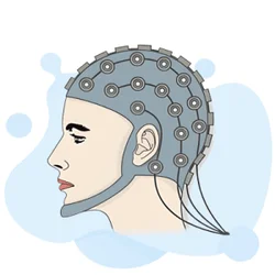
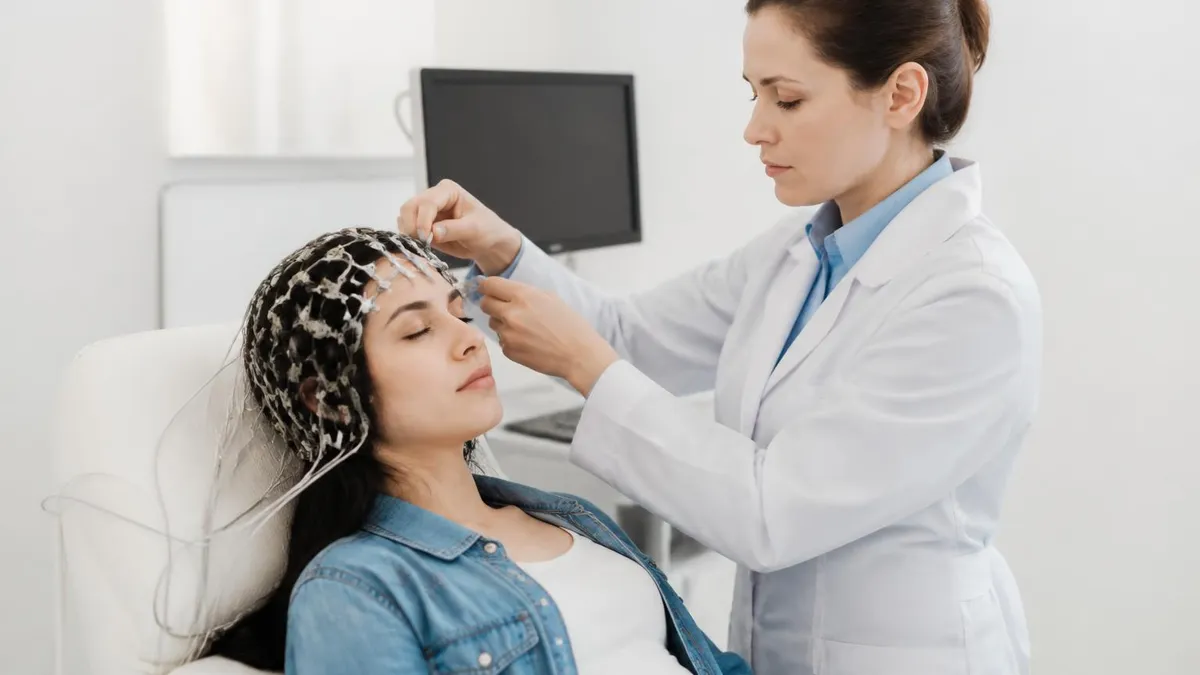
An EEG test — short for electroencephalogram — is one of the most important neurological diagnostic tools available today, and its effectiveness depends almost entirely on something most patients never think about: the leads. EEG leads are the small electrodes and their attached wires that rest on your scalp during the recording. They detect the tiny electrical signals your brain cells generate constantly, translating neural activity into the wavy lines that neurologists read to diagnose epilepsy, sleep disorders, brain injuries, and dozens of other conditions. Understanding eeg leads helps you walk into the testing room with confidence rather than anxiety.
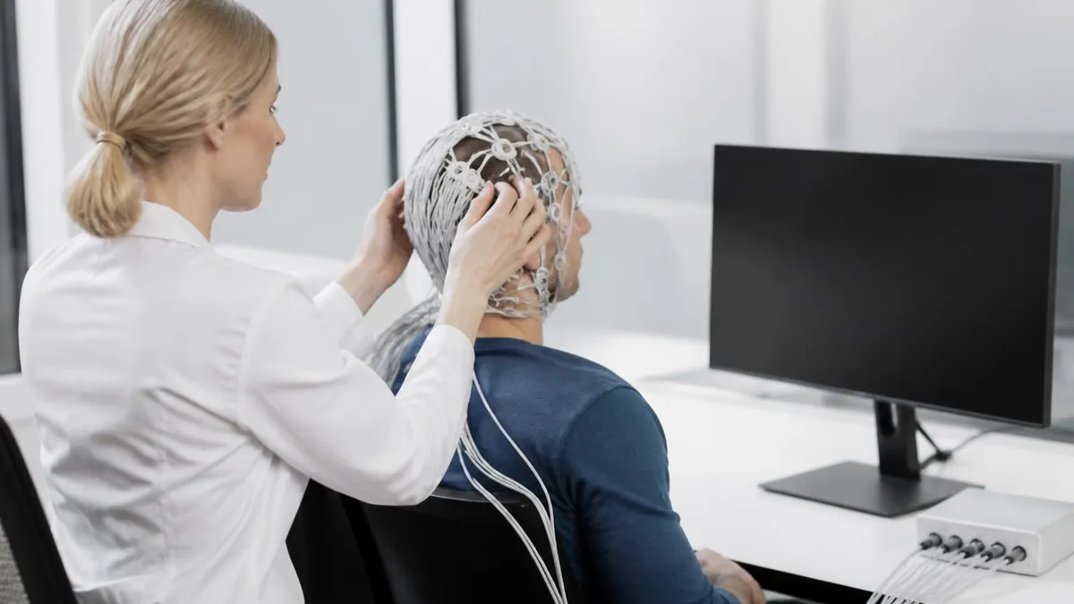
The phrase "EEG test" encompasses the entire procedure — preparation, electrode application, the recording session itself, and the subsequent interpretation by a trained neurologist or EEG technologist. When patients ask "what is an EEG test," they often picture a frightening mesh of wires, but the reality is far more routine. The typical outpatient EEG takes between 20 and 60 minutes, causes no pain, and carries no meaningful side effects for the vast majority of patients. Knowing what the leads actually do demystifies the whole experience.
EEG leads function as passive sensors. They do not send electricity into your brain — they only listen. Each lead picks up voltage differences at its specific scalp location and reports those differences back to an amplifier. Because brain activity is measured in microvolts, the amplifiers must be extraordinarily sensitive, which is why good electrode contact and minimal movement are so critical during the recording. A loose lead creates artifact noise that can obscure genuine brain signals and force a repeat session.
In a standard clinical EEG, between 19 and 25 electrodes are applied following a standardized placement scheme called the International 10-20 System. This system divides the skull into measured intervals — 10% and 20% of the total front-to-back or left-to-right skull distance — so that electrode positions are reproducible across patients, labs, and countries. This standardization is what allows neurologists in Boston to compare results with colleagues in Berlin, using the same lead labels and the same spatial reference points.
For patients preparing for an EEG medical test, the most important practical facts are these: wash your hair the night before or morning of the test, do not apply conditioner, gel, or hairspray, and follow any sleep-deprivation instructions from your ordering physician. Sleep-deprived EEGs are frequently ordered for seizure workups because drowsiness and light sleep lower the threshold for epileptiform discharges, making abnormalities easier to detect. Arriving rested when you were told to stay awake can invalidate the entire test.
The cost and accessibility of EEG testing varies widely across the United States. EEG test cost typically ranges from $200 to $700 for a routine outpatient study at a hospital outpatient department, though the final bill after insurance can be dramatically different. Ambulatory EEGs — worn at home for 24 to 72 hours — cost more upfront but often capture events that a brief in-lab recording misses entirely. We will cover cost breakdowns, lead types, preparation, and interpretation in the sections that follow.
Whether you are a patient preparing for your first study, a nursing student reviewing neurodiagnostics, or an EEG technologist candidate studying for certification, this guide gives you a thorough grounding in how EEG leads work and why proper electrode technique is the foundation of every reliable EEG recording. The sections below move from anatomy to application to interpretation, giving you a complete picture of this essential neurological test.
EEG Test by the Numbers

The International 10-20 Electrode System
Placed over the frontal lobes, frontal leads (Fp1, Fp2, F3, F4, F7, F8, Fz) capture activity linked to attention, executive function, and eye movement artifact. They are critical for detecting frontal lobe epilepsy and frontal intermittent rhythmic delta activity (FIRDA).
Temporal electrodes (T3/T7, T4/T8, T5/P7, T6/P8) sit over the temporal lobes and are among the most clinically important leads. Temporal lobe epilepsy is the most common focal epilepsy syndrome, and sharp waves in this region are a hallmark finding.
Central electrodes (C3, C4, Cz) overlie the sensorimotor cortex. They are especially important in sleep studies because vertex sharp waves and sleep spindles, key markers of N2 sleep, appear prominently at Cz during polysomnography and routine overnight recordings.
Parietal (P3, P4, Pz) and occipital (O1, O2, Oz) leads capture posterior dominant rhythm — the alpha wave that should appear at 8–13 Hz in a relaxed, eyes-closed adult. Slowing or asymmetry of this rhythm is one of the most reliable markers of posterior cortical dysfunction.
Linked earlobe or mastoid reference electrodes provide the electrical baseline against which all scalp leads are compared. Choosing the right reference is a critical technical decision because a poorly placed or noisy reference electrode can introduce artifact into every single channel of the recording simultaneously.
Understanding how EEG leads actually capture brain signals requires a brief detour into neurophysiology. Neurons communicate through electrochemical gradients — ions flowing in and out of cell membranes generate tiny electrical currents. When thousands of cortical neurons fire in synchrony, their summed currents are large enough to propagate through the brain tissue, cerebrospinal fluid, skull, and scalp, where EEG electrodes can detect them. The signals are extraordinarily small — typically 10 to 100 microvolts — requiring amplification on the order of one million times before they can be displayed or stored.
Each EEG channel records the voltage difference between two electrode sites. In a referential montage, every scalp electrode is compared against a common reference — often linked earlobes, the mastoid process, or a computed average of all electrodes. In a bipolar montage, adjacent electrodes are compared in chains running front-to-back or left-to-right across the scalp. Neurologists routinely switch between montages when reviewing a recording because different montages highlight different aspects of the same underlying electrical activity. A sharp wave that appears maximal at T4 in one montage might appear to spread when viewed in another.
Electrode impedance is one of the most critical technical variables in EEG recording. Impedance — the opposition to alternating current — must be kept below 5,000 ohms (5 kΩ) at every lead to ensure a clean signal.
Technologists check impedance values at the start of every study and will abrade the scalp gently with a prep gel, apply conductive paste or gel, and sometimes use a blunt-tipped prep stick to remove skin oils and dead cells that drive impedance up. High-impedance electrodes act like poor antennas — they pick up more environmental electrical noise (60 Hz interference from power lines, for instance) than genuine brain signal.
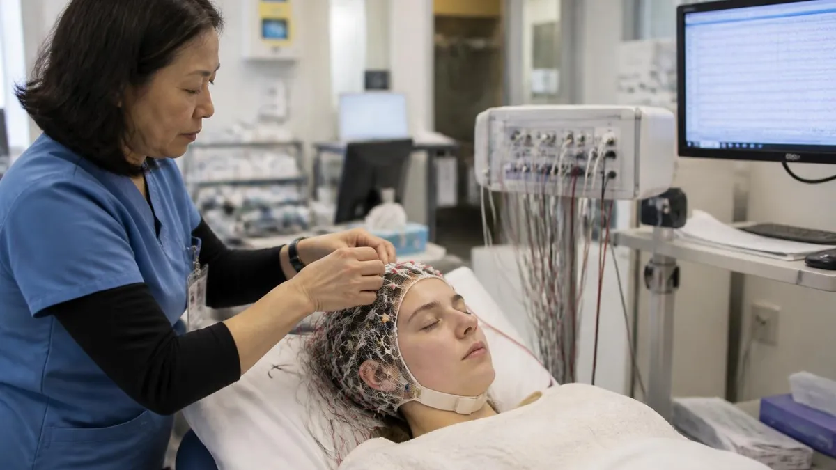
The type of electrode used also affects signal quality. Disposable, pre-gelled Ag/AgCl (silver-silver chloride) cup electrodes are the standard for routine clinical EEGs. Their electrochemical properties produce a stable half-cell potential that minimizes slow baseline drift. For long-term monitoring such as ICU EEG or ambulatory studies, subdermal needle electrodes or collodion-affixed disk electrodes may be used because they stay securely in place for 24 to 72 hours without falling off. Gel electrodes are preferred for short studies; adhesive methods suit extended recordings.
Digital EEG systems sample each channel at rates between 256 and 1,024 Hz. Higher sampling rates are essential when recording high-frequency oscillations (HFOs) — brief bursts above 80 Hz that are emerging biomarkers for epileptic tissue in surgical candidates. Standard clinical recording uses 256 Hz sampling, which is more than adequate for the 0.5–70 Hz frequency range of conventional clinical EEG. The recorded data is stored digitally and can be reviewed with adjustable filters, gain settings, and display speeds — a major advantage over the paper records of previous decades.
Artifact recognition is an indispensable skill for anyone reading or reporting an EEG test. Muscle artifact — high-frequency, irregular bursts generated by scalp and jaw muscles — is the most common contaminant and can mimic epileptiform activity in inexperienced hands. Eye movement artifact creates characteristic slow waves at the frontal leads (Fp1, Fp2) every time the patient blinks or looks around.
Cardiac artifact appears as a sharp spike repeating at the heart rate — easily confirmed by correlating with an EKG channel, which most modern EEG systems include. Proper electrode placement combined with patient coaching to relax and minimize movement reduces artifact to a manageable level.
For EEG technologists preparing for the ABRET R.EEG.T. examination, deep familiarity with electrode placement, impedance management, montage design, and artifact identification is non-negotiable. These technical fundamentals account for a substantial portion of the certification exam and underpin every interpretive decision the reading neurologist makes. A technically flawed recording with high-impedance leads and excessive artifact may be uninterpretable — wasting the patient's time and the ordering physician's clinical question — while a clean, well-montaged study provides diagnostically actionable information within minutes of the recording starting.
Types of EEG Medical Tests Explained
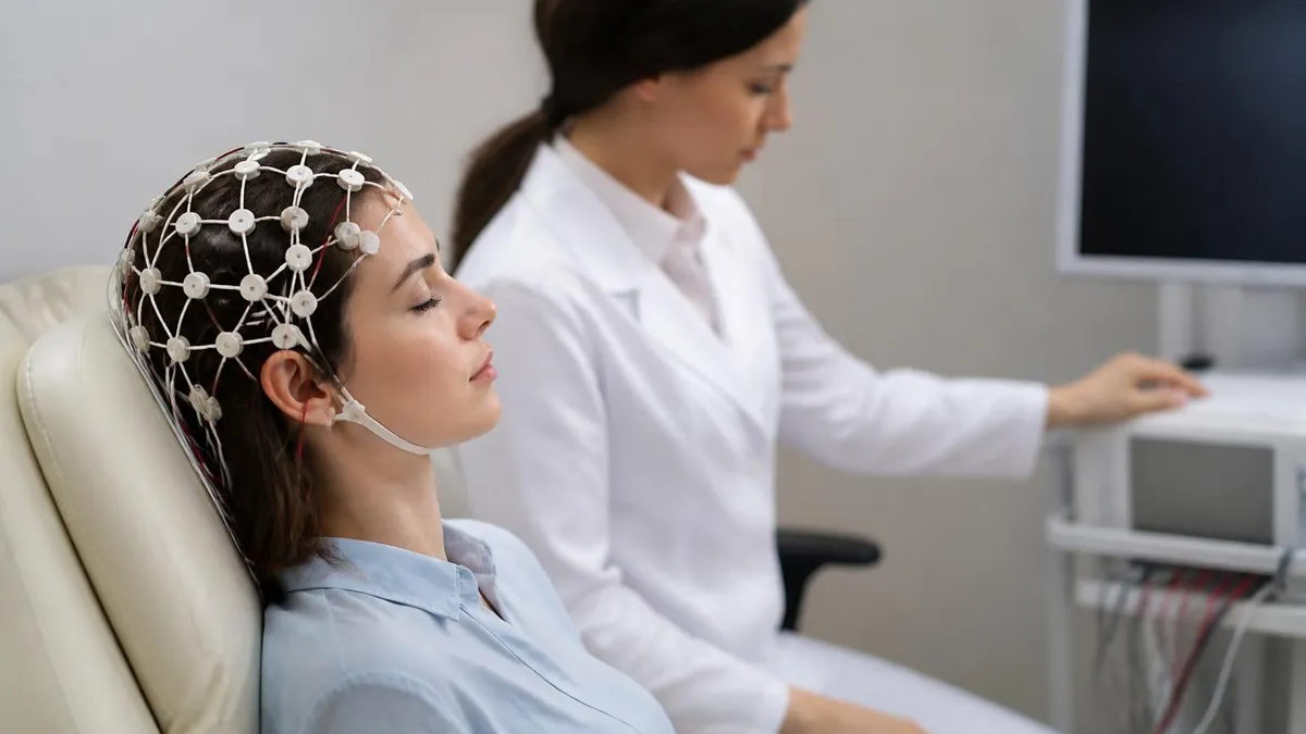
A routine EEG is the standard outpatient study ordered for first-time seizure evaluation, headache workup, or altered mental status. The recording lasts 20 to 40 minutes, includes resting wakefulness, hyperventilation for 3 minutes, and photic stimulation at frequencies from 1 to 30 Hz. These activation procedures increase the yield of epileptiform discharges by challenging the brain's excitatory-inhibitory balance. Most patients can return to normal activities immediately after the test.
A single routine EEG has a sensitivity of approximately 29 to 55 percent for epilepsy detection. Sensitivity rises to around 80 to 90 percent when three or more studies are performed, or when sleep is captured. For this reason, many neurologists order a sleep-deprived EEG as the second study rather than a repeat routine, since drowsiness dramatically increases the likelihood of capturing epileptiform activity in susceptible patients. The EEG test cost for a routine outpatient study typically falls between $200 and $400 before insurance adjustments.

EEG Test: Benefits and Limitations
- +Non-invasive — electrodes sit on the scalp, nothing penetrates skin in standard studies
- +No radiation exposure, making it safe for children, pregnant women, and repeated studies
- +Provides real-time temporal resolution down to milliseconds, capturing rapid brain events
- +Portable equipment allows bedside ICU monitoring and home ambulatory recording
- +Relatively low cost compared to MRI or fMRI for functional brain assessment
- +Activation procedures like hyperventilation and photic stimulation increase diagnostic yield without drugs
- −Limited spatial resolution — cannot pinpoint deep brain sources without intracranial electrodes
- −Highly susceptible to artifact from muscle movement, eye blinks, and electrical interference
- −A normal EEG does not rule out epilepsy — sensitivity of a single routine study is only ~50%
- −Sleep-deprived studies require patients to stay awake all night, which can be difficult or unsafe
- −Interpretation requires years of specialized training — subtle findings are easy to miss or misread
- −Hair products, scalp conditions, and thick hair can impede electrode contact and degrade signal quality
EEG Test Preparation Checklist
- ✓Wash your hair with shampoo the night before or morning of the test — no conditioner, oils, or sprays.
- ✓Follow your physician's instructions exactly regarding sleep deprivation if a sleep-deprived EEG is ordered.
- ✓Continue taking all regular medications unless your neurologist specifically instructs you to hold them.
- ✓Eat a normal meal before your appointment — low blood sugar can alter brain wave patterns.
- ✓Avoid caffeine for at least 8 hours before the test if your physician has recommended it.
- ✓Wear comfortable, loose-fitting clothing and avoid turtlenecks or other garments that obstruct neck access.
- ✓Arrive 10–15 minutes early to complete paperwork and allow the technologist to gather your history.
- ✓Bring a list of all current medications including supplements, dose, and frequency for the technologist.
- ✓Inform the technologist of any skin conditions, scalp lesions, or recent head injuries before electrode placement.
- ✓Relax during the recording — excessive muscle tension creates artifact that can obscure real brain signals.
A Normal EEG Does Not Mean No Epilepsy
Up to 50% of people with confirmed epilepsy will have a normal routine EEG on their first study. Epileptiform discharges are not present 100% of the time — they occur interictally (between seizures) in an unpredictable pattern. If your EEG comes back normal but your neurologist still suspects seizures based on your history, a repeat study, sleep-deprived recording, or ambulatory EEG is the appropriate next step, not a dismissal of your symptoms.
EEG test cost in the United States is highly variable and depends on the type of study, the facility, your insurance coverage, and your geographic region. A routine outpatient EEG at a hospital outpatient department typically generates a facility charge between $300 and $700 and a separate professional interpretation fee of $100 to $250. Patients with high-deductible health plans may pay the full amount before their deductible is met, while those with comprehensive insurance may owe only a copay of $30 to $75. Understanding these numbers helps you ask the right questions before scheduling.
EEG test price for ambulatory studies runs higher. The equipment rental and technologist setup time for a 24-hour ambulatory recording typically costs $500 to $900 before insurance. A 72-hour study may run $900 to $1,500. Some insurance plans require prior authorization for ambulatory studies, treating them as a step-up from routine EEG rather than a first-line option. If your neurologist orders an ambulatory study, ask your insurance company about authorization requirements before the appointment to avoid surprise billing.
For patients without insurance, self-pay rates are often negotiable. Federally Qualified Health Centers (FQHCs) and community health centers offer EEGs on sliding-scale fees based on income. Academic medical centers sometimes offer reduced rates for patients willing to participate in research protocols. Several free-standing neurodiagnostic labs have also emerged in major cities, operating with lower overhead than hospital systems and passing the savings to patients in the form of lower self-pay rates — sometimes as low as $150 to $200 for a routine study.
Long-term video-EEG monitoring in an inpatient epilepsy monitoring unit (EMU) represents the most expensive end of the spectrum. A 5-day EMU admission can generate charges of $15,000 to $40,000, the majority of which is the facility bed-day rate rather than the EEG itself. However, for surgical candidates, the EMU stay is an investment — accurate seizure localization can guide curative surgery with seizure-freedom rates exceeding 70% for ideal candidates with mesial temporal lobe epilepsy. The lifetime cost savings from eliminating medication and managing fewer breakthrough events typically exceeds the cost of the monitoring stay many times over.
When comparing facilities, it is worth asking whether the facility uses digital EEG equipment with remote review capability. Modern digital systems allow neurologists to review studies from home or a remote office, which has expanded coverage for smaller hospitals that cannot afford full-time on-site neurologists. Remote review is clinically equivalent to on-site review for routine studies, but you should confirm that the reviewing physician is board-certified in clinical neurophysiology or holds the ABRET CLTM credential for long-term monitoring cases.
Insurance coding matters more than most patients realize. EEG studies are billed under CPT codes that specify duration, type, and whether physician interpretation is included. Routine EEG without sleep is CPT 95816; with sleep it is 95819. Prolonged EEG monitoring codes (95950–95953) apply to ambulatory studies. If a claim is denied, ask your neurologist's billing department to confirm the correct code was used and to submit a peer-to-peer review if the denial was based on medical necessity — many initial denials are overturned on appeal when the clinical indication is properly documented.
For EEG technologists working in outpatient and hospital settings, understanding reimbursement and cost structures helps you communicate with patients who express concern about the financial burden of recommended testing. Knowing that prior authorization may be needed for ambulatory studies, and that the technologist setup note documenting clinical indication is often the basis for insurance approval, underscores why thorough intake documentation is both a clinical and administrative responsibility. A well-documented clinical indication note can be the difference between an approved claim and an unnecessary denial that delays the patient's diagnosis.

Do not stop taking anti-seizure medications before an EEG unless your neurologist has explicitly instructed you to do so in writing. Abrupt medication withdrawal can trigger status epilepticus — a medical emergency. In an epilepsy monitoring unit, medication tapering is done under continuous supervision precisely because breakthrough seizures are an expected part of the monitoring protocol. Self-managed medication changes at home before a routine outpatient EEG are dangerous and should never be attempted.
EEG test side effects are minimal for the overwhelming majority of patients, which is one of the procedure's greatest clinical advantages. The electrodes themselves cause no discomfort beyond the mild pressure of the cap or the cold sensation of the conductive gel. The prep gel used to reduce scalp impedance may cause temporary mild redness at electrode sites that resolves within an hour or two. Some patients with sensitive skin develop slight irritation from the electrode adhesive, but true allergic reactions to standard EEG materials are rare.
The most commonly reported adverse event from an outpatient EEG is not from the leads themselves but from the activation procedure of hyperventilation. Breathing deeply and rapidly for three minutes lowers arterial carbon dioxide (PCO₂), causing cerebral vasoconstriction. Most patients feel lightheaded, tingly in their fingers and lips, or slightly dizzy during this phase — all of which are expected physiological responses that resolve within one to two minutes of returning to normal breathing. Patients with severe pulmonary disease, sickle cell disease, or significant cardiovascular disease may have hyperventilation modified or omitted.
Photic stimulation — the flashing strobe light used at the end of most routine EEGs — carries a very small risk of triggering a photically induced seizure in susceptible individuals. This risk is highest in patients with juvenile myoclonic epilepsy (JME) and certain childhood epilepsy syndromes with known photosensitivity. However, because the stimulation is performed in a controlled environment with a trained technologist present, any triggered event is immediately recognized and managed. The clinical benefit of identifying photosensitivity far outweighs the small procedural risk, and most protocols stop stimulation at the first sign of a clinical or electrical response.
For patients undergoing sleep-deprived EEGs, the side effects are more related to the sleep deprivation itself than to the procedure. Fatigue, impaired concentration, irritability, and reduced reaction time are expected after a night without sleep. Patients should not drive to or from a sleep-deprived EEG appointment.
Most labs require a responsible adult to transport the patient, and technologists are trained to ask about transportation at intake. Falling asleep during the recording after a sleep-deprived night is not a problem — it is often exactly what the neurologist hopes will happen, since the transition from wakefulness to drowsiness is a high-yield period for capturing epileptiform activity.
Collodion — the acetone-based adhesive used to secure electrodes in ambulatory and long-term monitoring studies — occasionally causes scalp irritation with prolonged contact. Technologists are trained to inspect for skin breakdown at each lead site during long-term studies, and electrodes causing significant irritation are relocated. Acetone is used to remove collodion at the end of the study; it is flammable and should not be used near open flames, which means patients with home oxygen must inform the lab before an ambulatory study is scheduled.
There are no radiation side effects from an EEG, no injection-related risks, and no recovery period. This profile makes it safe for use in neonates in the NICU, in elderly patients with multiple comorbidities, and in pregnancy — all populations where MRI may be challenging or inadvisable in certain circumstances. The absence of meaningful side effects is why an EEG is almost always the first neurodiagnostic test ordered for new-onset seizures, before more expensive or invasive testing is considered.
For those studying for the ABRET R.EEG.T. examination or related neurodiagnostic certifications, understanding both the procedural and physiological basis of EEG test side effects is testable content. Questions about hyperventilation physiology, contraindications to photic stimulation, and the management of activation-procedure-induced events appear regularly in certification exams. Reviewing activation procedure protocols alongside electrode placement and signal interpretation gives certification candidates a complete picture of the EEG technologist's scope of practice and responsibilities.
For anyone preparing for EEG certification or simply wanting to understand the EEG medical test more deeply, building a systematic study approach is more effective than passive reading. Start with the anatomy: learn the lobar organization of the brain and the function of each major cortical region. Then map the 10-20 electrode positions onto those regions — knowing that T3 (or T7 in the revised nomenclature) sits over the left anterior temporal lobe helps you understand why temporal sharp waves at that electrode suggest left temporal lobe epilepsy rather than a frontal or parietal process.
Next, learn the normal EEG variants before tackling abnormalities. The posterior dominant rhythm, mu rhythm, lambda waves, 14-and-6 Hz positive bursts, small sharp spikes (SSS), and wicket spikes are all normal or benign variants that are frequently mistaken for abnormalities by inexperienced readers. The reverse is also true — some genuinely abnormal patterns are subtle enough to be dismissed as artifact or normal variant by readers unfamiliar with their characteristics. Systematic review of normal variants is the single fastest way to improve your interpretive accuracy and avoid false-positive readings.
When you have a solid grasp of normal EEG, move into the classification of epileptiform discharges. Understand the morphological criteria that define a spike (duration less than 70 milliseconds), a sharp wave (70 to 200 milliseconds), and a slow wave. Learn the difference between focal and generalized discharges and understand how to use field analysis — looking at which electrodes show the discharge and with what polarity — to localize a generator. These concepts are tested heavily on the R.EEG.T. and CNIM examinations and are also the core of clinical EEG interpretation.
Practice questions are an indispensable supplement to reading. Textbook knowledge tells you what to look for; practice questions reveal whether you can actually apply that knowledge under time pressure and with the distractor options that real exam writers use. The most effective study approach combines reading (for concept acquisition), image review (for pattern recognition), and practice questions (for application and retention). Spacing your practice sessions over weeks rather than cramming the night before is strongly supported by cognitive science research on durable learning.
EEG technologists also benefit from understanding the clinical context of the tests they perform. When a technologist knows that the patient was referred for evaluation of staring spells with post-ictal confusion, they can be more alert during the recording for subtle behavioral changes that might accompany an electrical event.
Noting that the patient briefly stopped responding to verbal commands at minute 14 of the recording, and documenting that in the technologist note, can be the critical piece of clinical correlation that allows the neurologist to call a recording diagnostic rather than inconclusive. The technologist is not just a lead-applier — they are a clinical observer whose notes become part of the permanent medical record.
Finally, approach EEG study as a continuous learning process rather than a finite task. New research on high-frequency oscillations, quantitative EEG, and connectivity measures is constantly expanding the clinical applications of EEG recording. Subspecialties such as intraoperative neurophysiological monitoring (IONM) and critical care EEG have developed their own bodies of expertise that build on but extend beyond routine clinical EEG. Staying current through the American Society of Neurophysiological Monitoring (ASNM), the American Clinical Neurophysiology Society (ACNS), and journals like the Journal of Clinical Neurophysiology ensures that your knowledge base keeps pace with the evolving field.
The combination of foundational electrode knowledge, normal variant recognition, epileptiform discharge classification, and activation procedure protocol mastery forms the core curriculum for any EEG professional. Whether you are a technologist, a neurology resident, a neurodiagnostics student, or a patient who simply wants to understand what those leads on your scalp are doing, the principles outlined in this guide give you the conceptual framework to engage with EEG as the sophisticated, clinically powerful tool that it truly is.
EEG Questions and Answers
EEG Electroencephalography Practice Test PDF (Free Printable 2026)
What Does an EEG Measure? Brain Waves, Electrical Activity, and What Your Results Reveal
EEG vs ECG vs EKG: Brain and Heart Test Differences
How to Read an EEG Test: A Complete Guide to Brain Wave Patterns and Results
EEG Waves Explained: Delta, Theta, Alpha, Beta, and Gamma Brain Rhythms
About the Author

Educational Psychologist & Academic Test Preparation Expert
Columbia University Teachers CollegeDr. Lisa Patel holds a Doctorate in Education from Columbia University Teachers College and has spent 17 years researching standardized test design and academic assessment. She has developed preparation programs for SAT, ACT, GRE, LSAT, UCAT, and numerous professional licensing exams, helping students of all backgrounds achieve their target scores.