EEG Device Guide: What Is an EEG Test, How It Works, and What to Expect
Learn what an EEG test is, how the eeg device works, how long it takes, what it costs, and what side effects to expect. Complete 2026 July guide. 🧠

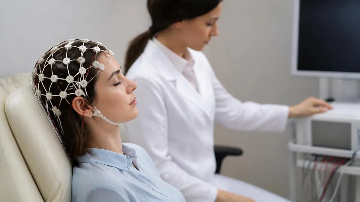
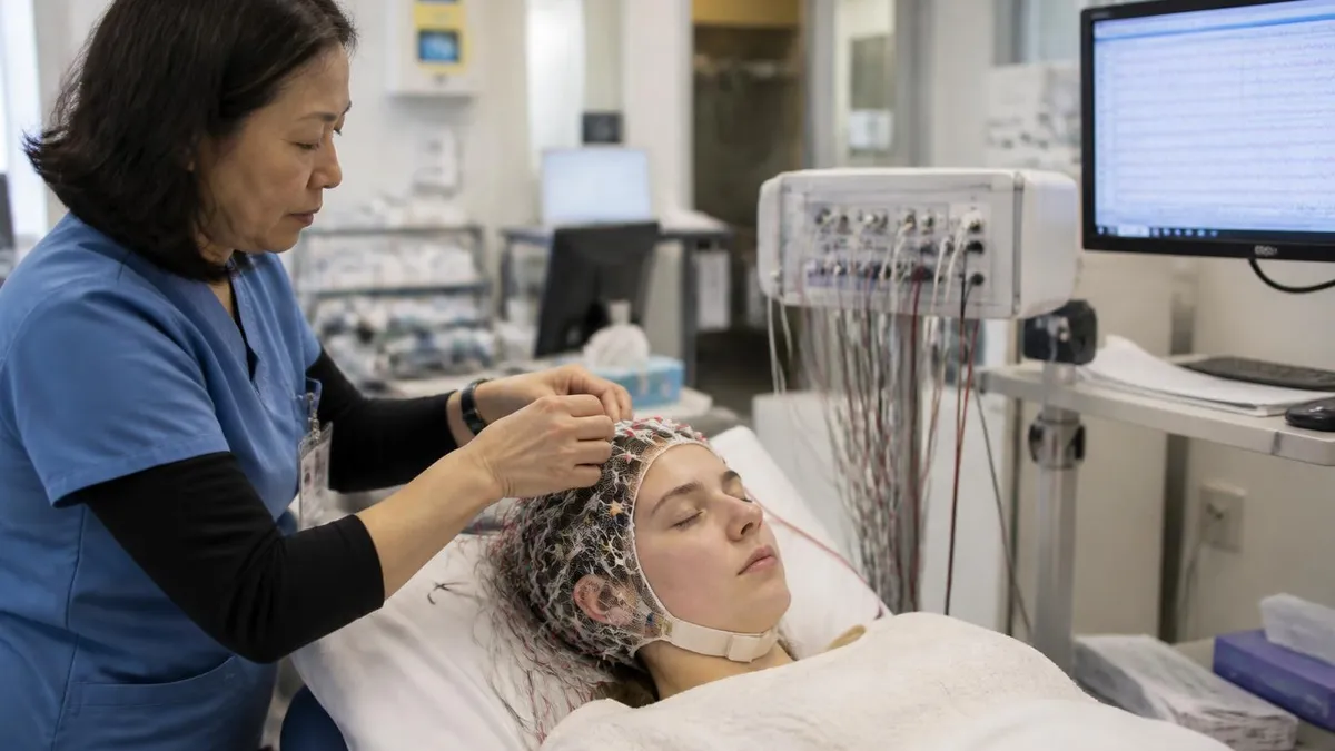
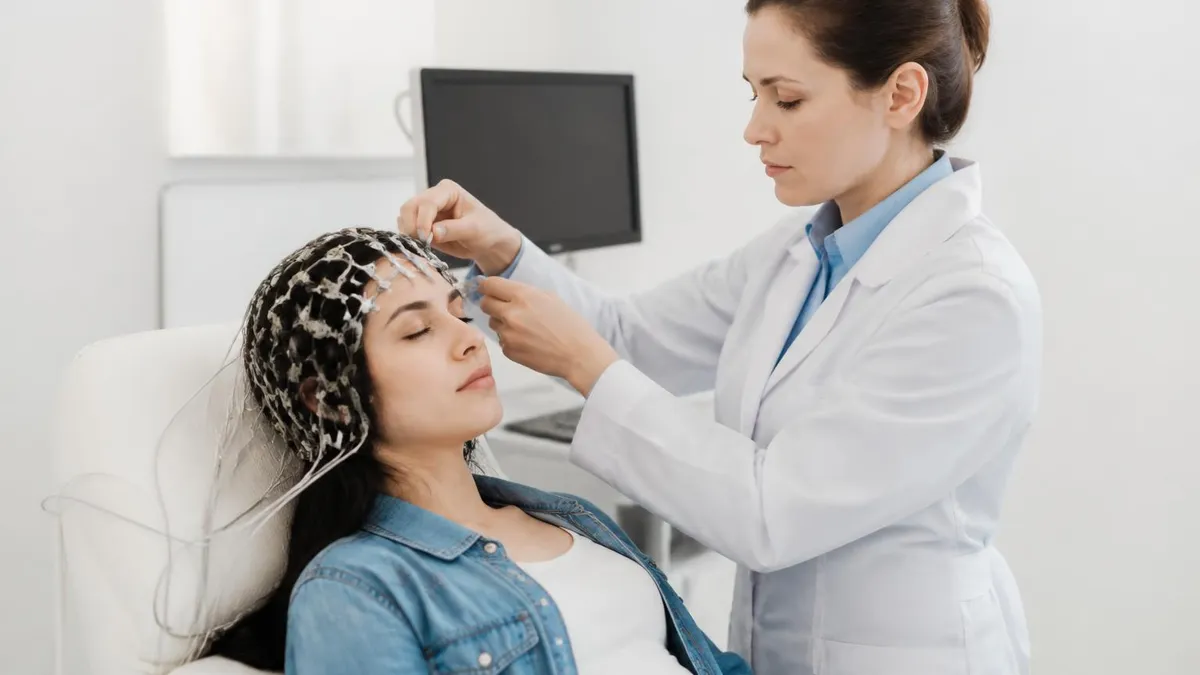
An EEG test — short for electroencephalogram — is one of the most important diagnostic tools in modern neurology. The eeg device works by placing small metal electrodes on the scalp to record the brain's spontaneous electrical activity over a period of time, typically between 20 minutes and several hours. Physicians order this eeg medical test when they need objective data about how the brain's neurons are firing, because abnormal patterns can point directly to conditions like epilepsy, sleep disorders, encephalopathy, or brain injury. Understanding what the test involves is the first step toward feeling prepared.
The question "what is an eeg test" comes up constantly for patients, students, and healthcare workers alike. At its core, the test is non-invasive and painless: electrodes attached to the scalp with a conductive gel or paste detect tiny voltage fluctuations produced by groups of neurons working in synchrony.
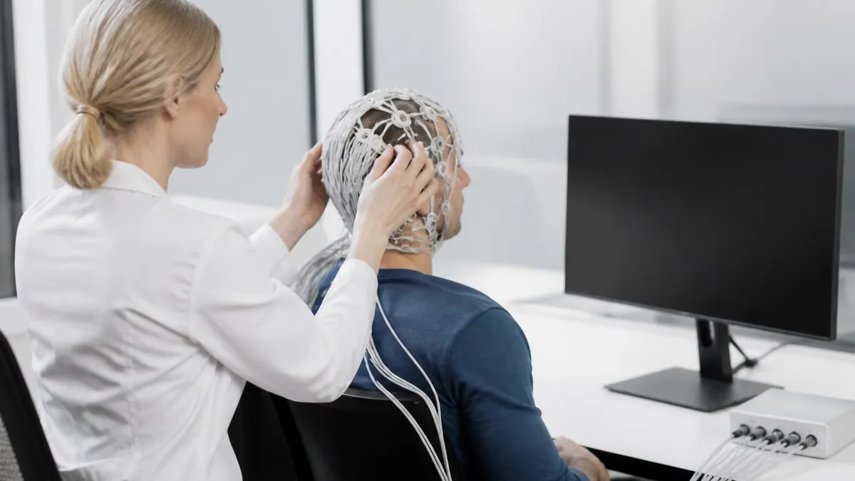
Those signals are amplified, digitized, and displayed as a series of wavy lines on a monitor — the classic EEG tracing. A trained EEG technologist ensures proper electrode placement while a neurologist later interprets the recording, looking for patterns that fall outside the normal range for the patient's age and level of alertness.
One of the most common follow-up questions patients ask after being scheduled for the test is "how long is an eeg test?" A routine outpatient EEG typically runs 20 to 40 minutes of actual recording time, though the full appointment — including electrode application, the recording itself, and electrode removal — often totals 60 to 90 minutes.
Sleep-deprived EEGs, which are used to capture abnormal activity that only emerges when the brain is fatigued, can extend to two or three hours. Ambulatory EEGs, worn at home on a portable recorder, may continue for 24 to 72 hours to catch intermittent events that would be missed in a short clinic session.
Cost is another major concern. The eeg test price in the United States ranges widely depending on the setting and the type of study ordered. A routine outpatient EEG at a hospital or neurology clinic generally bills between $200 and $700 before insurance adjustments. The eeg test cost after insurance is often just a copay or a percentage of that amount, though patients without insurance coverage may pay the full billed rate.
Video-EEG monitoring performed during an inpatient epilepsy monitoring unit stay can cost thousands of dollars per day, reflecting the continuous nursing and technical supervision involved. Always request an itemized estimate from the billing department before your appointment.
Safety and side effects are top of mind for many patients. The eeg test side effects are minimal because the procedure only records electrical activity — it does not send any current into the brain. The most common complaint is minor scalp irritation or temporary redness at the electrode sites, which resolves within a few hours.
Some patients find the conductive gel difficult to wash out of their hair. In rare cases, activation procedures such as hyperventilation or photic stimulation (strobe light exposure) can provoke a brief seizure in individuals with known seizure disorders; these procedures are performed in a controlled clinical environment precisely so the team is ready to respond.
For EEG students and technologists preparing for certification, the ABRET R. EEG T. examination tests mastery of the very skills involved in producing a high-quality recording: electrode placement according to the international 10-20 system, recognizing artifacts, performing activation procedures, and understanding the physiological basis of normal and abnormal waveforms. Practice questions covering EEG abnormal epileptiform patterns, ambulatory studies, and activation procedures are essential preparation tools. This guide is designed to help both patients navigating a new diagnosis and technologists deepening their clinical knowledge.
Throughout this article you will find detailed explanations of how the eeg device functions mechanically, what different EEG waveforms mean, what the test costs across different settings, what to do the night before your appointment, and how to interpret your results in conversation with your doctor. Whether you are a patient, a caregiver, a nursing student, or a practicing technologist studying for board exams, the sections below offer concrete, evidence-based information organized for quick reference.
EEG Test by the Numbers

How the EEG Device Works: Step by Step
Scalp Preparation
Electrode Application
Baseline Recording
Activation Procedures
Electrode Removal and Cleanup
Neurologist Interpretation
Understanding the different types of EEG tests helps patients and clinicians choose the right study for a given clinical question. The routine outpatient EEG is the starting point for most evaluations. It lasts approximately 20 to 40 minutes of recording time and is performed with the patient lying or sitting comfortably in a dimly lit room. Routine studies are excellent for capturing ongoing abnormalities such as generalized spike-and-wave discharges in absence epilepsy or focal slowing over an infarcted region, but they miss events that occur only rarely or only during sleep.
Sleep-deprived EEGs are ordered when a routine study is normal despite strong clinical suspicion of epilepsy. Asking the patient to stay awake all night or sleep only a few hours before the appointment raises the probability of detecting interictal epileptiform discharges — spikes, sharp waves, and spike-wave complexes that occur between actual seizures. The physiological drowsiness and light sleep that often follow sleep deprivation independently activate epileptiform activity, particularly temporal lobe and juvenile myoclonic epilepsy patterns. Patients should arrange a ride home because drowsiness after the test makes driving unsafe.
Ambulatory EEG monitoring allows continuous recording outside the hospital using a lightweight digital recorder worn on a belt or in a small backpack. The patient goes about daily activities — working, driving is sometimes permitted depending on seizure status, sleeping at home — while the device captures up to 72 hours of brain activity. This is particularly valuable for documenting rare events and correlating patient-reported symptoms with EEG changes. Ambulatory studies generally cost more than routine EEGs but far less than inpatient video-EEG monitoring.
Video-EEG monitoring, conducted in a specialized epilepsy monitoring unit, combines continuous EEG with synchronized video recording. Neurologists use this combination to precisely characterize seizure semiology — the clinical behaviors visible on video — and correlate them with the electrographic seizure pattern. This is the gold standard for pre-surgical evaluation of patients with drug-resistant epilepsy and for distinguishing epileptic seizures from non-epileptic events. Stays of three to ten days are typical, and anti-seizure medications may be temporarily reduced to provoke breakthrough seizures under safe, supervised conditions.
High-density EEG (HD-EEG) uses 64, 128, or 256 electrodes instead of the standard 21, providing dramatically improved spatial resolution. Source localization algorithms applied to HD-EEG data can pinpoint the cortical generator of an epileptiform discharge to within a centimeter or two, helping neurosurgeons plan resections. Research applications of HD-EEG extend into cognitive neuroscience, brain-computer interface development, and neurofeedback therapy. The electrodes used in HD-EEG systems are typically arranged in a geodesic net or elastic cap that applies all sensors simultaneously, reducing setup time considerably.
Neonatal EEG is a subspecialty that requires additional training because the developing brain produces waveform patterns dramatically different from those seen in adults or older children. Neonatal seizures often have subtle clinical manifestations and may only be detected electrographically. Continuous amplitude-integrated EEG (aEEG) monitoring is now standard in neonatal intensive care units, providing a compressed trend display that allows bedside nurses to monitor background activity even without full EEG training. Full conventional neonatal EEG interpretation remains the responsibility of a physician with specialized expertise in neonatal neurophysiology.
Intraoperative neurophysiological monitoring (IONM) represents another specialized application of EEG technology, used during neurosurgical procedures to detect ischemia or retraction injury in real time. EEG slowing or attenuation during carotid endarterectomy, for example, alerts the surgical team to impaired cerebral perfusion and may prompt shunt placement. While the recording principles are the same as in a diagnostic EEG, the noisy operating room environment and the need for rapid, confident interpretation under pressure make IONM a demanding subspecialty within clinical neurophysiology.
EEG Test Cost: What You Will Pay in 2026
Patients without health insurance face the full billed rate for an EEG, which varies considerably by facility and geography. A routine outpatient EEG at a hospital outpatient department typically bills $400 to $700, while the same test at an independent neurology clinic or freestanding imaging center may bill $200 to $400. Some facilities offer cash-pay discounts of 20 to 40 percent if payment is arranged before the appointment, so it always pays to ask about self-pay rates before scheduling.
For longer or more complex studies, the eeg test cost climbs substantially. A sleep-deprived EEG may bill $500 to $900, and ambulatory EEG monitoring for 24 to 72 hours can reach $1,000 to $2,500 depending on the recorder rental and interpretation fees. Video-EEG inpatient monitoring runs $2,000 to $5,000 per day including room, nursing, and technical services. Community health centers and academic medical centers often have financial assistance programs; patients experiencing hardship should request a financial counselor referral at the time of scheduling.
Advantages and Limitations of EEG Testing
- +Non-invasive and painless — no needles, radiation, or sedation required for most studies
- +Excellent temporal resolution captures brain activity millisecond by millisecond
- +Relatively affordable compared to MRI or PET scanning for initial neurological evaluation
- +Portable ambulatory systems allow recording during normal daily activities at home
- +Immediately available results in emergencies such as status epilepticus monitoring
- +Activations like hyperventilation can provoke latent abnormalities within the test window
- −Poor spatial resolution — standard 21-electrode EEG cannot precisely localize a deep source
- −Only records cortical surface activity; subcortical structures are largely invisible
- −Highly sensitive to artifacts from eye blinks, muscle tension, electrode movement, and electrical interference
- −A normal EEG does not rule out epilepsy — interictal discharges are not always present
- −Interpretation requires specialized training; misreads can lead to incorrect diagnoses
- −Conductive gel is messy and can be time-consuming to wash out of the patient's hair
How to Prepare for Your EEG Test
- ✓Wash your hair the night before with regular shampoo and do not apply any conditioner, oil, or styling product.
- ✓Ask your doctor whether to continue or hold your current anti-seizure or other medications before the test.
- ✓Avoid caffeine for at least 8 hours before the appointment if instructed by your physician.
- ✓If a sleep-deprived EEG is ordered, stay awake all night or limit sleep to 4–5 hours as directed.
- ✓Eat a normal meal before the test — low blood sugar can affect brain activity and test quality.
- ✓Arrive at least 15 minutes early to complete paperwork and allow extra time for electrode setup.
- ✓Bring a list of all current medications, including supplements, so the technologist can document them.
- ✓Wear comfortable clothing with a loose-fitting collar so electrodes can be placed without difficulty.
- ✓Arrange a ride home if you had a sleep-deprived study or if drowsiness is expected afterward.
- ✓Inform the technologist of any scalp conditions, recent head injuries, or claustrophobia before setup begins.
Up to 50% of people with epilepsy have a normal first routine EEG
A single routine EEG captures only 20 to 40 minutes of brain activity, and interictal epileptiform discharges are not continuously present in most patients. If clinical suspicion remains high after a normal EEG, physicians typically order a repeat study, a sleep-deprived EEG, or prolonged ambulatory monitoring. The clinical history and examination remain the foundation of the epilepsy diagnosis — the EEG provides supporting evidence, not a standalone yes-or-no answer.
Interpreting EEG results begins with recognizing the normal patterns expected for a patient's age and state of alertness. In an awake adult with eyes closed, the dominant rhythm over the posterior head regions is the alpha rhythm — smooth sinusoidal waves at 8 to 13 Hz with amplitudes of 20 to 70 microvolts. Alpha attenuates immediately when the patient opens their eyes or begins a mental task, a phenomenon called alpha blocking. The absence of a normal alpha rhythm over one hemisphere can itself be a significant finding, suggesting structural or functional pathology beneath those electrodes.
Beta activity, defined as frequencies above 13 Hz, is normally present over the frontal and central regions and reflects active cortical processing. Excess beta, particularly when diffuse and high in amplitude, is most commonly a medication effect — benzodiazepines, barbiturates, and some other sedative-hypnotic drugs dramatically increase beta activity. Focal beta enhancement or asymmetry can occasionally indicate a structural lesion. Recognizing drug effects on the EEG is an essential skill for any technologist or interpreter, because failure to account for medications is a common source of interpretive errors.
Theta activity (4–7 Hz) and delta activity (below 4 Hz) are normal during drowsiness and deeper stages of sleep but abnormal when seen in a fully awake adult. Focal slow-wave activity — theta or delta confined to one region — strongly suggests a structural lesion in the underlying cortex or white matter, such as a tumor, stroke, or cortical dysplasia. Diffuse slowing, on the other hand, suggests generalized cerebral dysfunction from metabolic encephalopathy, toxic insult, or widespread inflammation. Quantifying the degree of slowing and its distribution guides the clinician toward the most likely underlying cause.
Epileptiform discharges are the EEG findings most specifically associated with epilepsy, though they can occasionally appear in people who have never had a seizure. Spikes are defined as waveforms lasting less than 70 milliseconds with a sharply contoured morphology; sharp waves have a similar appearance but last 70 to 200 milliseconds. Both are abnormal when they clearly stand out from the background and are followed by a slow-wave component. The location of these discharges provides crucial information: temporal lobe spikes suggest mesial temporal lobe epilepsy, while generalized 3 Hz spike-wave complexes are the hallmark of childhood absence epilepsy.
An actual seizure recorded on EEG — an ictal discharge — appears as a rhythmic evolving pattern that builds in amplitude and frequency, spreads across electrodes, and eventually terminates, often followed by postictal slowing. The precise morphology, onset location, and propagation pattern of ictal discharges are used to classify seizures and epilepsy syndromes. For example, a unilateral temporal onset with spread to the ipsilateral frontal region and eventual bilateral involvement is characteristic of temporal lobe epilepsy with secondary generalization, which has specific implications for both medication selection and surgical candidacy.
Sleep architecture is clearly visible on EEG and provides a second level of useful information beyond epilepsy. Sleep spindles — bursts of 12 to 14 Hz activity generated by thalamocortical circuits — appear in stage N2 and are a reassuring indicator of intact thalamic function. K-complexes, slow waves, and the high-voltage delta of stage N3 follow in sequence. REM sleep is characterized by a low-voltage mixed-frequency pattern resembling wakefulness, accompanied by REMs visible in the eye-movement channels. Abnormalities of sleep architecture can reflect a wide range of conditions from obstructive sleep apnea to neurodegenerative disease affecting thalamocortical circuitry.
Artifacts are non-brain signals that contaminate the EEG tracing and must be distinguished from true cerebral activity. Muscle artifact from scalp and neck muscles produces high-frequency, irregular bursts that can obscure underlying brain signals. Eye blink artifact causes large deflections in the frontal electrodes that mirror the slow movements of the corneoretinal potential.
EKG artifact appears as a regular rhythm corresponding to the cardiac cycle, particularly in patients with large QRS complexes. Electrode artifact from a loose or poorly applied electrode produces channel-specific noise. Recognizing and annotating artifacts during recording is one of the core competencies assessed on the ABRET R. EEG T. certification examination.

Although the EEG test side effects are generally minimal, patients who experience a seizure during activation procedures — or who feel unusually confused, have prolonged head pain, or notice neurological symptoms after the test — should notify the technologist or a nearby medical professional immediately. Patients scheduled for sleep-deprived studies should not drive themselves home, as severe drowsiness significantly increases the risk of motor vehicle accidents. If you experience prolonged redness, swelling, or skin breakdown at electrode sites, contact your doctor's office for guidance on wound care.
Pursuing a career as an EEG technologist is a compelling path for individuals interested in neuroscience, patient care, and medical technology. Registered EEG Technologists (R. EEG T.) are credentialed by the American Board of Registration of Electroencephalographic and Evoked Potential Technologists (ABRET), which has administered certification examinations since 1961. The credential is widely recognized by hospitals, outpatient neurology centers, and epilepsy monitoring units across the United States and is increasingly required by employers as a condition of employment or advancement. Board certification demonstrates mastery of the technical, physiological, and patient care competencies that underpin high-quality EEG recording.
To sit for the R. EEG T. examination, candidates must meet specific educational and experience prerequisites. ABRET currently requires a minimum of a high school diploma combined with completion of an accredited EEG technology program, or an equivalent combination of education and supervised clinical hours. Most accredited programs are offered at community colleges, vocational schools, or hospital-based schools of clinical neurophysiology and take one to two years to complete. Programs accredited by the Commission on Accreditation of Allied Health Education Programs (CAAHEP) provide structured didactic and clinical training that aligns directly with examination content areas.
The R. EEG T. examination itself covers electrode application, amplifier settings and montages, normal and abnormal adult and neonatal EEG patterns, activation procedures, artifacts, patient safety, and the legal and ethical responsibilities of the technologist. The exam consists of approximately 150 multiple-choice questions administered at computer-based testing centers across the country. Candidates typically prepare for three to six months using a combination of textbooks, practice question banks, and supervised clinical experience. Passing the examination confers the R. EEG T. credential, which must be maintained through continuing education and periodic renewal.
Beyond the R. EEG T., ABRET offers several advanced credentials for experienced practitioners. The Certificate of Added Qualification in Long-Term Monitoring (CAQ-LTM) is designed for technologists who specialize in ambulatory EEG and epilepsy monitoring unit work. The Certificate of Added Qualification in Neurocritical Care (CAQ-NCC) recognizes expertise in continuous EEG monitoring for critically ill patients. Registered Evoked Potential Technologists (R. EP T.) credential covers visual, auditory, and somatosensory evoked potential testing, which is closely allied with EEG in many clinical neurophysiology departments. Each advanced credential has its own eligibility requirements and examination process.
Salary prospects for credentialed EEG technologists are solid and continue to grow as demand for neurological diagnostics increases with an aging population. According to the Bureau of Labor Statistics and multiple salary survey sources, the median annual salary for cardiovascular and diagnostic medical technologists in the United States is approximately $64,000 to $68,000, with experienced EEG specialists in inpatient or epilepsy monitoring settings often earning $70,000 to $85,000.
Geographic variation is substantial — technologists in California, New York, and Massachusetts typically earn 20 to 35 percent more than the national median, reflecting both higher cost of living and concentrated demand in major medical centers.
Work settings for EEG technologists are varied and offer flexibility in scheduling and specialization. Hospital outpatient neurology clinics and freestanding diagnostic centers provide predictable daytime schedules with limited on-call obligations. Epilepsy monitoring units, pediatric hospitals, and neurological intensive care units offer more acute and specialized work, often including evening and weekend coverage.
Traveling EEG technologist positions — short-term contracts at facilities experiencing staffing shortages — command premium hourly rates and provide rapid exposure to diverse patient populations and recording environments. Intraoperative neurophysiological monitoring, performed in operating rooms during spine, vascular, and cranial neurosurgeries, is a growing subspecialty with some of the highest compensation in the field.
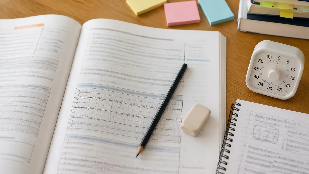
Students preparing for the R. EEG T. examination benefit enormously from targeted practice with questions that mirror the examination's style and content distribution. The ABRET examination blueprint emphasizes electrode placement and the 10-20 system, recognition of normal and abnormal waveforms across the lifespan, artifact identification, and patient management. Practice tests that focus specifically on abnormal epileptiform patterns, activation procedures, and ambulatory monitoring prepare candidates for the most challenging and frequently tested content areas. Reviewing rationales for both correct and incorrect answers solidifies the underlying clinical reasoning rather than mere memorization of isolated facts.
Practical preparation for an EEG certification examination begins with an honest self-assessment of strengths and weaknesses across the major content domains. Most candidates find electrode placement and the mathematics of the 10-20 system straightforward after repetitive practice but struggle initially with the pattern recognition skills needed to identify abnormal waveforms quickly and confidently. Developing those pattern recognition skills requires systematic exposure to a large variety of EEG samples — normal studies across different ages and states, common artifact patterns, and the full spectrum of abnormalities from mild diffuse slowing to status epilepticus.
Building a structured study schedule in the eight to twelve weeks before the examination maximizes retention and avoids the common pitfall of passive re-reading.
Divide the content blueprint into weekly modules: spend the first two weeks on electrode application, amplifier physics, and digital EEG systems; weeks three and four on normal adult and pediatric EEG patterns; weeks five and six on abnormal patterns including epileptiform discharges and encephalopathy; week seven on neonatal EEG; week eight on evoked potentials and activation procedures; and the final weeks on mock examinations under timed conditions. Adjust the schedule based on your self-assessment results after each module.
Active recall outperforms passive review by a substantial margin for medical examination preparation. Rather than rereading your notes or textbook chapters, close the book and write down everything you remember about the topic, then check for gaps. Use spaced repetition flashcard software to schedule review of difficult concepts at increasing intervals — the algorithm ensures you revisit material just before you would otherwise forget it, maximizing long-term retention with the minimum time investment. Many successful candidates report spending 60 to 80 percent of their study time on active recall and practice questions rather than reading.
Practice questions should be used diagnostically as well as for content coverage. After each practice session, categorize your wrong answers by content area and track error rates over time. A persistently high error rate in a specific area — say, neonatal EEG patterns or photic driving responses — signals that more targeted review is needed there. Seek out additional resources for your weak areas: textbook chapters, ABRET study guides, online EEG atlas resources, and clinical mentorship from credentialed technologists who can walk you through real tracings and explain their reasoning in real time.
Time management during the actual examination deserves specific attention. With approximately 150 questions in a defined time window, most candidates have roughly 90 seconds per question on average. Questions with EEG images or montage diagrams typically require more time, so plan to move through purely factual knowledge questions efficiently to preserve time for image-based items.
If you encounter a question you cannot answer confidently, flag it and move on rather than spending five minutes on a single item. Return to flagged questions after completing the rest of the examination; sometimes a later question provides context that helps resolve an earlier uncertainty.
On the day of the examination, prioritize sleep, a nutritious breakfast, and arriving at the testing center with ample time to spare. Fatigue and hunger measurably impair cognitive performance, and the stress of arriving late compounds both problems. Bring the required identification documents and any authorization paperwork sent by ABRET. The testing center environment is typically quiet and well-controlled, but bring earplugs if you are sensitive to ambient noise. Once seated, take two or three slow deep breaths before beginning, scan the first few questions to calibrate difficulty, and trust the preparation you have done over the preceding weeks.
After the examination, regardless of outcome, reflect on the experience constructively. Candidates who pass should maintain their credential through ABRET's continuing education requirements and consider pursuing an advanced CAQ credential as their clinical specialization deepens. Candidates who do not pass on the first attempt receive a score report identifying performance by content area, which provides a precise roadmap for targeted remediation before the next attempt. The EEG field rewards dedication and continuous learning: the most respected technologists are those who never stop asking questions about the physiology behind the patterns they record every day.
EEG Questions and Answers
EEG vs ECG vs EKG: Brain and Heart Test Differences
EEG Electroencephalography Practice Test PDF (Free Printable 2026)
What Does an EEG Measure? Brain Waves, Electrical Activity, and What Your Results Reveal
How to Read an EEG Test: A Complete Guide to Brain Wave Patterns and Results
EEG Waves Explained: Delta, Theta, Alpha, Beta, and Gamma Brain Rhythms
About the Author

Educational Psychologist & Academic Test Preparation Expert
Columbia University Teachers CollegeDr. Lisa Patel holds a Doctorate in Education from Columbia University Teachers College and has spent 17 years researching standardized test design and academic assessment. She has developed preparation programs for SAT, ACT, GRE, LSAT, UCAT, and numerous professional licensing exams, helping students of all backgrounds achieve their target scores.