
ACLS Rhythm Identification: Complete 2026 October Study Guide & Practice Test
📚 Master ACLS rhythm identification with expert explanations, ECG tips, and free practice tests. Prep for 2026 October certification with confidence.
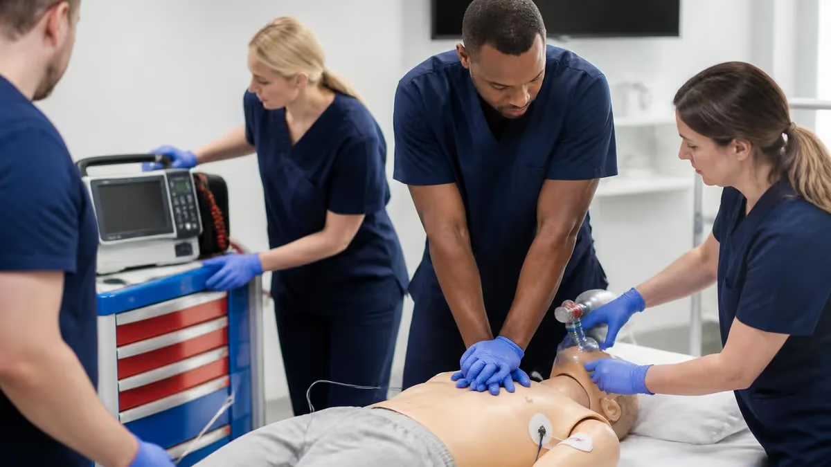
Mastering ACLS rhythm identification is the single most important clinical skill you will develop during your Advanced Cardiovascular Life Support certification. Every resuscitation algorithm — from ventricular fibrillation to pulseless electrical activity — begins with one question: what does the rhythm look like?
Providers who can rapidly and accurately read an ECG strip are better equipped to choose the correct intervention, administer the right drug, and direct their team with authority. If you are preparing for your 2026 ACLS exam, rhythm recognition is where the majority of test questions are focused, and it is where your preparation time will pay off most directly.
The American Heart Association's 2025 ACLS guidelines place ECG interpretation at the core of every major cardiac arrest and peri-arrest protocol. Whether you are working through a shockable rhythm like ventricular fibrillation or navigating a bradyarrhythmia that requires transcutaneous pacing, you must be able to identify the rhythm before you can act. This guide walks you through every rhythm category you are likely to encounter on your certification exam and at the bedside, giving you a systematic framework that reduces errors under pressure.
Many candidates underestimate how much deliberate ECG practice it takes to build true pattern recognition. Reading a rhythm strip is not purely memorization — it involves training your eye to spot rate, regularity, P-wave morphology, PR interval, QRS width, and QT duration almost simultaneously. The good news is that consistent practice with realistic strips, especially timed practice-test conditions, accelerates this pattern recognition dramatically. Within two to three weeks of focused study, most candidates report significant improvements in both speed and accuracy.
This article is structured as both a content review and a practical study plan. We cover the major rhythm categories tested on the ACLS written exam, explain the clinical reasoning behind each treatment algorithm, and point you toward the best free practice quizzes available on PracticeTestGeeks. Along the way, you will find stat summaries, study checklists, and tips from providers who passed their exam on the first attempt. Understanding acls rhythm identification in the context of pharmacology will also help you connect the dots between what you see on the monitor and what medication you reach for next.
One common mistake candidates make is studying rhythm strips in isolation without connecting them to the clinical context. On the actual ACLS exam, questions often describe a patient scenario — "the patient is unresponsive with no pulse, monitor shows a coarse, irregular waveform" — and ask you to identify the rhythm and the next best action. That means you need to be fluent not only in naming rhythms but also in linking them to the correct algorithm branch. We will emphasize that integration throughout this guide so your study is clinically grounded from the start.
Another key point: the written exam tests both recognition and management. You may be shown a rhythm and asked which medication is indicated, or given a drug and asked which rhythm it treats. This means your rhythm study must be paired with pharmacology and algorithm review. We have designed this guide to support that integrated approach, referencing the relevant drugs and protocols wherever rhythm recognition intersects with treatment decisions. By the end of this article, you will have a clear, prioritized study plan that targets the highest-yield topics for your 2026 ACLS exam.
Finally, do not overlook the value of practice tests as a diagnostic tool. Before you deep-dive into content review, take one of the free ACLS cardiac rhythm practice quizzes below to identify your weak spots. Most candidates are surprised to find that their errors cluster around one or two rhythm categories — typically AV blocks or wide-complex tachycardias — rather than being spread evenly. Knowing your weak spots lets you allocate your study time efficiently and enter your certification session with real confidence rather than general familiarity.
ACLS Rhythm Identification by the Numbers

Core ACLS Rhythm Categories You Must Know
Ventricular fibrillation and pulseless ventricular tachycardia are the two rhythms requiring immediate defibrillation. Both are chaotic or rapid ventricular rhythms that produce no effective cardiac output. Early defibrillation is the single most important intervention for survival.
Asystole (flatline) and pulseless electrical activity (PEA) require high-quality CPR and identification of reversible causes via the Hs and Ts. Defibrillation is not indicated and can delay effective resuscitation if applied incorrectly.
Supraventricular tachycardia, atrial fibrillation, atrial flutter, and ventricular tachycardia with a pulse each have specific management pathways. Stability is determined by hemodynamic compromise: hypotension, altered consciousness, or chest pain.
Sinus bradycardia, junctional rhythms, and first-, second-, and third-degree AV blocks are frequently tested. Management hinges on whether the patient is symptomatic and whether the block is at the AV node or infranodal.
Return of spontaneous circulation (ROSC) rhythms, accelerated idioventricular rhythm, and agonal rhythms appear in scenario-based questions. Recognizing these prevents misinterpretation and inappropriate intervention in the post-arrest phase.
Reading an ECG strip systematically is the foundation of reliable rhythm identification, and the best providers use the same mental checklist every single time — whether it is 3 a.m. in a code or a quiet afternoon classroom session. A consistent approach eliminates the most common errors, which almost always occur when a provider skips a step because the answer seems obvious at first glance. The standard five-step method covers rate, rhythm regularity, P waves, PR interval, and QRS duration, and each step narrows down the differential before you reach your final interpretation.
Start with rate. A normal heart rate falls between 60 and 100 beats per minute. Rates above 100 define tachycardia; rates below 60 define bradycardia. On a standard ECG strip running at 25 mm per second, each large box equals 0.20 seconds and each small box equals 0.04 seconds. The quickest rate calculation method for regular rhythms is the 300-divide rule: count the number of large boxes between two consecutive R waves and divide 300 by that number. For irregular rhythms, count the number of QRS complexes in a 6-second strip and multiply by 10.
Next, assess rhythm regularity by measuring R-to-R intervals across the strip. A regular rhythm has consistent, equal intervals. Irregular rhythms fall into two subtypes: irregularly irregular (no discernible pattern, as in atrial fibrillation) and regularly irregular (a repeating pattern of irregular intervals, as in second-degree AV block type I). This distinction is clinically significant because it immediately points you toward different diagnostic categories and different treatment algorithms.
P-wave analysis is the step that distinguishes atrial from junctional and ventricular rhythms. Ask three questions: Are P waves present? Do they have a consistent, upright morphology in lead II? Is there one P wave before every QRS? Absent P waves combined with an irregular rhythm strongly suggest atrial fibrillation. Inverted P waves in lead II suggest a junctional origin. P waves that bear no fixed relationship to QRS complexes — AV dissociation — are the hallmark of complete heart block.
The PR interval is normally 0.12 to 0.20 seconds (3 to 5 small boxes). A prolonged PR interval that is consistent across beats defines first-degree AV block — benign in isolation but significant as a marker of conduction system disease. A PR interval that progressively lengthens until a P wave fails to conduct (dropped QRS) defines Mobitz type I (Wenckebach), a typically benign nodal block. A constant PR interval with intermittent non-conducted P waves defines Mobitz type II, a dangerous infranodal block that can progress to complete heart block without warning.
QRS duration is the final critical measurement. A narrow QRS (less than 0.12 seconds, or 3 small boxes) indicates normal ventricular conduction through the His-Purkinje system, confirming that the impulse originated above the bundle branches. A wide QRS (0.12 seconds or greater) indicates either a bundle branch block, an aberrantly conducted supraventricular beat, or a rhythm of ventricular origin.
When a wide-complex tachycardia is present, the default assumption on the ACLS exam is ventricular tachycardia until proven otherwise, because treating VT as SVT with aberrancy can be life-threatening. Understanding this principle is central to safe patient care, and it is heavily tested across all versions of the ACLS written examination.
Putting these five steps together takes practice but becomes automatic with repetition. When you work through the free practice quizzes on this site, apply the full five-step method to every strip rather than guessing based on first impression. Candidates who develop this discipline consistently outperform those who rely on gestalt pattern recognition alone, because the systematic approach catches the subtle variants — like fine ventricular fibrillation masquerading as asystole, or slow VT that looks deceptively like a sinus rhythm — that trip up even experienced providers on the exam.
ACLS Practice Test Questions
Prepare for the ACLS Advanced Cardiovascular Life Support Practice exam with our free practice test modules. Each quiz covers key topics to help you pass on your first try.
ACLS Cardiac Rhythms & ECG Interpretation
ACLS Exam Questions covering ACLS Cardiac Rhythms & ECG Interpretation. Master ACLS Test concepts for certification prep.
ACLS Pharmacology & Medications
Free ACLS Practice Test featuring ACLS Pharmacology & Medications. Improve your ACLS Exam score with mock test prep.
ACLS Best Practices & Methodologies
ACLS Mock Exam on Best Practices & Methodologies. ACLS Study Guide questions to pass on your first try.
ACLS Communication & Stakeholder Relations
ACLS Test Prep for Communication & Stakeholder Relations. Practice ACLS Quiz questions and boost your score.
ACLS Core Concepts & Fundamentals
ACLS Questions and Answers on Core Concepts & Fundamentals. Free ACLS practice for exam readiness.
ACLS Data Analysis & Reporting
ACLS Mock Test covering Data Analysis & Reporting. Online ACLS Test practice with instant feedback.
ACLS Documentation & Record Keeping
Free ACLS Quiz on Documentation & Record Keeping. ACLS Exam prep questions with detailed explanations.
ACLS Industry Regulations & Compliance
ACLS Practice Questions for Industry Regulations & Compliance. Build confidence for your ACLS certification exam.
ACLS Leadership & Team Management
ACLS Test Online for Leadership & Team Management. Free practice with instant results and feedback.
ACLS Professional Standards & Ethics
ACLS Study Material on Professional Standards & Ethics. Prepare effectively with real exam-style questions.
ACLS Quality Assurance & Improvement
Free ACLS Test covering Quality Assurance & Improvement. Practice and track your ACLS exam readiness.
ACLS Risk Management & Assessment
ACLS Exam Questions covering Risk Management & Assessment. Master ACLS Test concepts for certification prep.
Shockable vs. Non-Shockable Rhythms: ACLS Algorithm Decision Points
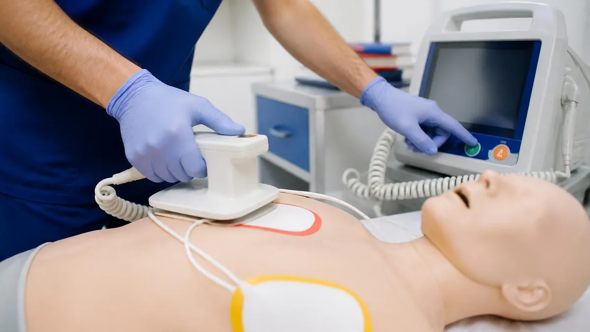
Ventricular fibrillation is characterized by chaotic, irregular waveforms with no identifiable P waves, QRS complexes, or T waves. The amplitude varies from coarse (large-amplitude waves, more likely to respond to defibrillation) to fine (small-amplitude waves, harder to distinguish from asystole). Pulseless VT presents as a wide-complex, regular tachycardia at rates typically above 150 bpm with no palpable pulse. Both rhythms receive the same ACLS treatment: immediate unsynchronized defibrillation at 200 joules (biphasic), followed by CPR resumption and epinephrine every 3 to 5 minutes. Amiodarone or lidocaine is added after the third shock for refractory VF or pulseless VT. Early high-quality CPR and rapid defibrillation remain the two interventions with the strongest evidence for survival benefit.
A common exam pitfall involves distinguishing coarse VF from a very rapid polymorphic VT such as torsades de pointes. Both appear irregular and wide on the monitor. The clinical context — a patient with a known prolonged QT interval, recent hypomagnesemia, or antiarrhythmic drug use — should raise your suspicion for torsades, which responds to intravenous magnesium sulfate. On the exam, if a scenario describes a patient with a prolonged QT and a twisting, polymorphic wide-complex tachycardia, magnesium is the answer. For standard coarse VF, defibrillation comes first, every time.
Systematic ECG Approach vs. Gestalt Pattern Recognition: Which Works Better for Exam Prep?
- +Five-step systematic method catches subtle variants that gestalt misses
- +Builds transferable clinical skills beyond the exam
- +Reduces anxiety by providing a reliable algorithm to fall back on
- +Consistent approach works even with unfamiliar or degraded strip quality
- +Easier to teach and standardize within a resuscitation team
- +Fewer errors on time-pressured exam questions when a method is internalized
- −Takes more cognitive effort early in training before it becomes automatic
- −Can feel slower at first compared to quick visual recognition
- −May create over-reliance on steps rather than integrating clinical context
- −Some exam questions are designed for rapid pattern recognition, not step-by-step analysis
- −Requires deliberate practice with many strip examples to fully internalize
- −Does not eliminate the need to memorize key diagnostic criteria for each rhythm
Pros and Cons at a Glance
| Pros | Cons |
|---|---|
| Five-step systematic method catches subtle variants that gestalt misses | Takes more cognitive effort early in training before it becomes automatic |
| Builds transferable clinical skills beyond the exam | Can feel slower at first compared to quick visual recognition |
| Reduces anxiety by providing a reliable algorithm to fall back on | May create over-reliance on steps rather than integrating clinical context |
| Consistent approach works even with unfamiliar or degraded strip quality | Some exam questions are designed for rapid pattern recognition, not step-by-step analysis |
| Easier to teach and standardize within a resuscitation team | Requires deliberate practice with many strip examples to fully internalize |
| Fewer errors on time-pressured exam questions when a method is internalized | Does not eliminate the need to memorize key diagnostic criteria for each rhythm |
ACLS Rhythm Identification Exam Prep Checklist
- ✓Identify VF and pulseless VT and state the correct defibrillation energy setting (200 J biphasic)
- ✓Distinguish coarse VF from fine VF and know when to check lead placement before calling asystole
- ✓List all five Hs and five Ts for reversible causes of PEA and asystole from memory
- ✓Differentiate first-degree, Mobitz I, Mobitz II, and third-degree AV block using PR interval and QRS criteria
- ✓State the first-line drug and dose for symptomatic bradycardia (atropine 0.5 mg IV)
- ✓Identify the rhythm that requires magnesium sulfate rather than standard defibrillation (torsades de pointes)
- ✓Calculate ventricular rate on a regular ECG strip using the 300-divide rule in under 10 seconds
- ✓Recognize SVT with aberrancy versus ventricular tachycardia and apply the default ACLS assumption
- ✓Describe the synchronized cardioversion energy sequence for unstable SVT and atrial flutter
- ✓Complete at least three full 25-question ECG practice quiz sets before your exam date
Wide-Complex Tachycardia = Ventricular Tachycardia Until Proven Otherwise
On the ACLS written exam and in real clinical practice, any wide-complex tachycardia in a patient with cardiac history should be treated as ventricular tachycardia until a definitive diagnosis rules it out. Treating VT as SVT with aberrancy by giving verapamil or diltiazem can cause hemodynamic collapse in a VT patient — this is a classic, heavily tested danger scenario. When in doubt, treat wide and fast as VT.
The highest-yield ECG patterns for the 2026 ACLS exam are not necessarily the most dramatic-looking rhythms — they are the rhythms most commonly misidentified under exam pressure. Ventricular fibrillation gets a lot of study time, but it is often the second-degree AV blocks and the wide-complex tachycardia differential that generate the most exam errors. Understanding exactly why these patterns are frequently confused is the key to answering them correctly when it counts.
Second-degree AV block type II (Mobitz II) is frequently confused with third-degree block, and the distinction is clinically critical. In Mobitz II, some P waves conduct and produce QRS complexes with a constant PR interval; others are simply blocked and produce no QRS. The atrial rate is regular, and the ventricular rate is some fraction of the atrial rate (2:1, 3:1, etc.). In third-degree block, there is complete AV dissociation — no relationship whatsoever between P waves and QRS complexes.
The atria and ventricles march independently. On an exam strip, count the P-to-P interval and the R-to-R interval separately: if they are different constants, you have complete heart block. If the PR interval is consistent on the beats that conduct, you have Mobitz II.
Accelerated idioventricular rhythm (AIVR) is a post-resuscitation rhythm that commonly appears after ROSC and is frequently misidentified as ventricular tachycardia. AIVR has a wide QRS, an accelerated rate (typically 60 to 100 bpm), and is usually benign and self-terminating. Treating it as VT with antiarrhythmics can suppress the escape rhythm and actually worsen the patient's hemodynamic status.
The key distinguishing feature is the rate: true VT is almost always above 100 bpm, often above 150 bpm, while AIVR occupies the 60 to 100 range. In the post-arrest scenario, always integrate the clinical picture — stable hemodynamics and a rate under 100 bpm strongly suggest AIVR, not VT.
Atrial flutter with 2:1 conduction is another rhythm that reliably catches candidates off guard. At a typical atrial flutter rate of 300 bpm with 2:1 conduction, the ventricular rate is approximately 150 bpm — a rate that can mimic sinus tachycardia at first glance. The giveaway is the sawtooth flutter waves, best seen in leads II, III, aVF, and V1.
When you see a narrow-complex tachycardia at exactly 150 bpm, flutter with 2:1 conduction should be your first differential, not sinus tachycardia. Applying adenosine in this scenario can be diagnostically useful: it temporarily increases AV block and unmasks the flutter waves, confirming the diagnosis without converting the rhythm.
Junctional tachycardia is less commonly tested but appears in rhythm identification question banks and occasionally on the actual exam. It originates in the AV node or bundle of His, producing a narrow-complex tachycardia with absent P waves or retrograde P waves that appear just before, within, or just after the QRS complex.
Because the rate is usually 100 to 180 bpm and the QRS is narrow, it resembles SVT. The distinction from AVNRT and other forms of SVT is not clinically urgent in most cases, since both respond to vagal maneuvers and adenosine, but knowing the ECG characteristics demonstrates comprehensive rhythm knowledge on the exam.
Wolff-Parkinson-White (WPW) pattern deserves special attention because it creates a dangerous trap: patients with WPW who develop atrial fibrillation can conduct rapidly down the accessory pathway, producing a wide-complex, irregularly irregular tachycardia with rates exceeding 250 bpm. This is hemodynamically dangerous and potentially fatal.
The critically important ACLS point is that AV nodal blocking agents — adenosine, beta-blockers, calcium channel blockers, and digoxin — are contraindicated in this scenario. They block the normal AV pathway, forcing all conduction down the accessory pathway and potentially triggering VF. The correct treatment for pre-excited AF in WPW is synchronized cardioversion if unstable, or procainamide/ibutilide if stable. This contraindication is a classic, high-stakes exam question.
Finally, pulseless electrical activity encompasses a wide range of organized electrical patterns — sinus rhythm, junctional rhythm, even a seemingly normal ECG — all without a detectable pulse. The danger of PEA on the exam is that candidates see organized electrical activity and assume there must be a pulse.
The scenario will always tell you there is no pulse; your job is to recognize that organized electricity does not equal mechanical contraction, start CPR immediately, and search aggressively for the underlying cause. PEA questions test whether you understand this fundamental distinction between electrical and mechanical cardiac activity, and whether you can link the clinical clues in the scenario to the correct reversible cause from the Hs and Ts list.

A common scenario-based exam trap: a patient is in ventricular fibrillation and IV access has not yet been established. The correct answer is always to defibrillate immediately — do not delay the shock to place an IV. Every minute without defibrillation in VF reduces survival odds by approximately 7 to 10 percent. IV access and medications come after the first shock, not before.
One of the most reliable predictors of first-attempt ACLS exam success is the number of distinct rhythm strips a candidate has reviewed before their test date. Research on medical certification exam performance consistently shows that candidates who practice with high-volume, varied question sets outperform those who read content alone.
This is especially true for rhythm identification, where pattern recognition is a perceptual skill built through repetition rather than a factual knowledge domain that can be absorbed by reading. The target is not just to recognize rhythms you have seen before — it is to recognize rhythms you have never seen before by applying a reliable analytical framework.
Building your rhythm library should be a structured process. Start with the four cardiac arrest rhythms — VF, pulseless VT, asystole, and PEA — until you can identify each in under five seconds. These are the highest-stakes rhythms in any code, and instant recognition is essential. Once those are solid, move to the tachyarrhythmias: SVT, atrial fibrillation, atrial flutter, and ventricular tachycardia with a pulse. Learn to differentiate narrow-complex from wide-complex at a glance, and practice the stable versus unstable determination using the clinical criteria of hypotension, altered mental status, chest pain, and signs of shock.
Next, dedicate focused time to the AV blocks. Print or download a set of strips specifically targeting first-degree, Mobitz I, Mobitz II, and third-degree block side by side, and practice labeling the PR intervals and identifying the pattern of conduction. Many candidates find flashcards with strips on one side and interpretation on the other to be particularly effective for this category. The goal is to differentiate Mobitz I from Mobitz II within two seconds of seeing the strip — a skill that takes most candidates five to ten practice sessions to develop but pays off enormously on the exam.
Integrate pharmacology review with every rhythm you study. For each rhythm, ask: what drug is indicated first-line? What is the dose? Are there contraindications? For VF and pulseless VT, that is epinephrine 1 mg every 3 to 5 minutes and amiodarone 300 mg for the first shock-resistant episode. For symptomatic bradycardia, it is atropine 0.5 mg.
For stable SVT, it is adenosine 6 mg rapid IV push followed by 12 mg. For unstable tachycardia of any type, it is synchronized cardioversion. These drug-rhythm pairings are the most commonly tested connections on the exam, and they will also appear in the team dynamics and scenario-based questions that require you to direct care during a simulated resuscitation.
Simulation-based practice is the gold standard preparation method for the practical skills component, but it also reinforces your rhythm recognition in a way that passive review cannot. When you work through a code scenario with a manikin and a monitor, you practice the entire decision loop: see rhythm, identify it, call the intervention, lead the team.
This active retrieval under mild stress builds the same neural pathways you will use on exam day and during a real resuscitation. Even without access to a high-fidelity simulator, you can replicate some of this benefit by working through written case scenarios that force you to make sequential management decisions as the rhythm evolves.
A common oversight in ACLS rhythm study is neglecting the post-resuscitation rhythms and the nuances of ROSC management. After return of spontaneous circulation, candidates need to recognize normal sinus rhythm, AIVR, ventricular ectopy, and the re-emergence of a sinus tachycardia driven by catecholamine release. These rhythms do not require aggressive treatment in most cases, but candidates who are not familiar with them may overtreat, suppressing the patient's compensatory response or interfering with the normal cardiac recovery sequence. Post-ROSC rhythm recognition is increasingly emphasized in updated ACLS guidelines and appears more frequently in recent exam question banks.
As your exam date approaches, shift from content review to high-intensity practice testing. Take full timed mock exams that mix rhythm identification with pharmacology and algorithm questions, mimicking the actual exam format. Review every question you answer incorrectly — do not just note the right answer, but understand why your initial reasoning was wrong. This targeted error analysis is the most efficient use of your final week of study. Candidates who combine strong content knowledge with deliberate, timed practice consistently achieve the highest scores and the greatest retention of clinical skills beyond the exam itself.
Practical preparation for the ACLS rhythm identification section begins long before exam week, and the candidates who score highest are those who treat their study plan as a progressive training program rather than a last-minute content review. The first practical tip is to establish a daily rhythm practice habit of 15 to 20 minutes using the free quiz sets available on PracticeTestGeeks.
Consistent short sessions build stronger long-term retention than infrequent marathon study sessions, particularly for perceptual skills like ECG interpretation. Set a specific time each day — morning coffee, lunch break, or pre-shift — and protect it as you would any other clinical commitment.
Use spaced repetition for the rhythms and drug doses you find most challenging. After each practice session, flag the questions you answered incorrectly and return to those specific strips within 24 to 48 hours. Then review them again three to five days later. This spacing effect dramatically improves retention compared to blocking all your review of a weak topic into a single study session. Many candidates create a simple spreadsheet or use flashcard apps to implement spaced repetition without requiring sophisticated software. The investment of a few extra minutes of organization pays dividends in exam performance.
When practicing with ECG strips, always verbalize your interpretation out loud or write it down before checking the answer. This active production step — forcing yourself to commit to an answer before seeing the answer key — is significantly more effective for learning than passive reading of correct answers.
It mirrors the conditions of the actual exam and of bedside clinical practice, where you must make a decision and communicate it to your team before you have the luxury of second-guessing. This technique also helps you identify exactly where your reasoning breaks down, making your error review more targeted and efficient.
Group your practice sessions thematically during the first two weeks, then shift to random mixed practice in the final week. Themed sessions — all AV blocks one day, all tachyarrhythmias the next — build depth in each category. But the ACLS exam does not organize questions by theme; it presents rhythms in a random sequence, just as they appear clinically.
Random interleaved practice in the final week trains your brain to rapidly context-switch between rhythm categories, which is exactly what you need to do under exam conditions. Candidates who practice only themed sessions often struggle with the cognitive switching demand of the actual exam.
Pay particular attention to rhythm strips that are slightly degraded in quality — noisy baseline, movement artifact, lead placement issues — because these appear regularly in the question banks and reflect real clinical scenarios where the monitor signal is imperfect.
Learning to identify the key features of a rhythm despite artifact requires you to focus on the most diagnostic elements: the R-to-R regularity, the QRS width, and the presence or absence of organized atrial activity. A rhythm that is clearly VF in a clean strip should still be identifiable as VF in an artifact-laden strip if you focus on the absence of any organized electrical pattern.
Partner with a study buddy or join an ACLS study group if possible. Teaching a rhythm to another person is one of the most powerful learning strategies available, because it forces you to articulate your reasoning clearly and exposes any gaps in your understanding that reading alone would not reveal.
Explaining why Mobitz II requires pacing and Mobitz I generally does not, or why adenosine is used for SVT but not for WPW-associated AF, deepens your conceptual understanding and improves your ability to reason through novel scenarios you have not seen before. The ACLS exam includes scenario variants designed specifically to test this kind of transferable reasoning.
On exam day, manage your time carefully during the ECG recognition questions. Do not spend more than 60 to 90 seconds on any single rhythm identification question. Apply your five-step method quickly: rate, regularity, P waves, PR interval, QRS width. If the answer is not immediately clear after one pass, eliminate the obviously wrong options and make your best educated decision.
The exam is not designed to reward agonizing — it rewards well-prepared, confident candidates who have internalized a reliable approach. Walk in with that foundation in place, and the rhythm identification section will be the portion of the exam where you earn points most efficiently.
ACLS Questions and Answers

ACLS Drug Doses: Complete Pharmacology Guide for 2026 Certification

How Much of a Safety Margin for Transcutaneous Pacemaker ACLS: Complete 2026 Guide to Algorithms, Protocols & Capture Thresholds

ACLS Course Near Me: How to Find Local Classes, Costs, Schedules & Fast-Track Options in 2026
ACLS Renewal Class: Complete 2026 Guide to Recertification Requirements, Costs, Formats & Pass Strategies

ACLS Drugs: Complete 2026 Guide to Medications, Doses, Indications & Algorithm Use
About the Author

Registered Nurse & Healthcare Educator
Johns Hopkins University School of NursingDr. Sarah Mitchell is a board-certified registered nurse with over 15 years of clinical and academic experience. She completed her PhD in Nursing Science at Johns Hopkins University and has taught NCLEX preparation and clinical skills courses for nursing students across the United States. Her research focuses on evidence-based exam preparation strategies for healthcare certification candidates.