ACLS Ventricular Fibrillation Algorithm: Complete 2026 July Study Guide
Master the ACLS ventricular fibrillation algorithm with step-by-step breakdowns, 💡 drug dosages, timing, and exam prep tips for 2026 July certification.

The ACLS ventricular fibrillation algorithm is the cornerstone of advanced cardiac life support training, and mastering it is non-negotiable for anyone seeking ACLS certification. Ventricular fibrillation (VF) represents one of the most time-critical cardiac emergencies a clinician will ever face — every second of delay in defibrillation reduces the chance of survival by roughly 7 to 10 percent. Understanding the precise sequence of interventions, drug dosages, and timing cycles outlined in the AHA algorithm is what separates a confident responder from a hesitant one. This guide walks through every step so you can internalize the protocol before your exam.
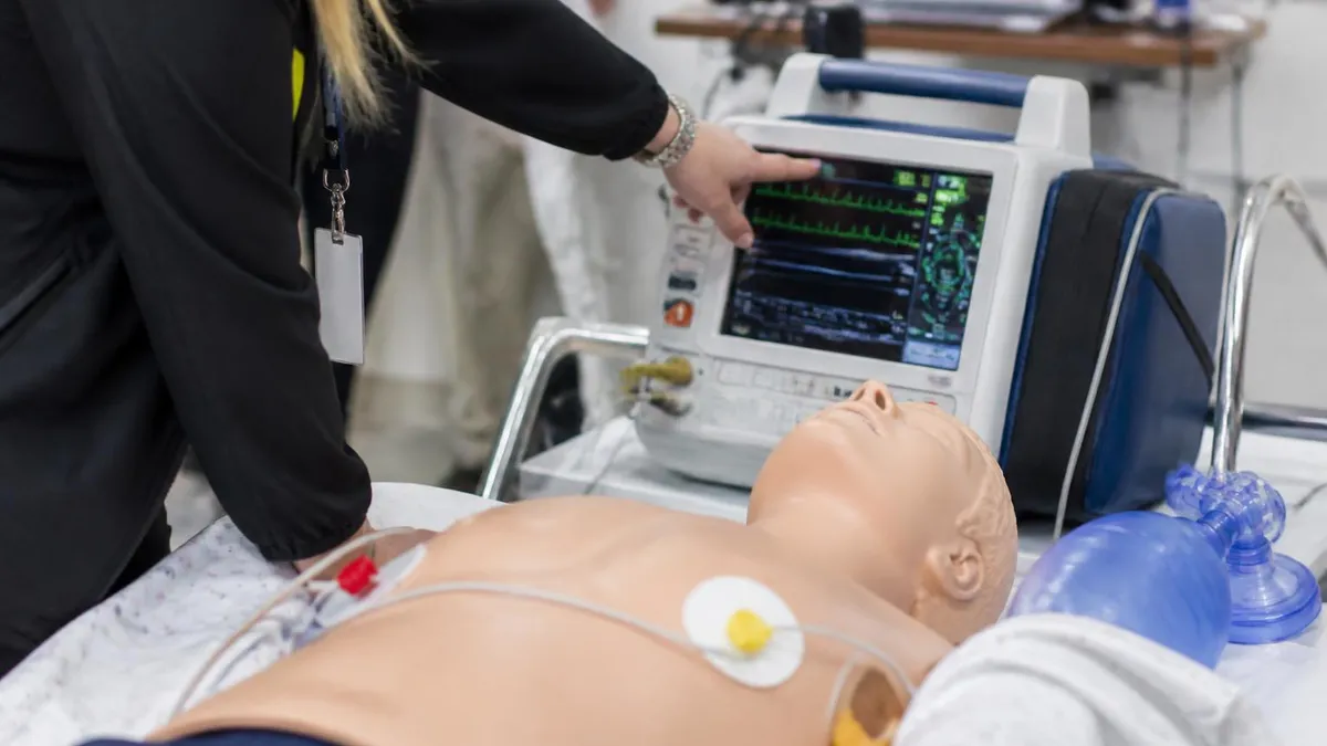
Ventricular fibrillation is a chaotic, disorganized electrical rhythm in which the ventricles quiver rather than contract effectively, producing no meaningful cardiac output. On an ECG, VF appears as an irregular, undulating baseline with no identifiable P waves, QRS complexes, or T waves. The rhythm is coarse when the amplitude is high (generally more responsive to defibrillation) and fine when the amplitude is low. Pulseless ventricular tachycardia (pVT) is treated identically to VF within the ACLS shockable rhythm pathway, which is why both rhythms share the same algorithm branch.
The 2020 AHA Guidelines reinforced a high-quality CPR-first approach while maintaining the primacy of early defibrillation. The algorithm is structured around 2-minute CPR cycles, with rhythm and pulse checks occurring at the end of each cycle rather than interrupting compressions unnecessarily. This rhythmic structure minimizes no-flow time — the period during which the heart receives no perfusion pressure — and is the single most influential factor in improving neurologically intact survival rates after cardiac arrest. Every provider on the resuscitation team must know exactly what happens at each two-minute checkpoint.
Drug therapy within the VF algorithm is administered during ongoing CPR, never as a reason to pause compressions. Epinephrine 1 mg IV/IO is given every 3 to 5 minutes beginning after the second shock is delivered. Amiodarone 300 mg IV/IO is the first-line antiarrhythmic for shock-refractory VF, with a second dose of 150 mg available if needed. Lidocaine 1 to 1.5 mg/kg IV/IO is an acceptable alternative antiarrhythmic when amiodarone is unavailable or contraindicated. Understanding when and why each drug is administered — not just memorizing doses — will help you answer ACLS exam questions more accurately.
Reversible causes, summarized by the Hs and Ts mnemonic, must be identified and corrected during any resuscitation attempt. The six Hs are hypovolemia, hypoxia, hydrogen ion excess (acidosis), hypo/hyperkalemia, hypothermia, and hypoglycemia. The five Ts are tension pneumothorax, tamponade (cardiac), toxins, thrombosis (pulmonary), and thrombosis (coronary). Failing to address an underlying cause is one of the most common reasons resuscitation efforts ultimately fail even when the algorithm is followed correctly. Every team leader should vocalize a systematic search for reversible causes early in the resuscitation.
Airway management during VF resuscitation requires a measured approach. The AHA guidelines de-emphasize early intubation in favor of continuous high-quality chest compressions. A bag-valve mask with a properly sized oral airway adjunct is appropriate for the first several minutes of resuscitation. When advanced airway placement (endotracheal tube or supraglottic airway) is performed, compressions should continue uninterrupted, and ventilations should be delivered at a rate of one breath every 6 seconds (10 breaths per minute) once the airway is secured. Capnography waveform monitoring is strongly recommended to confirm placement and assess CPR quality.
For candidates preparing for their certification exam, the shockable rhythm pathway is tested repeatedly through scenario-based questions and rhythm strip interpretation. Practicing with timed drills, memorizing energy levels for biphasic defibrillators (typically 120 to 200 J for the first shock, manufacturer-recommended for subsequent shocks), and understanding the rationale behind each intervention will build the mental fluency needed on test day. If you want a structured curriculum that reinforces algorithm mastery, consider reviewing the full acls ventricular fibrillation algorithm content pathway before your exam date.
ACLS Ventricular Fibrillation by the Numbers

ACLS Ventricular Fibrillation Algorithm: Step-by-Step
Recognize VF/pVT and Call for Help
Deliver First Defibrillation Shock
Resume CPR and Establish IV/IO Access
Rhythm Check → Shock 2 → Give Epinephrine
Rhythm Check → Shock 3 → Give Amiodarone
Treat Reversible Causes (Hs and Ts)
Understanding drug dosages and their timing within the ACLS ventricular fibrillation algorithm is one of the most heavily tested areas on the certification exam. Medications are administered during active CPR cycles — never as a justification for pausing compressions — and providers must be able to recall doses instantly under pressure. The two categories of drugs used in the VF/pVT pathway are vasopressors and antiarrhythmics, each serving a distinct physiological purpose in the resuscitation sequence.
Epinephrine remains the foundational vasopressor in ACLS despite decades of debate about its long-term outcomes. Its alpha-1 adrenergic effects cause peripheral vasoconstriction, which increases aortic diastolic pressure and, consequently, coronary perfusion pressure — the driving force for blood flow to the myocardium during CPR. The dose is 1 mg IV/IO, delivered every 3 to 5 minutes throughout the resuscitation. High-dose epinephrine (up to 0.2 mg/kg) was studied extensively and is not recommended for routine use; it may worsen post-resuscitation myocardial dysfunction despite improving short-term ROSC rates.
Vasopressin 40 units IV/IO was once listed as an alternative to epinephrine for the first or second dose, but the 2020 AHA Guidelines removed vasopressin from the adult algorithm because it offers no advantage over epinephrine and adds complexity. Providers should use epinephrine exclusively unless institutional protocols specify otherwise. On ACLS examinations, questions about vasopressin often test whether candidates know it has been removed from the current guidelines — a common source of errors for candidates studying from older materials.
Amiodarone is the preferred antiarrhythmic for VF and pVT that persists after three shocks. The mechanism involves blocking sodium, potassium, and calcium channels as well as alpha- and beta-adrenergic receptors, making it a broadly acting antiarrhythmic drug. The first dose is 300 mg IV/IO given as a rapid push; a second dose of 150 mg may follow for continued refractory VF. Side effects in the acute setting include hypotension and bradycardia, which become relevant considerations during post-ROSC care but are generally acceptable trade-offs during active resuscitation.
Lidocaine serves as the alternative antiarrhythmic when amiodarone is not available or the patient has a known contraindication. The initial dose is 1 to 1.5 mg/kg IV/IO, with a second dose of 0.5 to 0.75 mg/kg available if needed. Lidocaine's mechanism centers on sodium channel blockade, suppressing abnormal automaticity and re-entry circuits in ventricular tissue.
While head-to-head evidence slightly favors amiodarone for survival to hospital admission, lidocaine remains a clinically appropriate choice and should be known thoroughly for the exam. Magnesium sulfate 1 to 2 g IV/IO is indicated specifically for torsades de pointes — it is not a routine antiarrhythmic for standard VF and should not be substituted for amiodarone or lidocaine in the shockable rhythm algorithm.
Sodium bicarbonate is another drug that appears on ACLS exams in the context of reversible causes. It is not part of the standard VF algorithm but may be indicated for specific circumstances: pre-existing metabolic acidosis, hyperkalemia, or tricyclic antidepressant overdose. Routine sodium bicarbonate administration during cardiac arrest is not recommended because it can paradoxically worsen intracellular acidosis and create a hyperosmolar state. When a question asks about bicarbonate use in VF, the answer almost always relates to a specific underlying cause rather than routine resuscitation practice.
Calcium chloride (500 to 1,000 mg IV) is reserved for hyperkalemia, hypocalcemia, calcium channel blocker overdose, or hypermagnesemia — not routine VF. Atropine, which was formerly used for asystole and PEA, has been removed from the cardiac arrest algorithm entirely and is no longer recommended for pulseless rhythms. These removals and replacements are the source of many high-yield exam questions, so candidates who study with outdated resources often make predictable errors. Staying current with the 2020 AHA guidelines ensures your drug knowledge is aligned with what the exam actually tests.
Rhythm Recognition, Defibrillation, and CPR Quality in VF
Ventricular fibrillation appears on the ECG as a completely chaotic, irregular baseline with no discernible P waves, QRS complexes, or T waves. The amplitude and frequency of the waveform vary continuously, producing a pattern that can look almost sinusoidal at times and completely erratic at others. Coarse VF — defined by waveform amplitude greater than 3 mm — generally indicates a more recently initiated arrest and tends to respond better to defibrillation than fine VF, which has lower amplitude and suggests prolonged ischemia of the ventricular myocardium.
Distinguishing VF from artifact is a critical skill that exam questions frequently probe. Motion artifact from compressions, a loose lead, or patient movement can mimic VF on a monitor. The clinical rule is straightforward: if the patient has no pulse and the monitor shows a chaotic rhythm, treat as VF. Conversely, if a patient appears to have VF on the monitor but is conscious and has a palpable pulse, the rhythm is artifact — stop compressions and reassess. Pulseless ventricular tachycardia produces a wide, regular monomorphic or polymorphic pattern and is treated identically to VF within the shockable pathway.
Amiodarone vs. Lidocaine for Shock-Refractory VF: Key Tradeoffs
- +Amiodarone has multi-channel blocking properties (Na, K, Ca, alpha, beta) that address multiple arrhythmia mechanisms simultaneously
- +Clinical trial data (ARREST, ALIVE trials) show improved survival to hospital admission with amiodarone vs. placebo or lidocaine
- +Amiodarone is the AHA first-line recommendation for shock-refractory VF and pVT, reflecting guideline authority
- +A second 150 mg dose of amiodarone provides an additional treatment option when VF persists after the first antiarrhythmic dose
- +Lidocaine has a longer track record, is widely stocked in crash carts globally, and providers may have more familiarity with its use
- +Lidocaine's narrower mechanism (sodium channel blockade only) may be preferable when a pure membrane-stabilizing effect is desired
- −Amiodarone can cause significant acute hypotension, particularly with rapid IV infusion during active resuscitation
- −Amiodarone's bradycardic effect becomes problematic in post-ROSC management and requires careful monitoring
- −Lidocaine has less robust evidence for improving survival to hospital discharge compared to amiodarone in randomized trials
- −Neither antiarrhythmic has demonstrated a statistically significant improvement in neurologically intact survival to hospital discharge in the largest trials
- −Both drugs require IV or IO access, which takes time to establish, delaying administration in the early resuscitation phase
- −Routine use of antiarrhythmics is considered after the third shock, meaning drug therapy is always secondary to early defibrillation and high-quality CPR
ACLS VF Algorithm Certification Prep Checklist
- ✓Memorize the two-minute CPR cycle structure and identify exactly when rhythm checks, shocks, and drug doses occur within each cycle
- ✓Know the first biphasic defibrillation energy range (120–200 J) and the monophasic default (360 J) without hesitation
- ✓Recite epinephrine dosing (1 mg IV/IO every 3–5 minutes) and identify when the first dose is given relative to the algorithm sequence
- ✓State amiodarone first-dose (300 mg) and second-dose (150 mg) amounts and confirm when antiarrhythmics enter the algorithm (after shock 3)
- ✓Identify lidocaine as the approved alternative antiarrhythmic (1–1.5 mg/kg IV/IO) when amiodarone is unavailable
- ✓List all six Hs and five Ts reversible causes and describe at least one intervention to correct each during active resuscitation
- ✓Describe the ECG appearance of coarse VF, fine VF, and pulseless ventricular tachycardia, and explain why all three follow the shockable pathway
- ✓Confirm that vasopressin and atropine are no longer included in the 2020 AHA cardiac arrest algorithm
- ✓Practice identifying ROSC on capnography (sudden ETCO2 rise to >35 mmHg) and describe the immediate transition to post-cardiac arrest care
- ✓Complete at least three full timed algorithm simulation drills — oral, written, and scenario-based — before your certification exam date
Compressions Before and After Every Shock — No Exceptions
The single most common technique error in ACLS VF scenarios is pausing CPR for more than 10 seconds to charge the defibrillator or assess the rhythm. AHA data show that every 10-second interruption in compressions reduces coronary perfusion pressure toward zero, requiring up to 60 seconds of compressions to rebuild. Pre-charge the defibrillator in the last 30 seconds of the CPR cycle so the only interruption is the brief shock delivery — then resume compressions within 5 seconds of shock delivery. This habit dramatically improves outcomes in both real codes and certification scenarios.
Post-cardiac arrest care begins the moment return of spontaneous circulation (ROSC) is achieved, and the quality of care in the first hour after ROSC is as important to neurological outcomes as the resuscitation itself. The immediate priorities after ROSC are optimizing oxygenation and ventilation, initiating hemodynamic stabilization, obtaining a 12-lead ECG to evaluate for ST-elevation myocardial infarction (STEMI), and activating targeted temperature management (TTM) protocols if indicated. Each of these interventions has robust evidence behind it and is tested on the ACLS certification exam.
Oxygenation targets after ROSC are precise: titrate inspired oxygen to maintain SpO2 between 94 and 99 percent. Hyperoxia — defined as a PaO2 above 300 mmHg — has been associated with increased mortality in post-arrest patients, likely due to oxygen free radical generation in reperfused brain tissue. Similarly, hypoxia (SpO2 below 94%) must be avoided because the post-arrest brain is exquisitely sensitive to secondary ischemic injury. Ventilation should target a PaCO2 of 35 to 45 mmHg; both hypocapnia (causing cerebral vasoconstriction) and hypercapnia (causing cerebral vasodilation and increased intracranial pressure) are harmful in the post-arrest period.
Targeted temperature management involves cooling the patient to a core body temperature of 32 to 36 degrees Celsius for 24 hours following ROSC in comatose survivors of cardiac arrest. The 2020 AHA Guidelines maintain TTM as a Class I recommendation for comatose adults after out-of-hospital cardiac arrest with an initial shockable rhythm (VF or pVT). The mechanism is thought to involve suppression of the metabolic cascade triggered by reperfusion injury, including excitotoxicity, inflammatory cytokine release, and apoptosis.
Active rewarming should be gradual — typically 0.25 to 0.5 degrees Celsius per hour — to avoid rebound hyperthermia, which is independently associated with poor neurological outcomes.
Hemodynamic optimization targets a mean arterial pressure (MAP) of at least 65 mmHg and a systolic blood pressure above 90 mmHg in the post-ROSC period. Vasopressor support with norepinephrine or dopamine is commonly required, and fluid resuscitation should be guided by dynamic measures of preload responsiveness rather than static CVP targets.
Cardiogenic shock is common after resuscitation from VF because the myocardium has been subjected to ischemia during arrest and may exhibit post-resuscitation myocardial dysfunction — sometimes called myocardial stunning — even when the underlying coronary anatomy is normal. An echocardiogram is invaluable in this early phase for assessing ventricular function and guiding management.
Coronary angiography with primary percutaneous coronary intervention (PCI) is indicated for all post-cardiac arrest patients with STEMI on the initial 12-lead ECG. For patients without STEMI who remain comatose, the timing of coronary angiography is more nuanced; the COACT and TOMAHAWK trials suggested that immediate angiography does not improve outcomes compared to angiography after neurological stabilization, but individualized clinical judgment remains essential. Communication with the cardiac catheterization laboratory team should occur early in any resuscitation from VF, since coronary occlusion is the most common precipitating cause of sudden cardiac arrest in adults over 40.
Neurological prognostication after cardiac arrest is a multidisciplinary process that should not begin before 72 hours of normothermia in patients who underwent TTM. Premature withdrawal of life-sustaining therapy based on early neurological examination is one of the most concerning sources of potentially avoidable death in post-arrest patients. The ACLS guidelines recommend a multimodal approach to prognostication that combines clinical examination, EEG findings, serum biomarkers (particularly neuron-specific enolase), somatosensory evoked potentials, and brain imaging (CT or MRI). No single finding is sufficient to declare poor prognosis with certainty in the immediate post-arrest period.
For ACLS exam candidates, post-cardiac arrest care questions typically involve applying the ROSC transition protocol: correct oxygen titration, 12-lead ECG interpretation, TTM indications, and blood pressure targets. These questions reward candidates who understand the physiological rationale behind each intervention rather than simply memorizing numbers. The post-arrest period is a direct continuation of the VF algorithm, and demonstrating a seamless transition from resuscitation to post-ROSC management is what the certification exam is designed to evaluate at the highest level of competency.

The 2020 AHA Guidelines made several significant changes to the cardiac arrest algorithms, including removing vasopressin and atropine from the pulseless arrest pathway, updating TTM temperature targets, and refining post-ROSC oxygenation thresholds. Candidates using study materials based on the 2015 or earlier guidelines will encounter questions where their memorized answers are incorrect. Always verify that your textbooks, practice tests, and review courses cite the 2020 AHA Guidelines update before your exam date.
Among the most common mistakes ACLS certification candidates make on their exams are errors related to algorithm sequencing — specifically, misidentifying when to administer drugs relative to shocks and CPR cycles. The algorithm's structure is logical but unforgiving: epinephrine is not given with the first shock, antiarrhythmics do not enter until after the third shock, and every intervention happens during CPR, not instead of it. Candidates who blur these boundaries consistently choose incorrect answers on scenario-based questions, even when they know the individual drug doses correctly.
A second frequent error involves confusing the shockable and non-shockable algorithm pathways. Ventricular fibrillation and pulseless ventricular tachycardia are shockable rhythms. Asystole and pulseless electrical activity (PEA) are non-shockable rhythms. The non-shockable pathway has no defibrillation step, uses epinephrine as the only medication, and relies entirely on CPR quality and reversible cause correction.
Many exam questions deliberately present a rhythm that looks like one but is actually the other — for example, artifact that mimics VF, or a very slow, wide PEA that could be confused with a pulseless brady rhythm. Developing strong rhythm interpretation skills eliminates most of these errors.
Timing errors around the two-minute CPR cycle represent a third high-frequency mistake. Some candidates assume that epinephrine should be given immediately when IV access is established, regardless of where they are in the cycle. The correct approach is to give epinephrine as soon as possible after establishing access, but only within the context of continuing CPR — the drug is pushed during compressions.
If IV access is established during the second shock cycle, give epinephrine then; do not wait for the next rhythm check. The every-3-to-5-minute interval simply means you should not re-dose in less than 3 minutes, not that you must wait exactly 3 minutes from the start of resuscitation.
Many candidates also underestimate the importance of the Hs and Ts on exam questions. A scenario that presents a patient in refractory VF who recently received succinylcholine is pointing toward hyperkalemia as a reversible cause — the correct answer involves calcium chloride and sodium bicarbonate, not simply continuing the standard algorithm.
A patient in cardiac arrest following trauma with decreased breath sounds on one side needs needle decompression for tension pneumothorax before any antiarrhythmic will have a chance to work. The algorithm is a framework, not a script, and exam questions regularly test whether candidates can identify when a clinical clue should trigger a departure from the standard sequence.
Airway management misconceptions also appear frequently on ACLS exams. The 2020 guidelines support a ventilation strategy that does not prioritize early intubation.
A common incorrect answer involves stopping CPR to perform laryngoscopy, or delivering ventilations at too high a rate after intubation (greater than 10 per minute in adults). Hyperventilation during cardiac arrest increases intrathoracic pressure, decreases venous return to the heart, and reduces coronary perfusion pressure — it is one of the most physiologically damaging things a provider can do during resuscitation, and it is a specific exam trap. The correct post-intubation rate is exactly 10 breaths per minute (one every 6 seconds), with compressions continuing without pause.
Team dynamics and communication errors, while not always emphasized in self-study, are tested on scenario-based ACLS skill stations. The 2020 guidelines reinforce a team-based resuscitation model with clearly defined roles: team leader, compressor, airway provider, IV/medication nurse, and recorder.
Closed-loop communication — where an order is repeated back verbatim and confirmed before execution — prevents drug dosing errors and ensures interventions are not duplicated or missed. On written exams, questions about team leadership and communication appear in the context of recognizing when someone should speak up about a potential error, when roles should be rotated, and how to debrief effectively after a resuscitation.
Finally, candidates often neglect the transition out of the resuscitation. When ROSC is achieved, the exam expects providers to immediately reassess airway, begin oxygen titration to SpO2 94–99%, obtain a 12-lead ECG, initiate TTM if appropriate, and transfer to intensive care.
When termination of resuscitation is indicated — after prolonged resuscitation without ROSC, absence of any organized rhythm, ETCO2 persistently below 10 mmHg — the exam expects providers to recognize this decision point with compassion and clinical clarity. Reviewing the acls ventricular fibrillation algorithm through a case-based lens, where you practice making these decisions in context, is the most effective way to eliminate algorithm errors before your certification exam.
Building the mental fluency to execute the ACLS ventricular fibrillation algorithm under pressure requires more than passive reading — it demands active, retrieval-based practice over multiple study sessions. Research on procedural learning consistently shows that spaced repetition (studying material over increasing intervals) produces far more durable retention than massed practice (cramming). For ACLS certification, this means spacing your study over at least two to three weeks rather than completing all preparation in a single weekend before the exam.
Start your preparation by drawing the entire VF algorithm from memory without looking at a reference. Begin with the initial recognition step, walk through each two-minute CPR cycle, note each rhythm check and shock, and annotate every drug dose and timing. Do this as a blank-page exercise, then compare your diagram to the official AHA algorithm.
The gaps you find — the steps you forgot or placed in the wrong order — are exactly the areas that need additional focused review. Repeat this exercise daily until your diagram matches the algorithm perfectly and you can produce it in under three minutes.
Use practice scenarios to build decision-making speed. Work through written ACLS scenarios where you are given a patient presentation, a rhythm strip, and a list of interventions performed so far, and you must identify the next correct action. This format closely mirrors the actual exam question structure.
Focus particular attention on the distractors — incorrect answer choices that would be appropriate in a different clinical context. Understanding why each wrong answer is wrong is as instructive as knowing why the correct answer is right, and this meta-level analysis is what distinguishes candidates who score in the high 80s from those who barely pass.
Form a study group with colleagues who are also preparing for ACLS certification. Verbalize the algorithm aloud while a partner tests you with rhythm strips and clinical scenarios. Teaching the algorithm to someone else — explaining not just what to do but why each step follows logically from the previous one — is one of the most effective consolidation strategies known in educational psychology.
When you can explain the physiological rationale for giving epinephrine after the second shock rather than the first, or why amiodarone is used for shock-refractory VF rather than adenosine, you have internalized the material at the level the exam expects.
Review ECG rhythm strips daily in the two weeks before your exam. Use an app, a flashcard deck, or an online rhythm library to expose yourself to VF, pVT, coarse versus fine VF, artifact, and the non-shockable rhythms (asystole, PEA). Time yourself — aim to correctly identify each rhythm in under 10 seconds, because that is approximately the time you have in a real cardiac arrest before bystanders expect a decision. If a particular rhythm consistently trips you up, spend an extra session specifically on that pattern until recognition becomes automatic rather than deliberate.
Attend a hands-on skills session if your certification program includes one (and most AHA-approved programs do). Physical repetition of chest compressions at the correct rate and depth, pad placement on a manikin, and practice delivering verbal orders in a simulated code scenario builds procedural memory that reading alone cannot create. Studies of resuscitation training consistently show that hands-on simulation improves both skill retention and algorithm adherence six months after training — far better outcomes than provider groups who completed only knowledge-based coursework.
On the day of your exam, review the algorithm one final time in the morning, then trust your preparation. The ACLS written exam is a multiple-choice assessment, and the vast majority of questions have one clearly correct answer based on current AHA guidelines. If you encounter a question where two answers seem plausible, ask yourself: which of these choices is explicitly supported by the 2020 AHA Guidelines?
Questions are written to test guideline compliance, not clinical judgment in edge cases. Candidates who answer based on personal clinical experience rather than guideline recommendations sometimes choose the wrong answer even when their experience-based reasoning is defensible. The exam tests the algorithm as written — know it cold, and you will pass with confidence.
ACLS Questions and Answers

ACLS Recertification Course — Complete Guide (2026)

ACLS Tachycardia Algorithm: 2020 AHA Step-by-Step Guide

ACLS Heart Rhythms: The Complete Guide to ECG Recognition, Interpretation, and Treatment Protocols

ACLS Certification Cost: Complete 2026 Price Guide for Initial Courses, Renewals, Online Options & Hidden Fees

HeartCode ACLS Answers: Complete 2026 Guide to Passing the Online Module With Confidence
About the Author

Registered Nurse & Healthcare Educator
Johns Hopkins University School of NursingDr. Sarah Mitchell is a board-certified registered nurse with over 15 years of clinical and academic experience. She completed her PhD in Nursing Science at Johns Hopkins University and has taught NCLEX preparation and clinical skills courses for nursing students across the United States. Her research focuses on evidence-based exam preparation strategies for healthcare certification candidates.