ACLS Tachycardia Algorithm: 2026 July AHA Step-by-Step Guide
🗨️ ACLS tachycardia algorithm 2026 July AHA: stable vs unstable, narrow vs wide QRS, cardioversion joules, adenosine, amiodarone doses. Step-by-step walkthrough.

The ACLS Tachycardia Algorithm at a Glance
When the monitor flashes a heart rate over 150 and the patient looks ashen, the clock starts ticking. The acls tachycardia algorithm is the framework that gets teams from "something is wrong" to a defensible plan in under two minutes.
Tachycardia is defined as a heart rate above 100 beats per minute, but the algorithm only really fires up when the rate climbs past 150. Below that, sinus tachycardia from fever, pain, hypovolemia, or anxiety is more likely than a primary arrhythmia. Above 150, the rhythm itself starts to drive the problem.
The 2020 AHA guidelines build the algorithm around two hinges: is the patient stable, and is the QRS narrow or wide? Four binary questions and the path picks itself.
This walkthrough mirrors what you will see on the cards, on the test, and at the bedside. We will move through assessment, the stable versus unstable fork, synchronized cardioversion doses, narrow QRS lane, wide QRS lane, and the drug doses that get tested most.
One note before we start: this is a teaching summary anchored to the 2020 AHA guidelines and the current ACLS provider manual. It is not a substitute for the official guidelines or local protocol. If your institution has modified the algorithm, the institutional version wins. Use this guide to drill the structure so the underlying logic stays sharp.
Why memorize an algorithm at all? Because in the first 30 seconds of a real event nobody pulls out a phone to check doses. You either know that adenosine starts at 6 mg or you don't. You either know that polymorphic VT does not get synchronized cardioversion or you waste precious seconds on a button that will not arm. The algorithm exists so the cognitive load drops and your hands can move.
There is also a teaching angle. Residents and new nurses watch how senior clinicians run a tachycardia and they pattern-match for life. If you can articulate the decision tree out loud as you work, you train the next generation while you treat the patient in front of you. The algorithm is communication too.
The Four Binary Questions
- Stable or unstable? Check for hypotension, altered mental status, shock, ischemic chest pain, or acute heart failure.
- Narrow or wide QRS? 0.12 seconds is the cutoff on the 12-lead.
- Regular or irregular? Look at the rhythm strip with your own eyes.
- Cause identified? Treat hypoxia, hypovolemia, ischemia, electrolytes before the algorithm.
Initial Patient Assessment
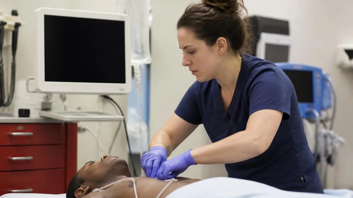
Before anything else, run the primary survey. Airway open? Breathing adequate? Circulation present with a pulse and a measurable blood pressure? Place the patient on oxygen if hypoxic, get IV or IO access, attach the monitor and pulse oximeter, and obtain a 12-lead ECG if it will not delay treatment.
Hunt for the underlying cause while you stabilize. Hypoxia, hypovolemia, electrolyte abnormalities, drug toxicity, acute coronary syndrome, pulmonary embolism, sepsis, and thyroid storm all push the rate up. The algorithm assumes you are doing this work in parallel.
If a fever is driving sinus tachycardia at 130, antipyretics and fluids will do more than adenosine. The point of assessment is to make sure the rate is the problem, not the symptom.
Vital signs deserve a closer look than they usually get. An automated cuff often misses or misreads in fast rhythms. Confirm with a manual cuff if anything seems off. End tidal CO2, if you have it, is a useful surrogate for cardiac output and will trend down before the blood pressure tanks.
Build a quick history while someone else gets the IV. Onset of symptoms, palpitations versus pre-syncope, prior episodes, structural heart disease, prior MI, cardiomyopathy, recent medication changes, electrolyte abnormalities, recreational drug use, and family history of sudden death. Each item shifts the differential and tightens the treatment plan before the 12-lead even prints.
Look at the patient too. Skin color, mental status, peripheral perfusion, neck veins, lung sounds. A tachycardia with cool mottled extremities and altered mental status is shock until proven otherwise. A tachycardia with warm dry skin and normal mentation buys you time to think.
Order labs in parallel: a basic metabolic panel for potassium and magnesium, a troponin if ischemia is on the table, a TSH if thyroid storm is plausible, a CBC if sepsis or anemia is suspected, and a D-dimer or CT-PA if PE is in the differential. Do not wait on results to start treatment, but the moment they are back, recalibrate.
Key Tachycardia Numbers
Stable vs Unstable: The First Branch
The first real decision is stability. The AHA defines instability with four signs, and any single one is enough: hypotension, acutely altered mental status, signs of shock, ischemic chest discomfort, or acute heart failure.
The phrasing matters. "Acutely" rules out the chronic confusion of a demented patient and the chronic hypotension of an athlete. You are asking whether the tachycardia is causing the deterioration right now.
If the patient is unstable and the tachycardia is the likely cause, the answer is synchronized cardioversion. Sedate if conscious and time allows, charge the defibrillator, hit the sync button, confirm the marker is on the R wave, and deliver the shock.
If sync fails to deploy on polymorphic VT or torsades, treat it as ventricular fibrillation and defibrillate unsynchronized at the maximum dose your device offers.
Stable does not mean safe forever. Reassess constantly. A patient who is stable at 180 can crash within minutes, especially with underlying ischemia or heart failure. Keep pads on, sedation drawn up, and a plan ready to switch to the unstable arm of the algorithm.
Sedation for cardioversion is a conversation. Etomidate 0.1 to 0.3 mg per kg, propofol 0.5 to 1 mg per kg, or midazolam 1 to 2 mg with fentanyl 50 to 100 mcg are all reasonable depending on hemodynamics. In a truly unstable patient with crashing pressures, you may shock with minimal or no sedation. The ethics committee will forgive you. The morgue will not.
Confirm the synchronization marker on the R wave before every shock. The sync function has to re-arm after each delivery; if you press the shock button and nothing happens, the most common reason is that sync did not re-engage. Some monitors handle this automatically, some do not. Know your device.
Synchronized Cardioversion Energy Doses
Start at 50 to 100 joules biphasic. Most commonly supraventricular tachycardia such as AVNRT or AVRT. Step up the energy if the first shock fails to convert. Confirm the sync marker is on the R wave before every delivery. Sedate the awake patient with etomidate or propofol if hemodynamics allow.
120 to 200 joules biphasic. Atrial fibrillation with rapid ventricular response, occasionally atrial flutter with variable conduction. Consider anticoagulation status and duration of AFib before elective cardioversion. Emergency cardioversion for unstable patient overrides anticoagulation concerns.
100 joules biphasic for monomorphic ventricular tachycardia. Assume VT in any wide regular tachycardia until proven otherwise, especially in patients with structural heart disease or prior MI. Step up the energy if the first shock fails. Be prepared to convert to defibrillation if rhythm degenerates.
Defibrillation dose, NOT synchronized. Polymorphic VT or torsades cannot be synchronized because there is no consistent R wave to sync to. Treat as VF: defibrillate at maximum joules. Give magnesium 1 to 2 g IV if torsades is suspected and fix electrolytes.
Narrow QRS Tachycardia
Narrow regular tachycardia in a stable adult is most often supraventricular tachycardia. The first move is vagal maneuvers. The modified Valsalva has the best conversion rate in trials.
If vagal fails, adenosine is next. The dose is 6 mg rapid IV push followed by a 20 ml saline flush. If there is no conversion in one to two minutes, give 12 mg. A third 12 mg dose is permitted.
Warn the patient about the brief pause, chest pressure, and impending doom that adenosine produces. It is short, but it is real, and an unwarned patient remembers it for years.
Adenosine has relative contraindications worth knowing. Active bronchospasm can flare. Heart transplant recipients are exquisitely sensitive — start at 3 mg. Dipyridamole or carbamazepine potentiate the effect; halve the dose. Caffeine and theophylline blunt it.
Narrow irregular tachycardia is almost always atrial fibrillation with rapid ventricular response. Adenosine will not convert it. Rate control is the goal: IV beta-blocker such as metoprolol 2.5 to 5 mg slow push, or a calcium channel blocker such as diltiazem 15 to 20 mg IV over two minutes.
Avoid calcium channel blockers in heart failure with reduced ejection fraction, and avoid both classes in pre-excited atrial fibrillation, where AV nodal blockade can drive the rate dangerously through an accessory pathway.
Atrial flutter sits in an awkward middle ground. The atrial rate is around 300, the ventricular response often locks at 150 with 2:1 conduction, which can be mistaken for SVT. Adenosine will not convert flutter, but the transient AV block it produces will expose the flutter waves on the rhythm strip, which is diagnostically useful. After diagnosis, rate control or cardioversion depending on stability and duration.
Multifocal atrial tachycardia is the third narrow irregular rhythm to know. Three or more distinct P wave morphologies, often in patients with severe COPD or pulmonary disease. Treatment is of the underlying lung disease and electrolyte repletion, not AV nodal blockade. Calcium channel blockers can help if rate control is needed.

Wide QRS Tachycardia
Wide complex tachycardia is the part that scares people, and it should. Until proven otherwise, wide regular tachycardia is ventricular tachycardia, especially in patients with structural heart disease, prior MI, or known low ejection fraction.
Treating VT as SVT with aberrancy is a classic, and dangerous, mistake. The default assumption is VT.
For stable wide regular monomorphic tachycardia, adenosine is reasonable as a diagnostic agent. If the rhythm breaks, it was probably SVT with aberrancy. If it does not, you have lost nothing.
If adenosine fails, move to an IV antiarrhythmic. Amiodarone is the workhorse: 150 mg IV over 10 minutes, repeat as needed, then 1 mg per minute infusion for six hours and 0.5 mg per minute for 18 hours.
Procainamide is 20 to 50 mg per minute IV until the arrhythmia is suppressed, hypotension develops, QRS widens more than 50 percent, or 17 mg per kg total. Sotalol 100 mg IV over five minutes is option three but is contraindicated in prolonged QT.
Wide irregular tachycardia is polymorphic VT or atrial fibrillation with pre-excitation. Polymorphic VT with prolonged QT is torsades de pointes: magnesium 1 to 2 g IV, fix electrolytes, defibrillate if collapse occurs. Polymorphic VT with normal QT suggests ischemia; treat the cause and use amiodarone.
Pre-excited AFib is treated with procainamide, ibutilide, or cardioversion — never AV nodal blockers. The accessory pathway in Wolff-Parkinson-White can conduct at one-to-one over 250 BPM and degenerate into ventricular fibrillation if the node is blocked.
Distinguishing VT from SVT with aberrancy on a 12-lead is its own art form. Brugada criteria and the Vereckei aVR algorithm exist for a reason. AV dissociation, fusion beats, capture beats, extreme axis deviation, and QRS over 160 ms in left bundle branch morphology all push toward VT. When in doubt, treat as VT — the consequence of missing it is worse than the consequence of giving amiodarone to SVT.
Age and history also matter. A 75-year-old with prior MI and a wide regular tachycardia is VT until proven otherwise — the positive predictive value is over 95 percent. A 25-year-old with no cardiac history and a wide regular tachycardia might still be VT, but the differential opens up to include antidromic AVRT and idiopathic VT.
NEVER give AV nodal blockers (adenosine, diltiazem, metoprolol, digoxin, amiodarone in some cases) for atrial fibrillation with pre-excitation (Wolff-Parkinson-White).
Blocking the AV node funnels impulses through the accessory pathway, which can conduct at one-to-one ratios over 250 BPM and degenerate into ventricular fibrillation.
Use procainamide, ibutilide, or synchronized cardioversion instead.
Drug Doses You Must Memorize
Drug doses get tested cold on the exam, so commit them to memory and rehearse them out loud. acls drugs are tested in context, and the algorithm rewards anyone who can recite the first dose without hesitating.
A practical tip on adenosine: the half-life is so short that the IV site matters. Push into the most proximal port you can find, ideally an antecubital or larger vein, and chase immediately with a 20 ml saline flush at the same time you elevate the arm.
Some clinicians use a stopcock with the syringe of adenosine and saline both connected, so the flush goes in milliseconds after the push. A peripheral IV in the hand will work, but the dose may need to be 12 mg to start because of dilution losses.
One last point on amiodarone. The 150 mg bolus over 10 minutes is for stable VT with a pulse. In pulseless VT or VF, the dose changes to 300 mg IV push, then 150 mg if needed. Do not confuse the two.
Procainamide has fallen out of favor in many centers, but the 2020 guidelines specifically mention it as a reasonable first-line option for stable wide regular tachycardia. PROCAMIO, a small randomized trial, suggested procainamide had a higher conversion rate and fewer adverse events than amiodarone for monomorphic VT. The drug is not dead, just out of fashion.
Sotalol is the third antiarrhythmic to memorize for the exam. 100 mg IV over five minutes, or 1.5 mg per kg. The IV formulation is not as widely stocked as the oral version, so check your pharmacy before the next code. Avoid in prolonged QT, severe renal impairment, and decompensated heart failure.
Magnesium deserves its own paragraph. For torsades de pointes, give 1 to 2 grams IV as a bolus, followed by an infusion of 1 to 2 grams per hour if recurrent. Check magnesium and potassium levels and replete aggressively — low levels of either prolong QT and perpetuate the arrhythmia.
Drug Doses by Indication
Indication: Stable narrow regular tachycardia (SVT). Also reasonable as a diagnostic agent in stable wide regular monomorphic tachycardia. Adenosine will not convert atrial fibrillation, atrial flutter, or polymorphic VT.
Dose: 6 mg rapid IV push with 20 ml saline flush. If no conversion in one to two minutes, give 12 mg. A third 12 mg dose is permitted but most clinicians stop after two failed pushes.
Half-life: Under 10 seconds. Push fast, flush immediately, ideally through a proximal antecubital IV. A peripheral IV in the hand may require 12 mg start dose because of dilution.
Warnings: Avoid in active bronchospasm and severe asthma. Halve the dose in heart transplant patients (start at 3 mg) and patients on dipyridamole or carbamazepine. Caffeine and theophylline blunt the effect.
Warn the patient about brief chest pressure, dyspnea, and sense of impending doom. These resolve in seconds but are distressing.
When to Call Electrophysiology
The algorithm has limits. Call electrophysiology early when the rhythm is unclear after a 12-lead, when the patient has Wolff-Parkinson-White with pre-excited AFib, when sustained VT recurs after first-line treatment, or when the patient is on chronic antiarrhythmics and the rhythm has changed.
Cardiology consult also belongs in the conversation early in pregnancy, in post-cardiac-surgery patients, and in anyone with a CIED that may be involved in the rhythm.
Some rhythms simply do not fit the textbook lanes. Fascicular VT looks narrow because the impulse uses the conduction system. Atrial tachycardia with 2:1 block can mimic SVT. Antidromic AVRT is wide regular but breaks with adenosine.
None of these are common, but they are the cases that end up in the EP lab, and getting the right person on the phone early shortens the patient's night.
Document everything. The rhythm strips before and after each intervention, the exact dose and route, the time of conversion, and the post-conversion 12-lead. This matters for the chart, for handoff, and for the case review that always follows a complicated tachycardia.
Post-arrest or post-conversion care is its own algorithm. Targeted temperature management for comatose survivors of VF/VT arrest. Coronary angiography for STEMI or unstable hemodynamics post-ROSC. Anticoagulation decisions for AFib that converted spontaneously or with electricity. None of this is in the tachycardia algorithm itself, but all of it follows from a successful resuscitation.

Tachycardia Algorithm Quick Checklist
- ✓Confirm HR > 150 BPM and that rate is causing symptoms
- ✓Primary survey: airway, breathing, circulation, oxygen if hypoxic
- ✓IV/IO access, monitor, pulse oximetry, 12-lead ECG
- ✓Identify and treat reversible causes (Hs and Ts)
- ✓Check for unstable signs: hypotension, altered mental status, shock, ischemia, acute HF
- ✓If unstable: synchronized cardioversion at rhythm-specific energy
- ✓If stable: assess QRS width (≤0.12s narrow, >0.12s wide) and regularity
- ✓Narrow regular: vagal maneuvers, then adenosine 6 mg, 12 mg, 12 mg
- ✓Narrow irregular: rate control with diltiazem or metoprolol
- ✓Wide regular: amiodarone 150 mg over 10 min (consider adenosine first)
- ✓Wide irregular: defibrillate if polymorphic; magnesium if torsades
- ✓Continuous reassessment; be ready to cardiovert if patient deteriorates
Making the Algorithm Automatic
Practice this algorithm until the branches feel automatic. The exam will not give you extra time to think, and the bedside gives you even less. The good news is that the structure is small.
Stable or unstable, narrow or wide, regular or irregular. Master the doses, rehearse the maneuvers, and the rest is just discipline.
Pair this material with regular hands-on simulation. Reading about adenosine is not the same as drawing it up at three in the morning with a saline flush in your off hand and a patient who is watching you. The algorithm tells you what to do. Practice tells your hands how.
Review the 2020 AHA changes annually. Algorithms get refined, doses get tightened, and the evidence base around magnesium, procainamide, and modified Valsalva continues to shift.
Stay current, and treat every shift on a tele unit as a chance to rehearse the branches in your head. When the real tachycardia rolls in, you will not be thinking about the algorithm. You will be running it.
A final word on team dynamics. Tachycardia codes are rarely solo. The team leader calls the rhythm and the dose. A second clinician pushes the drug or runs the defibrillator. A third manages airway and vitals. A fourth records times and doses. Roles get fuzzy in real events, but the algorithm gives everyone the same vocabulary. "Narrow regular, stable, 6 of adenosine" is a sentence the entire team understands.
Close the loop on every order. The person pushing adenosine repeats back "6 milligrams adenosine" and confirms after delivery. The person at the defibrillator calls "charging to 100 joules, sync on" and confirms before the shock. Closed-loop communication is the lowest-tech, highest-yield safety intervention in resuscitation, and it is free. Use it.
Adenosine vs Cardioversion for Stable SVT
- +Adenosine: fast, reversible, no sedation needed
- +Adenosine: diagnostic value for unclear narrow regular rhythms
- +Adenosine: cheap and universally available
- +Adenosine: short half-life means quick reassessment
- −Cardioversion: requires sedation if patient is awake
- −Cardioversion: small risk of triggering AFib or other arrhythmias
- −Adenosine: causes brief but distressing chest pressure and dyspnea
- −Adenosine: ineffective in AFib, AFlutter, and most VTs
ACLS Questions and Answers
Related ACLS Articles

ACLS Drugs: Complete 2026 Guide to Medications, Doses, Indications & Algorithm Use

ACLS Drug Doses: Complete Pharmacology Guide for 2026 Certification

ACLS Guidelines 2026: Complete AHA Update on Algorithms, Drugs, CPR Quality & Post-Arrest Care

ACLS Study Guide: Complete 2026 Certification Prep with Algorithms, Drugs & Practice Tests
About the Author
Registered Nurse & Healthcare Educator
Johns Hopkins University School of NursingDr. Sarah Mitchell is a board-certified registered nurse with over 15 years of clinical and academic experience. She completed her PhD in Nursing Science at Johns Hopkins University and has taught NCLEX preparation and clinical skills courses for nursing students across the United States. Her research focuses on evidence-based exam preparation strategies for healthcare certification candidates.