ACLS Bradycardia and Tachycardia Algorithms: Complete 2026 June Provider Guide
✏️ Master the 2026 June ACLS bradycardia and tachycardia algorithms: atropine, pacing, adenosine, cardioversion doses, rhythms, and decision steps for boards.

Key Numbers Every ACLS Provider Must Know
ACLS Bradycardia and Tachycardia Algorithms: Complete 2026 Provider Guide
The bradycardia and tachycardia algorithms sit at the heart of every ACLS advanced cardiovascular life support response. Cardiac arrest gets the headlines, but most ACLS scenarios you actually run in the ED, ICU, or rapid response team involve a patient who is still alive yet decompensating from a rhythm disturbance. Knowing when to give atropine versus when to pace, when to push adenosine versus when to shock — those are the decisions that separate a recovered patient from a code blue.
Both algorithms share the same opening move. Assess airway, breathing, and circulation. Put the patient on a monitor. Get a 12-lead ECG. Secure IV or IO access. Apply oxygen if hypoxic with a target SpO2 above 94%. Then ask the only question that matters: is the patient stable or unstable? That answer drives everything that follows on the rhythm algorithms.
An unstable rhythm is defined the same way for both ends of the spectrum. Look for hypotension with systolic BP under 90 mmHg, acutely altered mental status, signs of shock such as cool clammy mottled skin and poor capillary refill, ischemic chest discomfort with new ST changes, or acute heart failure with pulmonary edema and JVD. Any one of these in the presence of an abnormal rate means the rhythm is the problem, not just a symptom. Treat the rhythm fast, then treat the cause.
Stable patients get monitoring, expert consultation, and pharmacologic therapy delivered in a controlled order. Unstable patients get electricity or potent IV drugs immediately — the algorithms do not let you stall. Most students and even seasoned providers get caught freezing on whether the patient is unstable enough to act. The 2020 AHA guidance is clear: if you can name a single sign of instability, treat as unstable. Hesitation is the most common reason megacode candidates fail their scenarios.
This guide walks you through every decision branch the American Heart Association built into the 2020 guidelines (still current for the 2026 testing cycle), the drug doses you need committed to memory, and the rhythm strips you will see on the written exam and in the resus bay. The doses, energies, and decision rules below are the exact numbers that appear on the AHA written test and the megacode scoring rubric. If you are still building foundation knowledge, our acls algorithm overview maps how this fits with cardiac arrest, post-arrest care, and the systematic approach.
Unstable signs (any one triggers immediate intervention):
- Hypotension (SBP <90 mmHg or signs of shock)
- Acutely altered mental status
- Signs of shock (cool, clammy, mottled, poor cap refill)
- Ischemic chest discomfort or new ST changes
- Acute heart failure (pulmonary edema, JVD)
Stable patient = time to think. Get a 12-lead, identify the rhythm, treat reversible causes, consult cardiology. Unstable patient = act now. Bradycardia → atropine or pace. Tachycardia → synchronized cardioversion.
Algorithm Snapshots: Pick the Branch That Matches Your Patient
HR <50 bpm with symptoms. Assess for instability. If stable, identify and treat the underlying cause (hypoxia, electrolytes, drug effect from beta-blockers, calcium channel blockers, or digoxin toxicity). If unstable: Atropine 1 mg IV bolus, repeat every 3-5 min, max 3 mg. If atropine fails or is ineffective, start transcutaneous pacing or a chronotropic infusion (dopamine 5-20 mcg/kg/min or epinephrine 2-10 mcg/min). Call for expert consult and prepare for transvenous pacing in refractory cases or high-grade AV block.
Bradycardia Algorithm: Step-by-Step Flow
Step 1: Identify
Step 2: Stable?
Step 3: Atropine
Step 4: Pace or Pressors
Step 5: Expert Consult
Bradycardia Algorithm: When the Heart Is Too Slow
Symptomatic bradycardia means a heart rate below 50 beats per minute that is producing clinical signs. A heart rate of 45 in a marathon runner at rest is normal physiology and needs nothing more than reassurance. A heart rate of 45 in a 78-year-old who just collapsed with hypotension and confusion is a code-level emergency. The algorithm exists to help you separate the two scenarios and act on the second one without hesitation.
Identify the rhythm on the monitor and on the 12-lead. Common bradyarrhythmias you will see include sinus bradycardia, sinus arrest with junctional escape, first-degree AV block, Mobitz Type I (Wenckebach) which is usually benign, Mobitz Type II second-degree block which is concerning, and third-degree complete AV block which is the highest-risk scenario. Each rhythm has a different probability of responding to atropine, and the algorithm assumes you can identify them quickly from a strip.
Atropine 1 mg IV is the first-line pharmacological treatment for symptomatic bradycardia. It works by blocking the vagal tone that slows the SA and AV nodes. Push it fast and follow with a 10 to 20 mL saline flush. You can repeat every 3 to 5 minutes up to a total of 3 mg. The 2020 AHA guideline change increased the initial dose from 0.5 mg to 1 mg — make sure the version you trained on matches the current standard. Our aha acls reference covers every guideline update from the most recent cycle.
When atropine will not work
Atropine is largely ineffective in two situations: high-grade AV block (Mobitz Type II second-degree or third-degree complete heart block) and the post-cardiac transplant patient (denervated heart). Do not waste time pushing repeat doses while the patient deteriorates. Move directly to transcutaneous pacing or a chronotropic infusion. This is one of the most-tested distinctions on the written exam and one of the most common real-world errors at the bedside.
Transcutaneous pacing pearls
Apply the pacing pads in either anterior-posterior or anterior-lateral position. Set the rate at 60 to 80 bpm. Start the milliamps low and titrate up by 5 to 10 mA at a time until you see electrical capture on the monitor (a pacer spike followed by a wide QRS complex). Then confirm mechanical capture by palpating a femoral or radial pulse that matches the pacer rate. Mechanical capture is the only proof the heart is actually responding.
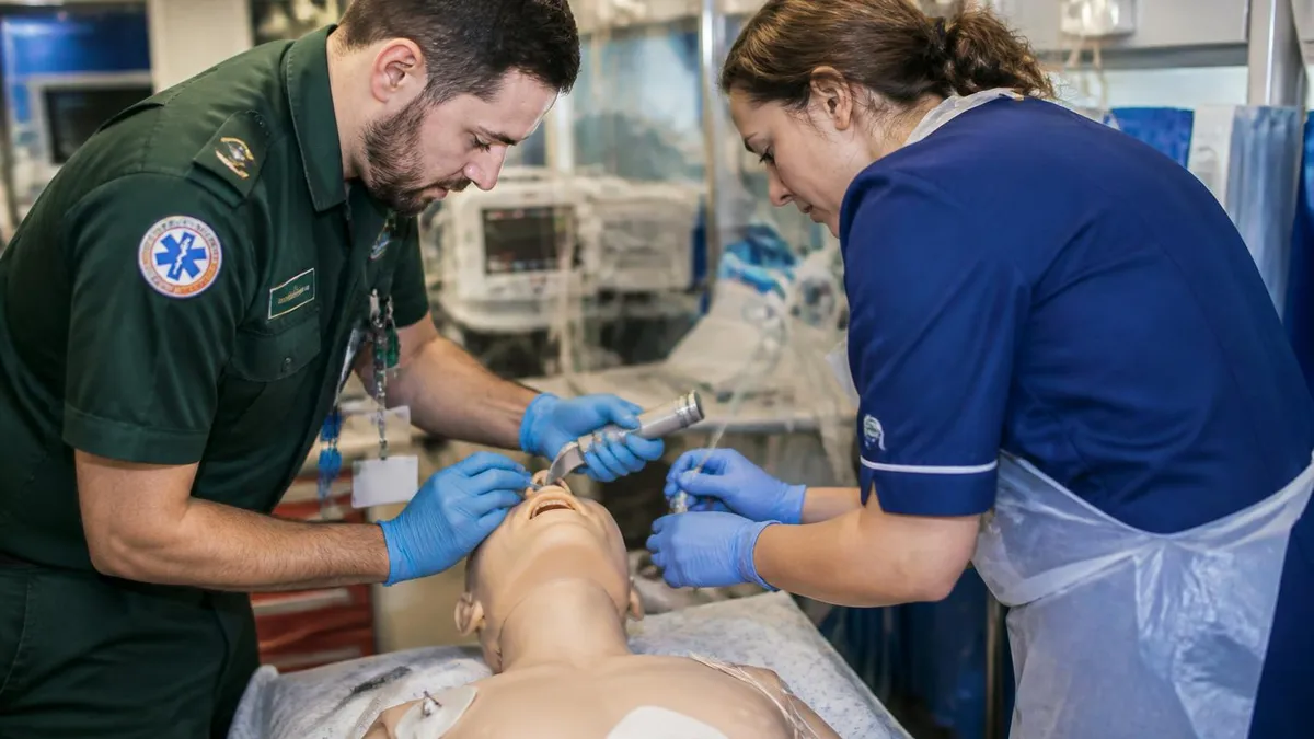
Pacing is painful for awake patients — give sedation with midazolam 1-2 mg IV or analgesia with fentanyl 25-50 mcg IV once the patient is stabilizing. Continue pacing until a transvenous pacer is placed or the underlying cause is reversed. If you are still preparing for certification, our acls classes near me guide explains how AHA-approved courses run the pacing megacode skills station.
Dopamine and epinephrine infusions
If pacing is not immediately available or fails to capture, start dopamine 5 to 20 mcg/kg/min titrated to BP and HR, or epinephrine 2 to 10 mcg/min titrated similarly. Both are positive chronotropes and inotropes. Epinephrine adds vasoconstriction at higher doses, which is useful when hypotension is the dominant feature. Dopamine is easier to start peripherally for short-term use, though central access is preferred for prolonged infusions.

Bradycardia Reversible Causes Checklist
- ✓Hypoxia — check SpO2, ABG, give oxygen, secure airway if needed
- ✓Hydrogen ion (acidosis) — ABG, treat underlying metabolic or respiratory cause
- ✓Hyperkalemia — STAT K+, give calcium chloride 1 g IV, insulin/D50, bicarb if acidotic
- ✓Hypothermia — core temperature, active rewarming, withhold drugs until >30°C
- ✓Tablets/toxins — beta-blocker (glucagon), CCB (calcium + high-dose insulin), digoxin (DigiFab)
- ✓Trauma — check for tension pneumothorax, hemorrhagic shock, neurogenic shock (high spinal)
- ✓Tamponade — bedside echo if available, pericardiocentesis if confirmed and unstable
Treat reversible causes in parallel
While you are working through atropine, pacing, and pressors, send a basic metabolic panel, magnesium, troponin, and a venous blood gas. The H's and T's still apply: hypoxia, hydrogen ion acidosis, hyperkalemia or hypokalemia, hypothermia, and the toxins category which covers most drug-induced bradycardias. Calcium chloride 1 g IV slow push reverses hyperkalemic conduction effects. Glucagon 3-10 mg IV is the antidote for beta-blocker overdose. High-dose insulin (1 unit/kg bolus then 0.5-1 unit/kg/hr) plus calcium and a glucose infusion treats calcium channel blocker overdose. Digoxin-immune Fab reverses dig toxicity.
The hypothermia exception
Severely hypothermic patients (core temp below 30°C) often present with bradycardia or asystole and will not respond to atropine, pacing, or chemical chronotropes until rewarmed. Avoid aggressive interventions in stable hypothermic bradycardia. Focus on active core rewarming, gentle handling, and supportive care. Cardiac drugs may accumulate in the cold patient and cause toxicity once rewarming starts and metabolism resumes. Knowing this distinction is a high-yield boards item.
Reading the bradycardia strip in 10 seconds
Look at three things on every strip before you make a treatment decision. First, the P-wave to QRS relationship — is every P followed by a QRS, are some Ps dropped, or is there complete dissociation between Ps and QRSs? Second, the PR interval — is it normal (under 200 ms), prolonged but constant (first-degree block), progressively lengthening before a dropped beat (Mobitz I), or normal until a sudden dropped beat with no warning (Mobitz II)? Third, the QRS width — narrow suggests junctional or higher origin, wide suggests ventricular escape and a much sicker conduction system.
If you can answer those three questions in 10 seconds, you can pick the right algorithm branch. A patient with sinus bradycardia at 42 bpm and a normal PR will likely respond to atropine. A patient with complete heart block, P-waves at 80 and ventricular escape at 30, will not respond to atropine and needs the pads on right now. Memorize the strip patterns by the time you walk into your acls certification exam.
Common bradycardia traps on the exam
Three scenarios trip up most candidates. First, asymptomatic bradycardia — a sleeping athlete at 38 bpm needs no intervention. Do not pace, do not push atropine. Second, bradycardia from inferior MI — the bradycardia itself is often vagally mediated and responds well to atropine, but the underlying MI is the real problem and gets reperfusion priority. Third, hypothermic bradycardia — covered above, rewarm before drugs. The exam loves these because they punish reflex protocol-following without thinking.
Five Drugs You Must Know Cold
First-line for symptomatic bradycardia. Blocks vagal tone at SA/AV nodes. Ineffective in high-grade AV block.
- Dose: 1 mg IV bolus
- Max: 3 mg total
- Interval: Every 3-5 min
Stops conduction at AV node. First-line for stable narrow-complex SVT after vagal maneuvers fail.
- First dose: 6 mg rapid IV
- Second/third: 12 mg x2
- Flush: 20 mL NS fast
Class III antiarrhythmic. Drug of choice for stable wide-complex tachycardia and VT.
- Loading: 150 mg IV over 10 min
- Repeat: May repeat once
- Drip: 1 mg/min x 6 hr, then 0.5 mg/min
Chronotrope and inotrope. Used as infusion when atropine fails in bradycardia or while preparing pacing.
- Dose: 5-20 mcg/kg/min
- Titrate to: MAP >65, HR >60
- Route: Central preferred
Chronotrope, inotrope, and vasopressor. Alternative to dopamine for refractory bradycardia.
- Infusion: 2-10 mcg/min
- Titrate to: HR and BP response
- Watch: Tachydysrhythmia
Specific treatment for torsades de pointes (polymorphic VT with prolonged QT).
- Dose: 1-2 g IV over 5-20 min
- Drip: 0.5-1 g/hr
- Indication: Torsades, hypoMg
Push fast or it will not work. Adenosine has a 10-second half-life. Use a 2-syringe technique: drug in one syringe, 20 mL saline flush in the other, both into the most proximal IV port. Push the drug, then immediately ram the flush, then raise the arm.
Expect transient asystole. A 3-10 second pause after adenosine is normal and tells you the drug worked. The rhythm strip will look terrifying. Stay calm and watch for the underlying rhythm to emerge.
Do not give in irregular wide-complex tachycardia. If you suspect pre-excited atrial fibrillation (WPW with a-fib), adenosine can accelerate conduction down the accessory pathway and degenerate into VF. Use procainamide or cardiovert instead.
Tachycardia Algorithm: When the Heart Is Too Fast
The tachycardia algorithm handles any heart rate above 100 bpm but the decision tree only activates at rates greater than 150 with symptoms or a sustained tachydysrhythmia. Below that range, sinus tachycardia is almost always physiological — the heart speeding up to compensate for fever, pain, sepsis, dehydration, hypovolemia, hypoxia, anxiety, or hyperthyroidism. Treat the cause, not the rate. Pushing a beta-blocker into a septic patient with HR 130 is a textbook way to crash the BP and earn a rapid response.
Once you confirm a pathological tachycardia, the first split is QRS width. A narrow QRS (under 120 ms) tells you the rhythm originates above the ventricles — sinus tach, SVT, PSVT, atrial fibrillation, atrial flutter, or multifocal atrial tachycardia. A wide QRS (over 120 ms) means the rhythm is ventricular (VT) or supraventricular with aberrancy. Until proven otherwise, treat wide-complex tachycardia as VT. The clinical cost of being wrong about SVT-with-aberrancy is small. The cost of treating real VT as SVT is fatal.
Narrow-complex stable tachycardia
Start with vagal maneuvers. The modified Valsalva — a semi-recumbent strain followed by passive leg raise — converts SVT in roughly 43% of patients in published trials, far better than the standard Valsalva (around 17%). Carotid sinus massage is an alternative in younger patients without bruits or cerebrovascular disease. Auscultate for bruits first and never massage both carotids simultaneously.
If vagal maneuvers fail, push adenosine 6 mg fast IV, then 12 mg if no conversion, then 12 mg one more time. Use the two-syringe technique with a 20 mL flush into the most proximal port. After three failed doses, switch strategies — diltiazem 15-20 mg IV (rate control for a-fib), metoprolol 5 mg IV, or expert consultation. Reviewing the rate-control algorithms is part of every acls course skills station.
Wide-complex stable tachycardia
Get a 12-lead. Look for AV dissociation, fusion or capture beats, very wide QRS (>160 ms), and concordance across the precordial leads — all favor VT over SVT with aberrancy. The Brugada and Vereckei criteria help formalize this differentiation if you have time and a clean strip.
For stable monomorphic VT, amiodarone 150 mg IV over 10 minutes is first-line. Alternatives include procainamide 20-50 mg/min until arrhythmia suppression, hypotension, QRS widens by more than 50%, or maximum 17 mg/kg; and sotalol 100 mg IV over 5 minutes. Avoid sotalol and procainamide in patients with prolonged QT or heart failure with reduced ejection fraction. For polymorphic VT with normal QT, treat ischemia and revascularize urgently. For polymorphic VT with long QT (torsades), give magnesium 1-2 g IV. Recertifying providers will recognize these patterns from acls renewal testing.

Cardioversion Energy Quick Reference
Unstable tachycardia of any width
Synchronized cardioversion. Sedate if the patient is awake — etomidate 0.1-0.3 mg/kg or ketamine 1-2 mg/kg are reasonable choices that preserve hemodynamics. Confirm sync mode is engaged on the defibrillator (you should see markers on each R wave). Energy selection: narrow regular SVT 50-100 J, narrow irregular a-fib 120-200 J biphasic, wide regular VT 100 J, polymorphic VT — defibrillate at maximum energy unsynchronized because the irregular morphology prevents reliable sync.
After each shock, reassess pulse, rhythm, mental status, and BP. Re-press SYNC before each subsequent shock because most defibrillators reset to unsynchronized mode automatically after a discharge. If the patient becomes pulseless at any point, you are now in the cardiac arrest algorithm — start CPR, deliver unsynchronized shock at max energy, and follow the VF/pulseless VT pathway covered in our acls and pals comparison guide.
Atrial fibrillation with RVR — a special case
A-fib with rapid ventricular response is the most common irregular narrow-complex tachycardia you will see. If the patient is stable, rate control with diltiazem 0.25 mg/kg IV (then 5-15 mg/hr infusion) or metoprolol 2.5-5 mg IV every 5 minutes is reasonable. If onset is clearly under 48 hours and the patient is stable, cardioversion (electrical or chemical) is an option. If onset is unclear or over 48 hours, anticoagulate first or get a TEE — cardioverting a clot-loaded left atrium is how strokes happen.
If the same patient becomes unstable, synchronized cardioversion overrides the anticoagulation discussion. Start at 120-200 J biphasic and escalate. The risk of an embolic stroke is real but smaller than the risk of imminent cardiovascular collapse. Document the decision clearly and start anticoagulation as soon as the patient is stable post-cardioversion.
The pulseless transition: when tachy becomes arrest
If at any point in the tachycardia algorithm your patient loses pulse, immediately transition. Pulseless VT and ventricular fibrillation are both treated identically: start high-quality CPR, deliver unsynchronized shock at maximum biphasic energy (200 J on most modern defibrillators, 360 J monophasic), epinephrine 1 mg IV every 3-5 minutes, and amiodarone 300 mg IV bolus after the second shock (then 150 mg after the fifth). The transition from tachycardia algorithm to arrest algorithm is often the difference between a save and a death. Practice this handoff on every megacode you run.
Post-conversion care
Once the rhythm converts to sinus, the work is not done. Get a 12-lead ECG immediately to look for ischemia, post-cardioversion ST changes, and any new conduction abnormalities. Send troponin, electrolytes, magnesium, and a TSH if a-fib was the rhythm. Start or continue anticoagulation per CHADS-VASc score for any patient with paroxysmal or persistent a-fib. Consider electrophysiology consultation for recurrent or refractory rhythms. Document the rhythm strip before and after, energy delivered, drugs given, and the patient response so the receiving team has a complete picture.
Practice with real questions
The algorithm walk-through is essential but rote memorization fades fast under stress. Run timed scenarios with a partner where one of you reads out a vital-sign change and a rhythm strip every minute and the other has to call the next step. Use the megacode skill stations in your acls training course as the rehearsal benchmark — if you can name the drug, dose, and energy without looking, you are ready for both the exam and the resus bay.
Synchronized Cardioversion vs Defibrillation: Know the Difference
- +Used for unstable tachycardia with a pulse — SVT, a-fib with RVR, atrial flutter, monomorphic VT
- +Shock is timed to the R wave to avoid the vulnerable T-wave period (R-on-T phenomenon)
- +Lower energies typically required (50-200 J depending on rhythm)
- +Press SYNC on the defibrillator each time — most units default OFF after a shock
- +Sedate if conscious — etomidate or ketamine work well in unstable patients
- +Watch the monitor for sync markers on every R wave before pressing shock
- −Used for pulseless VT, ventricular fibrillation, and polymorphic VT (torsades)
- −Delivered immediately without R-wave timing — the rhythm has no organized R waves anyway
- −Maximum energy — 200 J biphasic or 360 J monophasic on first and subsequent shocks
- −No sedation needed — patient is unconscious or pulseless
- −Part of the cardiac arrest algorithm with CPR, epinephrine, and amiodarone
- −Polymorphic VT in a patient WITH a pulse still gets unsynchronized shock — sync cannot lock on
Pre-Cardioversion Checklist (Memorize This)
- ✓Confirm the patient is unstable (hypotension, AMS, ischemic chest pain, shock, or acute HF)
- ✓Apply defibrillator pads in anterior-lateral or anterior-posterior position
- ✓Connect ECG leads — pads alone may not give the defib a clean R-wave signal
- ✓Press the SYNC button — verify sync markers appear on each R wave
- ✓Select energy based on rhythm: narrow regular 50-100 J, narrow irregular 120-200 J, wide regular 100 J
- ✓Sedate the conscious patient — etomidate 0.1-0.3 mg/kg or ketamine 1-2 mg/kg IV
- ✓Charge to selected energy, announce CLEAR loudly, ensure no contact with patient or bed
- ✓Press and HOLD discharge — synced shock has a small delay until the next R wave
- ✓Reassess rhythm, pulse, BP, and mental status after every shock
- ✓Re-press SYNC before each subsequent shock — most defibs reset to unsynchronized mode automatically
ACLS Questions and Answers

ACLS Algorithm: Master Cardiac Arrest, Bradycardia, and Tachycardia Protocols

ACLS and PALS: Differences, Who Needs Both, and How to Get Certified

ACLS Classes Near Me: How to Find AHA-Approved Courses Locally

AHA ACLS: American Heart Association Advanced Cardiovascular Life Support Guide
ACLS Certification: How to Get Certified, Recertified & Stay Current

ACLS Renewal: Online vs In-Person, Cost, AHA Providers, and What to Do If Expired
About the Author
Registered Nurse & Healthcare Educator
Johns Hopkins University School of NursingDr. Sarah Mitchell is a board-certified registered nurse with over 15 years of clinical and academic experience. She completed her PhD in Nursing Science at Johns Hopkins University and has taught NCLEX preparation and clinical skills courses for nursing students across the United States. Her research focuses on evidence-based exam preparation strategies for healthcare certification candidates.
Join the Discussion
Connect with other students preparing for this exam. Share tips, ask questions, and get advice from people who have been there.
View discussion (5 replies)