
Wound Care for Cellulitis on Legs: Complete Treatment and Recovery Guide
Master wound care for cellulitis on legs. Learn treatment steps, dressings, warning signs & recovery tips. 🎯 Evidence-based guide for patients & clinicians.

Wound care for cellulitis on legs is a critical clinical skill that affects millions of Americans every year. Cellulitis is a common but potentially serious bacterial skin infection that causes redness, swelling, warmth, and pain — and when it occurs on the lower extremities, managing the wound correctly can mean the difference between a straightforward recovery and a dangerous, spreading infection. Understanding the principles of proper wound care wound care protocols for this condition helps both patients and healthcare providers reduce complications, prevent hospitalization, and promote faster healing of affected tissue.
Cellulitis typically results from bacteria — most often Streptococcus pyogenes or Staphylococcus aureus — entering through a break in the skin. On the legs, common entry points include cuts, insect bites, surgical incisions, athlete's foot, and areas of chronic edema or poor circulation. Once bacteria penetrate the dermis and subcutaneous tissue, they trigger an intense inflammatory response. Without timely and appropriate wound care, the infection can spread rapidly along fascial planes, potentially leading to bacteremia, sepsis, or necrotizing fasciitis.
The first step in managing cellulitis wound care is accurate assessment. Clinicians should mark the border of erythema with a skin marker at initial evaluation and document the date and time. This practice allows providers to objectively track whether the infection is spreading, stable, or resolving over the first 24 to 48 hours of treatment. If redness advances beyond the marked line, escalation of antibiotic therapy or hospital admission may be warranted. Patients managed at home should be instructed to repeat this marking technique and contact their provider if the border extends.
Proper wound care products play an essential role in managing the skin surface during active cellulitis. The affected skin is often tense, shiny, and fragile, making gentle cleansing and moisture balance critical priorities. Mild saline or gentle wound cleansers should be used to irrigate any open lesions without causing further trauma. Harsh antiseptics like undiluted hydrogen peroxide or betadine can impair healthy tissue regeneration and are generally discouraged for routine wound bed preparation in modern evidence-based practice.
Compression therapy is a cornerstone of wound care for cellulitis on legs, particularly when edema is significant. Improving the affected limb above the level of the heart reduces hydrostatic pressure, decreases tissue fluid accumulation, and limits bacterial spread through lymphatic channels. Patients should be instructed to improve the leg as much as possible throughout the day and night during the acute phase. Compression bandaging, once the acute infection has begun to resolve, can further support lymphatic drainage and reduce recurrent edema that makes the skin vulnerable to re-infection.
Selecting the right wound care dressings for any open lesions associated with cellulitis requires careful consideration of wound moisture levels, depth, and exudate volume. Highly exudative wounds benefit from absorbent dressings such as foams or alginates that manage drainage without allowing maceration of the periwound skin. For wounds with minimal exudate, hydrocolloids or thin films may be appropriate to maintain a moist healing environment. A manuka honey wound care approach can be particularly useful for wounds with biofilm concerns or areas of localized necrosis adjacent to the cellulitis zone, given its demonstrated antimicrobial and pH-balancing properties.
Understanding the complete picture of wound care wound care for cellulitis also means recognizing when outpatient management is insufficient. Red flag indicators that demand immediate escalation include rapidly expanding erythema, bullae formation, skin necrosis, crepitus (which suggests gas-forming organisms), high fever, rigors, hypotension, or any signs of systemic sepsis. Immunocompromised patients, those with diabetes, peripheral vascular disease, or chronic venous insufficiency face higher complication risks and often require more aggressive initial management. Having a wound care nurse coordinate care in these complex cases significantly improves outcomes and ensures continuity of dressing changes, patient education, and antibiotic monitoring.
Cellulitis Wound Care by the Numbers

Step-by-Step Wound Care Treatment Timeline for Cellulitis
Initial Assessment & Marking (Day 1)
Initiate Antibiotic Therapy (Day 1–2)
Wound Cleansing & Dressing (Day 1–7)
Elevation & Edema Control (Ongoing)
Reassessment & Transition (Day 5–10)
Prevention & Long-Term Management
Choosing the correct wound care dressings for cellulitis-associated wounds requires a systematic approach rooted in wound bed preparation principles. The goal is always to create and maintain an optimal healing environment — one that is moist enough to support cell migration, absorbent enough to prevent maceration of surrounding skin, and protective enough to guard against secondary contamination. On the legs, where venous and lymphatic pressures are already improved, dressing selection becomes even more consequential because pooling exudate can dramatically worsen the periwound condition and delay recovery.
Foam dressings are among the most versatile wound care products for managing moderate-to-heavily exudating wounds in cellulitis patients. Silicone-bordered foam dressings adhere gently to fragile periwound skin without causing trauma at removal, which is particularly important for elderly patients or those on anticoagulants who bruise easily. These dressings can remain in place for two to four days depending on exudate volume, reducing the frequency of painful dressing changes. When selecting foam thickness, choose thicker profiles for wounds with higher drainage and thinner profiles as healing progresses and exudate decreases.
Alginate dressings derived from seaweed are highly absorbent and convert to a gel upon contact with wound fluid, making them ideal for wounds with significant exudate or minor bleeding. They should be secured with a secondary dressing since they do not adhere independently. One clinical advantage of alginates in cellulitis wound management is their ability to conform to irregular wound contours — common in ulcerative or bullous cellulitis where tissue necrosis has created uneven wound beds. Always irrigate alginate residue thoroughly at each dressing change to prevent accumulation that could build bacterial growth.
Hydrocolloid dressings offer an occlusive, self-adhesive option for low-to-moderate exudate wounds in patients recovering from cellulitis. Their waterproof outer layer provides an excellent bacterial barrier during bathing or physical activity. However, hydrocolloids are contraindicated over actively infected wounds because the occlusive nature can accelerate anaerobic bacterial growth. Clinicians should reserve hydrocolloids for wounds confirmed to be in the proliferative phase of healing, not during the inflammatory phase when bacterial load may still be improved and drainage is unpredictable.
The role of antimicrobial dressings — including silver-impregnated and iodine-based products — deserves careful consideration in cellulitis wound care. Silver dressings exert broad-spectrum antimicrobial activity at the wound surface and are particularly valuable when biofilm is suspected or confirmed. They are indicated for wounds with signs of critical colonization such as increased pain, delayed healing, friable granulation tissue, or malodor.
Silver dressings should not replace systemic antibiotics but can serve as a powerful adjunct, especially for wounds with recalcitrant infection or in patients where systemic therapy options are limited. You can explore more about wound care supplies to find the right products for each stage of cellulitis healing.
Wound irrigants also play a significant role in cellulitis wound management. Sterile normal saline remains the gold standard for wound irrigation due to its physiological compatibility and lack of cytotoxicity. Commercial wound cleansers containing surfactants are acceptable alternatives that may help lift debris and biofilm from the wound surface more effectively than saline alone. Irrigation pressure matters: too low and debris is not removed, too high and fragile granulation tissue is damaged. A 19-gauge needle attached to a 35-mL syringe delivers approximately 8 psi — the target pressure recommended by most wound care guidelines for effective yet atraumatic irrigation.
Secondary dressings and wound securing techniques are just as important as the primary dressing choice. Tubular retention garments, rolled gauze, and self-adhesive bandages all have roles depending on wound location on the leg, patient mobility, and exudate level. Clinicians should avoid adhesive tape directly on fragile, edematous skin because tape-related skin tears are a significant source of new entry points for bacteria — which can trigger new episodes of cellulitis in exactly the area being treated. Instead, use non-adhesive tube bandages or wrap-style dressings that distribute pressure evenly across the leg without creating focal points of skin stress.
Wound Care Dressings: Comparing Options for Cellulitis Stages
During the acute inflammatory phase of cellulitis — typically the first 48 to 72 hours — dressing choices should prioritize high absorption, gentle adhesion, and antimicrobial support. Foam dressings with silver or iodine components are ideal for managing heavy exudate while reducing bacterial burden at the wound surface. Non-adherent contact layers placed directly on the wound bed protect fragile tissue during removal and reduce pain, which improves patient compliance with dressing change schedules.
Open bullae that have ruptured or been drained should be covered with non-adherent antimicrobial dressings changed at least once daily. Intact bullae are generally left undisturbed unless they are large, tense, or in a location where pressure will cause involuntary rupture. Documenting the size, location, and fluid character of bullae at each visit provides objective data for tracking infection trajectory and treatment effectiveness throughout the acute phase of cellulitis management.

Outpatient vs. Inpatient Wound Care for Cellulitis: Weighing the Options
- +Outpatient care reduces healthcare costs significantly — average savings of $3,000–$8,000 per episode compared to hospitalization
- +Home-based wound care allows patients to maintain normal daily routines, reducing psychological and functional disruption
- +Outpatient antibiotic therapy with oral agents achieves equivalent outcomes to IV therapy in non-severe cases
- +Wound care nurse home visits provide professional monitoring without requiring the patient to travel while in pain
- +Reduced hospital-acquired infection risk — keeping cellulitis patients out of hospitals lowers MRSA exposure probability
- +Telemedicine follow-up options allow border progression to be monitored remotely through smartphone photos, improving access in rural areas
- −Rapid progression can be missed if outpatient follow-up intervals are too long or patient self-monitoring is inconsistent
- −Patients with diabetes or immunosuppression may deteriorate quickly despite appearing clinically stable at initial assessment
- −Oral antibiotic adherence is a persistent challenge — non-completion of the full course dramatically increases recurrence and resistance risk
- −Home wound care requires patient dexterity and caregiver support that not all patients have available
- −IV antibiotic therapy at home (OPAT) requires specialist coordination, venous access, and pharmacy support that may not be locally available
- −Insurance coverage for wound care center near me services and home health nursing visits varies widely and can create access barriers
Daily Wound Care Checklist for Cellulitis on the Legs
- ✓Measure and record the erythema border daily using the skin-marker technique to track progression or regression objectively.
- ✓Cleanse open wound areas gently with sterile saline or an approved wound cleanser before each dressing change.
- ✓Select and apply the appropriate primary dressing based on current exudate level, wound depth, and infection status.
- ✓Secure dressings with non-adhesive bandage materials to avoid tape-related skin tears on fragile, edematous tissue.
- ✓Improve the affected leg above heart level for a minimum of 30 minutes three to four times daily to reduce edema.
- ✓Take oral antibiotics at the exact prescribed times each day — never skip doses even when symptoms appear to be improving.
- ✓Monitor for warning signs including fever above 101°F, rapidly expanding redness, new bullae, crepitus, or worsening pain.
- ✓Inspect the periwound skin for maceration, excoriation, or satellite lesions and apply a skin barrier protectant if needed.
- ✓Document wound dimensions, color, odor, exudate character, and patient pain score at each dressing change session.
- ✓Contact your wound care provider or go to the emergency department immediately if systemic symptoms such as rigors or confusion develop.
The 48-Hour Border Rule Changes Everything
If the erythema border has NOT advanced beyond the initial marking within 48 hours of starting appropriate antibiotics, outpatient wound care management is almost always appropriate and safe to continue. Conversely, any expansion beyond the marked border — even modest — in a febrile patient is a reliable trigger for escalation to intravenous therapy or hospital admission, regardless of how the patient reports feeling subjectively.
Special populations present unique wound care challenges in the setting of lower leg cellulitis, and understanding these nuances is essential for any clinician working in wound care or preparing for wound care certification. Patients with diabetes mellitus represent one of the highest-risk groups: improved blood glucose impairs neutrophil chemotaxis and phagocytosis, disrupts collagen synthesis, and promotes peripheral neuropathy that can mask pain — the most reliable early warning symptom of spreading infection. In diabetic patients, cellulitis can progress to deep tissue infection or osteomyelitis with alarming speed, often before the patient or caregiver recognizes the severity.
Chronic venous insufficiency and lymphedema create a biologically hostile environment in the lower leg that dramatically amplifies cellulitis risk and complicates wound care. When venous hypertension causes chronic interstitial edema, the skin and subcutaneous tissue become progressively fibrotic, poorly perfused, and nutrient-depleted. This creates a state of local immunosuppression where bacteria can establish infection more easily and the inflammatory response needed to contain the infection is weakened. Wound care in these patients must address the underlying hemodynamic problem through compression therapy in addition to managing the acute infection itself.
Obesity is an independent risk factor for lower leg cellulitis recurrence. Adipose tissue has relatively poor vascular supply, meaning antibiotic concentrations in the interstitium of obese patients may be subtherapeutic despite standard dosing. Additionally, skin folds create warm, moist environments where fungal and bacterial colonization thrive. Wound care nurses working with obese patients should pay particular attention to skin fold assessment, use moisture-wicking dressings or barrier products in skin fold areas, and advocate for weight-adjusted antibiotic dosing discussions with the prescribing physician when standard therapy appears inadequate.
Patients with peripheral arterial disease (PAD) require a fundamentally different wound care wound care approach because compression therapy — the cornerstone of venous cellulitis management — can be dangerous or even limb-threatening in the setting of arterial insufficiency. An ankle-brachial index (ABI) below 0.8 indicates significant arterial compromise, and compression above 20 mmHg is generally contraindicated. Wound care providers should confirm ABI measurements before initiating any compression therapy in cellulitis patients with risk factors for arterial disease including smoking history, hypertension, hyperlipidemia, or claudication symptoms.
Immunocompromised patients — including those receiving chemotherapy, biologic agents for autoimmune disease, or long-term corticosteroids — face dramatically improved risks of atypical organisms causing cellulitis. Standard antibiotic regimens targeting streptococci and staphylococci may be insufficient if organisms such as Pseudomonas aeruginosa, Acinetobacter, or fungal pathogens are responsible for the infection. Wound care providers working with immunocompromised patients should have a low threshold for obtaining wound cultures and consulting infectious disease specialists to guide antibiotic selection. Wound cultures from intact skin surfaces are unreliable; cultures from aspirates or punch biopsies of the leading edge provide the most clinically useful microbial data.
Cellulitis in patients with recurrent episodes warrants a systematic investigation of predisposing factors that may be addressable. Tinea pedis — athlete's foot — is identified as the underlying entry point in up to 70% of recurrent lower leg cellulitis cases in some studies. Treating tinea pedis aggressively with topical antifungals, maintaining careful foot hygiene, and using moisture-wicking socks can significantly reduce the bacterial portal of entry. Similarly, chronic leg ulcers or poorly controlled venous disease should be addressed with a comprehensive wound management program that includes wound care center near me referrals for specialist evaluation and multidisciplinary intervention.
Post-erysipelas scarring and lymphatic damage represent long-term sequelae of recurrent cellulitis that alter the wound care approach indefinitely. Each episode of cellulitis causes further lymphatic vessel damage, progressively worsening the lymphedema that predisposes to future episodes — a vicious cycle that can be interrupted only through rigorous preventive measures. Wound care teams that document this cycle clearly and advocate for early lymphedema therapy referral play a critical role in breaking the pattern of recurrent hospitalization and progressive limb damage that characterizes poorly managed, recurrent lower leg cellulitis in high-risk populations.

Seek emergency care immediately if cellulitis is accompanied by any of the following: rapidly expanding redness that crosses the marked border within hours, skin turning dark purple or black (suggesting necrosis), a crackling sensation under the skin (crepitus indicating gas-forming organisms), fever above 103°F, confusion or altered mental status, blood pressure below 90/60 mmHg, or severe pain disproportionate to the wound's appearance. These signs may indicate necrotizing fasciitis or sepsis — both life-threatening emergencies requiring surgical and intensive care intervention.
Wound care certification provides clinicians with the validated competency framework needed to deliver high-quality, evidence-based care for complex conditions like lower leg cellulitis. The most recognized credentials in the United States include the Wound, Ostomy and Continence Nursing Certification (WOCNCB) designation and the certifications offered by the American Board of Wound Management (ABWM). These credentials signal to employers, patients, and interdisciplinary teams that the holder has demonstrated comprehensive knowledge of wound physiology, assessment methodologies, product selection, and care coordination — all of which are directly applicable to cellulitis management.
For nurses and advanced practice providers considering wound care certification, understanding the exam content domains is critical for focused preparation. Major topic areas include wound healing physiology, etiology-specific wound management (covering venous, arterial, diabetic, and pressure wounds), infection and biofilm management, debridement techniques, nutrition in wound healing, and documentation practices. Cellulitis falls within the infection management domain, and exam questions frequently address antibiotic stewardship, wound culture techniques, escalation criteria, and the integration of systemic treatment with local wound care interventions.
The wound care nurse (WCN) plays an indispensable role in coordinating comprehensive cellulitis care across the continuum — from acute hospital settings to outpatient wound care centers and home health services. In hospital settings, wound care nurses perform formal wound assessments, recommend appropriate dressing protocols to the medical team, educate floor nurses on application techniques, and serve as the clinical bridge between bedside nursing staff and wound care physicians or podiatrists. Their expertise directly reduces complications such as wound infection escalation, pressure injuries acquired during cellulitis hospitalization, and inappropriate dressing use that can delay healing.
Wound care center referrals are a critical resource for patients with complex or recurrent cellulitis who need more intensive management than a primary care setting can provide. Wound care centers typically offer interdisciplinary teams including physicians, nurses, physical therapists, and dietitians who collaborate on comprehensive care plans.
Advanced services available at wound care centers include negative pressure wound therapy for deep tissue wounds, biofilm-targeted debridement, advanced biological dressings, and vascular assessment with on-site ABI measurement. Patients with three or more cellulitis episodes per year, those with lymphedema, and those with chronic leg ulcers should be considered for wound care center referral.
The growing role of telehealth in wound care certification practice is transforming how clinicians monitor cellulitis between in-person visits. Smartphone-based wound documentation apps allow patients to photograph wound borders and submit images to wound care nurses for remote assessment. This technology enables earlier detection of border progression, reduces unnecessary emergency department visits, and extends the reach of certified wound care specialists into rural and underserved communities. Wound care nurses who complete additional telehealth training are increasingly positioned as the primary clinical contact point for cellulitis patients managed in community settings.
Exploring hydrogel in wound care and other advanced dressing modalities is a natural extension of formal wound care education. Hydrogel dressings, for example, are particularly valuable for dry wound environments — such as when cellulitis has resolved and the residual wound bed is desiccated or when eschar softening is needed before autolytic debridement can proceed. Understanding the full spectrum of wound care products and their evidence base is precisely what distinguishes a certified wound care specialist from a generalist clinician, and it directly translates to better outcomes in conditions as varied and clinically demanding as lower leg cellulitis.
For healthcare professionals aspiring to wound care certification, practice questions and simulated exam environments are among the most effective preparation tools. The wound care certification exam tests not only factual recall but also clinical reasoning — the ability to integrate patient history, wound assessment findings, comorbidity profiles, and product knowledge into sound, individualized care decisions. Regular practice with exam-style questions helps candidates identify knowledge gaps, build testing stamina, and develop the decision-making frameworks needed to excel both in the exam room and at the bedside of complex wound care patients.
Practical wound care management for cellulitis on the legs begins long before the acute infection occurs — it starts with patient education about risk reduction. Clinicians should teach all at-risk patients to inspect their lower legs and feet daily using a hand mirror if flexibility is limited. Early identification of minor skin breaks, insect bites, or fungal infections and prompt treatment of these entry points can prevent the bacterial invasion that initiates cellulitis. Patients with sensory neuropathy need particular reinforcement of this message since pain — their most reliable warning signal — is blunted or absent.
Skin moisturization is a deceptively simple but clinically powerful wound prevention strategy. Intact, well-hydrated skin provides a far more effective barrier against bacterial penetration than dry, cracked, or fissured skin. Thick emollient creams such as those containing petrolatum, dimethicone, or ceramides should be applied generously to the lower legs and feet after bathing, when the skin is still slightly damp and absorption is maximal.
Patients should be advised to avoid applying moisturizer between the toes, where increased moisture can promote fungal growth — the very organisms that create the most common bacterial entry points for cellulitis in the foot and lower leg.
Nutrition plays a frequently underappreciated role in wound care wound care for cellulitis recovery. Adequate protein intake is essential for immune function, collagen synthesis, and tissue repair. Patients who are malnourished or protein-deficient heal more slowly, respond less robustly to antibiotic therapy, and are at greater risk of recurrent infection. Wound care providers should routinely screen cellulitis patients — particularly elderly, hospitalized, or homebound individuals — for malnutrition using validated tools such as the Malnutrition Universal Screening Tool (MUST) or the Mini Nutritional Assessment (MNA), and make appropriate dietary or supplementation referrals when deficits are identified.
Pain management is an integral component of cellulitis wound care that directly affects patient compliance with other treatment elements. The inflammatory process in active cellulitis causes significant discomfort that can make leg elevation, dressing changes, and mobility challenging. Scheduled non-opioid analgesics such as acetaminophen and NSAIDs provide adequate pain control for most patients and should be timed to peak effect approximately 30 minutes before dressing changes to reduce procedural pain. When pain is severe or uncontrolled, reassessing for complications such as deep tissue extension, abscess formation, or compartment syndrome is mandatory before simply escalating analgesic therapy.
Documentation standards in wound care are not merely administrative requirements — they are clinical safety tools that protect patients and providers alike. For cellulitis specifically, serial documentation of erythema borders with measurements, photographs, pain scores, antibiotic administration records, and dressing type and application dates creates an objective clinical record that allows any provider on the care team to understand disease trajectory at a glance. This continuity of documentation is particularly important when patients transition between care settings — from emergency department to hospital ward, from hospital to home health, or from urgent care to wound care center follow-up.
The wound care icd 10 coding for cellulitis requires precision because the specific code affects reimbursement, care authorization, and quality reporting. The primary ICD-10 codes used for lower leg cellulitis include L03.115 (cellulitis of right lower limb), L03.116 (cellulitis of left lower limb), and L03.119 (cellulitis of lower limb, unspecified). When cellulitis is complicated by abscess formation, the appropriate code shifts to L03.211 or L03.212.
Wound care nurses and advanced practice providers who understand ICD-10 coding conventions for wound-related diagnoses contribute directly to accurate billing, appropriate resource allocation, and facility-level quality measurement programs that drive system-wide improvements in wound care outcomes.
Ultimately, excellence in wound care for cellulitis on legs requires integrating clinical knowledge, product expertise, patient education, and systems-level thinking into every encounter. Whether you are a student preparing for wound care certification, an experienced wound care nurse managing complex patients, or a caregiver supporting a loved one through a cellulitis episode, the principles remain consistent: assess thoroughly, treat systematically, prevent aggressively, and escalate promptly when warning signs appear.
The patients who benefit most are those whose providers combine evidence-based wound care wound care techniques with genuine attentiveness to the whole-person factors — nutrition, mobility, psychology, and social support — that shape healing outcomes.
Wound Care Questions and Answers
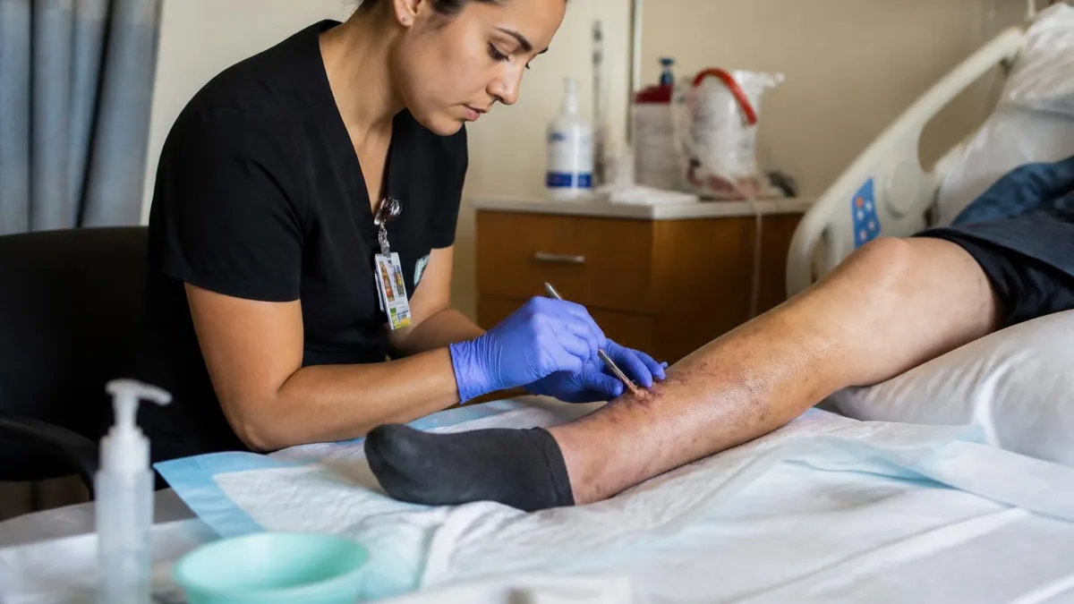
Hydrogel in Wound Care: How It Works, When to Use It, and Types in 2026
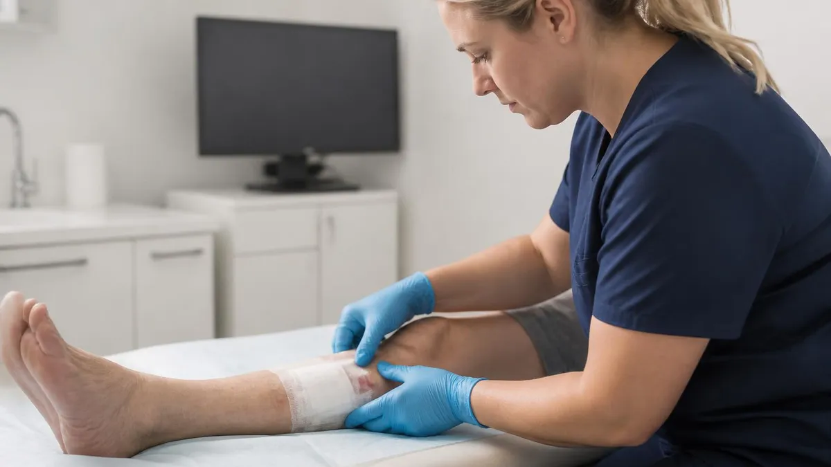
After Surgery Wound Care: Complete Recovery Guide for Patients & Caregivers

Wound Care Courses: Your Complete Guide to Training, Certification, and Career Advancement
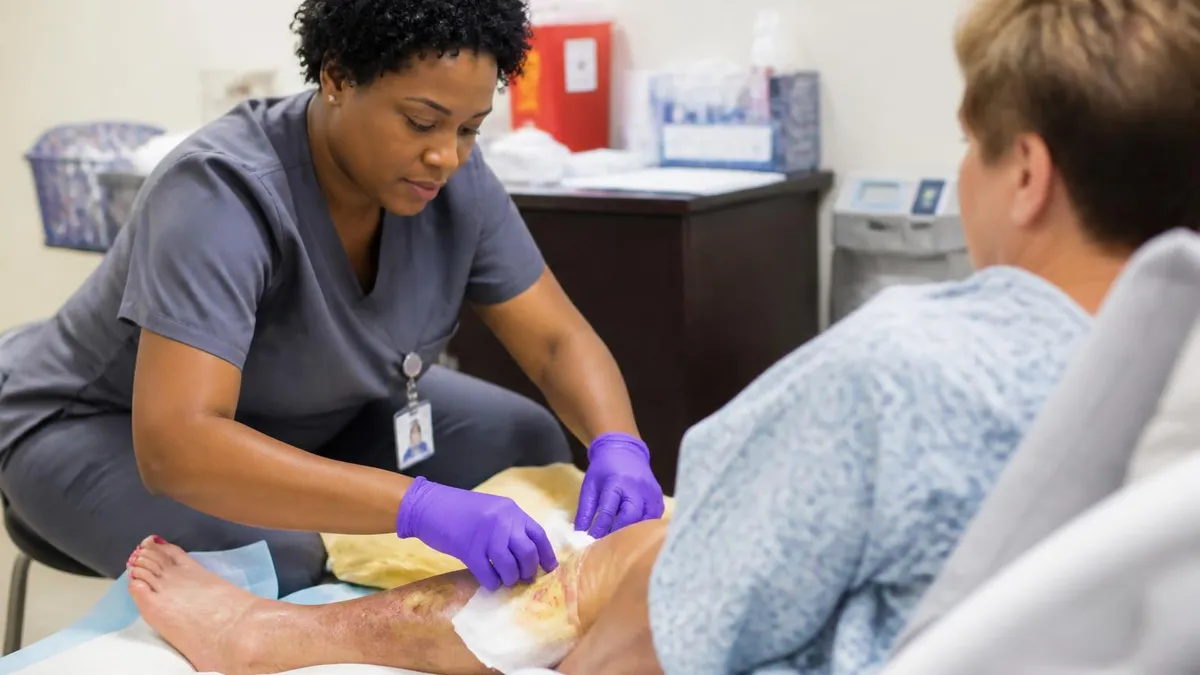
Wound Care NP Jobs: Career Guide for Nurse Practitioners in Wound Management
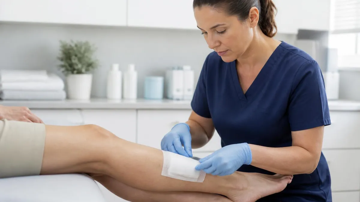
Wound Care Certification for LPN: Complete 2026 Guide to Requirements, Exams & Career Growth
About the Author

Registered Nurse & Healthcare Educator
Johns Hopkins University School of NursingDr. Sarah Mitchell is a board-certified registered nurse with over 15 years of clinical and academic experience. She completed her PhD in Nursing Science at Johns Hopkins University and has taught NCLEX preparation and clinical skills courses for nursing students across the United States. Her research focuses on evidence-based exam preparation strategies for healthcare certification candidates.