Wound Care Treatments: Dressings, Debridement, and NPWT — A Clinical Reference
📗 Wound care treatments explained: moist healing, dressing types, debridement methods, NPWT, wet-to-dry, and special cases like diabetic foot ulcers.
Wound care has changed a lot over the past forty years. The old textbook idea — pack it, dry it out, change it twice a day — is mostly gone in modern practice. What replaced it is a layered approach built around moist wound healing, careful debridement, and dressings matched to the wound's stage and exudate level. You'll see this on every wound care certification exam, and you'll see it at every bedside.
This guide walks through the main treatment categories you need to know. We'll cover the dressings, the debridement methods, negative pressure therapy, the still-common wet-to-dry technique (and why it's mostly inappropriate now), and special cases — diabetic foot ulcers, sacral pressure injuries, skin tears, and puncture wounds. The goal isn't a substitute for clinical training. It's a structured reference so you can connect the dots between exam content and real practice.
Why does it matter? Because chronic wounds affect roughly 6.5 million Americans, and the wrong dressing — or the wrong debridement choice — can stall healing for months. The right treatment, picked by someone who understands the underlying tissue, often cuts healing time in half. That's the difference a credentialed wound care clinician makes.
Wound Care by the Numbers

The moist wound healing principle
Almost every modern treatment starts here. In 1962, George Winter published a paper showing that wounds covered with a polyethylene film healed roughly twice as fast as wounds left to scab over. That single finding overturned a century of practice. Cells need moisture to migrate. Epithelial cells, in particular, can't crawl across a dry crust — they have to dig under it, which slows things down and often fails.
So what does moist actually mean? Not soaking wet. Not dry. A balanced wound bed has enough fluid to keep the tissue hydrated but not so much that the surrounding skin macerates. Most modern dressings are designed to manage that balance: absorb extra exudate, donate moisture if the bed is dry, and keep a stable microenvironment underneath.
When you pick a dressing, you're really answering three questions. How wet is the wound right now? How deep is it? Is there infection or a heavy bacterial load? The answers point you toward one of a handful of dressing families.
Why Moist Wound Healing Wins
Winter's 1962 study showed covered wounds re-epithelialize roughly twice as fast as wounds left to scab. Modern dressings translate that finding into clinical practice — they keep the wound bed at the moisture level cells need to migrate, while protecting periwound skin from maceration. If your dressing is leaving the bed bone-dry or visibly soaking through, the dressing choice is wrong for that wound at that moment.
Dressing categories at a glance
Wound dressings get marketed in dozens of brand names, but they fall into a small number of functional groups. Each one matches a different exudate level and wound type. The trick is to stop thinking in brands and start thinking in categories — alginate, hydrocolloid, foam, hydrogel, transparent film, antimicrobial silver, and collagen. Each has a job.
Alginates are seaweed-derived and great at soaking up heavy drainage. Hydrocolloids form a gel as they absorb fluid and stay on for several days, which makes them useful for pressure injuries with mild exudate. Foams handle moderate to heavy drainage and protect from pressure. Hydrogels donate water to dry, sloughy wounds. Films are vapor-permeable covers — perfect for shallow, low-exudate areas. Silver dressings add antimicrobial coverage when bioburden is high. Collagen products stimulate fibroblast activity in stalled wounds.
Dressing Categories and Their Jobs
Calcium alginate fibers absorb 15-20x their weight in exudate. Use on heavy-draining wounds; gel as they absorb.
Occlusive wafers that form gel against the bed. Stay on 3-5 days. Mild to moderate exudate, partial-thickness wounds.
Polyurethane absorbent pads with adhesive borders. Moderate-to-heavy drainage, pressure injuries, peri-stomal areas.
Water-based gels that donate moisture to dry wounds. Use for dry slough, eschar softening, painful wounds.
Silver ion release controls bioburden. Use for critically colonized or infected wounds; not a substitute for systemic antibiotics.
Vapor-permeable adhesive sheets. Shallow, low-exudate wounds, IV site protection, secondary cover over packing.
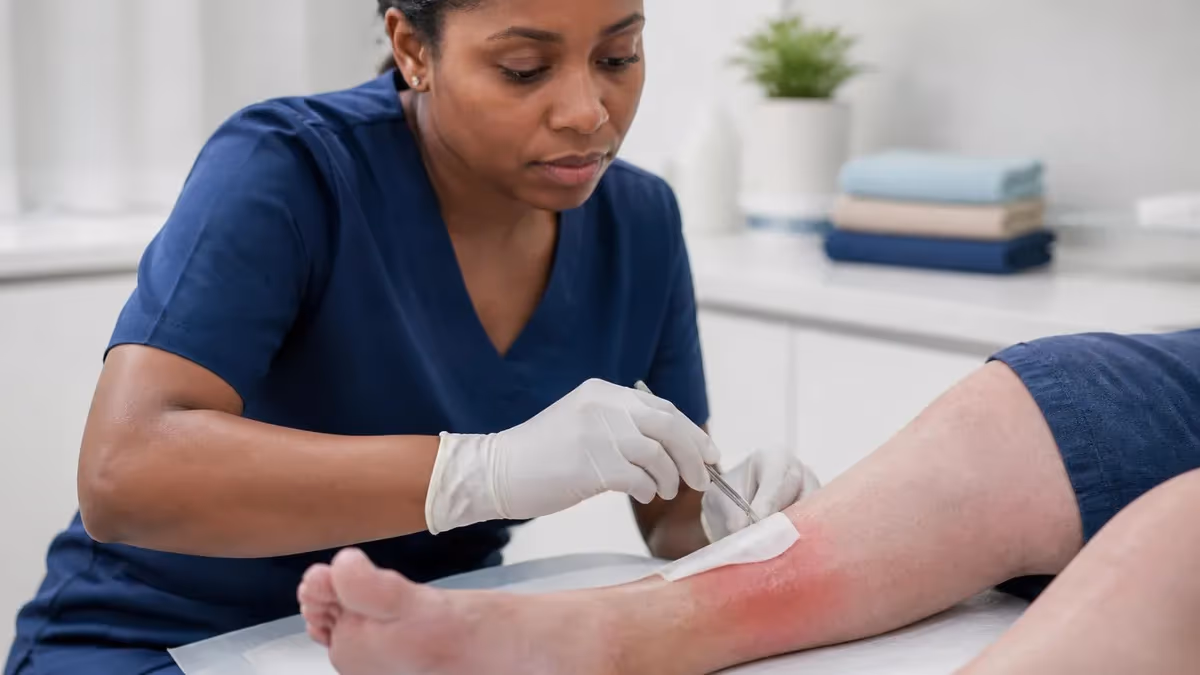
Debridement: removing what shouldn't be there
A wound bed full of necrotic tissue won't heal. Granulation can't form on slough. Antibiotics can't reach bacteria buried under eschar. So before anything else works, you usually have to remove the dead and devitalized tissue — that's debridement. There are four main methods, and choosing between them depends on the wound, the patient, and your scope of practice.
Sharp debridement uses a scalpel, scissors, or curette to cut away dead tissue. It's fast and selective. Only certified clinicians (physicians, advanced practice nurses with training, certified wound specialists in many states) perform it. Autolytic debridement uses the body's own enzymes — you cover the wound with a moisture-retentive dressing and let the wound liquefy its own slough.
Slow but painless. Enzymatic debridement applies a topical preparation (collagenase is the common one) to digest necrotic protein. Faster than autolytic, gentler than sharp. Mechanical debridement means physical removal — irrigation, monofilament pads, or the old wet-to-dry technique. It's nonselective, meaning it strips healthy tissue along with the dead.
Most chronic wounds get a mix. A clinician might sharp-debride the bulk of the eschar at the first visit, then use enzymatic or autolytic methods between visits to keep the bed clean. Pure mechanical debridement is largely outdated for chronic wounds because it damages the granulation tissue you're trying to grow.

The Four Debridement Methods
Scalpel, scissors, or curette removes nonviable tissue. Fastest and most selective. Performed by physicians, advanced practice clinicians, or certified wound specialists (within state scope). Used when rapid removal matters — infected wounds, large eschar burdens, before NPWT placement. Painful for some patients; topical or local anesthetic may be needed.
Negative pressure wound therapy (NPWT)
You may know it as a wound vac. NPWT applies controlled sub-atmospheric pressure — usually 75 to 125 mmHg — to a sealed wound bed. The system uses a foam or gauze filler, a sealed adhesive drape, a suction tube, and a portable pump that collects exudate in a canister. It changed wound care for complex cases.
What does the suction actually do? Several things at once. It pulls excess interstitial fluid out of the wound, which reduces edema and improves perfusion. It mechanically contracts the wound edges, shortening the distance new tissue has to bridge. It increases blood flow and triggers granulation tissue formation. And it provides a closed, controlled environment that reduces dressing change frequency.
NPWT works best on large, deep, exudating wounds — dehisced surgical wounds, traumatic injuries, diabetic foot ulcers, large pressure injuries. It's contraindicated on wounds with exposed major vessels, untreated osteomyelitis, malignancy in the wound, or unexplored fistulas. Dressing changes typically happen every 48 to 72 hours, and patients often go home with portable units.
Do not apply negative pressure wound therapy over exposed major blood vessels or organs, malignancy within the wound bed, untreated osteomyelitis, necrotic tissue with eschar, or unexplored fistulas. Use cautiously near actively bleeding sites, in patients on anticoagulants, and where adhesive contact dermatitis is a known issue. Always confirm the indication with the prescribing clinician before placement.
The wet-to-dry dressing question
Wet-to-dry dressings used to be standard. You'd moisten gauze with saline, pack the wound, let it dry, then rip it out — debriding by force. It worked, after a fashion, and it's cheap. But here's the catch: it strips healthy granulation tissue along with the slough, causes significant patient pain, dries the wound bed (the opposite of what we want), and increases infection risk because gauze fibers shed into the wound.
Modern wound care guidelines (WOCN, AAWC, EWMA) actively discourage routine wet-to-dry use for chronic wounds. There are still niche cases — short-term mechanical debridement of grossly contaminated acute wounds, certain limited settings without access to better dressings — but in most facilities the technique has been replaced by selective debridement plus modern moisture-balancing dressings. Expect exam questions to test whether you understand why it's outdated, not just that it is.
Wound Care Step-by-Step Procedure
- ✓Hand hygiene, gather supplies, position patient and explain steps
- ✓Remove old dressing carefully — note exudate amount, color, odor
- ✓Assess wound bed: size, depth, undermining, tissue type, periwound condition
- ✓Cleanse with normal saline or wound cleanser; irrigate at 4-15 psi for cavity wounds
- ✓Debride as appropriate — within your scope and per the care plan
- ✓Apply skin prep barrier to periwound skin if at risk for maceration or adhesive trauma
- ✓Select primary dressing matched to exudate, depth, and bioburden
- ✓Pack cavity wounds loosely with a moist filler; never overstuff
- ✓Apply secondary cover dressing; secure without tension on fragile skin
- ✓Document wound dimensions, tissue types, dressing used, and patient tolerance
Special case: diabetic foot ulcers
Diabetic foot ulcers behave differently from other wounds. The patient often has neuropathy, so they don't feel injury developing. Peripheral arterial disease may limit perfusion. Hyperglycemia impairs neutrophil function. Add the constant pressure of walking, and you get a wound that resists healing even with good local care.
Treatment is a stack of interventions. Offloading is the single most important — total contact casting, removable cast walkers, or specialty footwear takes pressure off the ulcer. Debridement (usually sharp, weekly) keeps the wound bed clean. Dressings are typically chosen for moisture balance and may include antimicrobial silver if bioburden is high. Diabetic foot ulcer wound care dressing protocols often layer foam over a contact layer to protect the bed without macerating the skin around it. NPWT, hyperbaric oxygen, and skin substitutes come in for wounds that stall after four weeks of standard care.
Glycemic control, smoking cessation, and vascular assessment aren't optional. They're part of the treatment. A textbook-perfect dressing can't fix an underlying perfusion problem.
Wet-to-Dry Dressings: A Modern Verdict
- +Inexpensive and uses materials available in any setting
- +Provides aggressive mechanical debridement in select acute cases
- +Familiar technique that staff in many facilities still know well
- +Can be useful as a short-term bridge before specialty wound care is available
- −Nonselective — strips healthy granulation along with slough
- −Painful for most patients during removal
- −Dries the wound bed, working against moist wound healing principles
- −Gauze fibers shed into the wound and can become a nidus for infection
- −No longer recommended as routine care by WOCN, AAWC, or EWMA guidelines
Sacral wound care
Sacral wounds are usually pressure injuries — Stage 1 through 4, plus unstageable and deep tissue injury. The sacrum sits over a bony prominence with thin tissue cover, and patients who can't reposition (bedbound, post-surgical, neurologically impaired) develop pressure injuries here more than anywhere else on the body. Sacral wound care is one part wound management, three parts pressure redistribution.
The wound itself gets a dressing matched to its stage. Stage 2 wounds often do well with hydrocolloid or foam. Stage 3 and 4 need cavity packing — alginate or hydrofiber rope, topped with a foam border. Heavy exudate may call for NPWT. Sloughy or necrotic bases need debridement before moist healing can take off.
But all of that fails without pressure redistribution. Turn every two hours. Use a low-air-loss or alternating pressure mattress for at-risk patients. Float the heels. Keep the sacrum clean and dry. Manage incontinence aggressively — moisture-associated skin damage is its own problem and often coexists with pressure injury.
Skin tears, puncture wounds, and other special cases
Not every wound needs an advanced dressing. Some need a different approach entirely.
Skin tears are common in older adults and in patients on chronic steroids. The epidermis separates from the dermis, often from minor trauma. Treatment focuses on realigning the flap if it's still viable, covering with a non-adherent contact layer (silicone foam works well), and avoiding adhesive removal trauma at the next change. Wound care skin tear protocols emphasize gentle products and longer wear times to minimize repeat injury.
Puncture wounds need different thinking. The opening is small but the tract can be deep and dirty. How to care for puncture wound presentations starts with irrigation — copious saline under low pressure — and an assessment of depth, contamination, and tetanus status. Don't close puncture wounds primarily; the risk of trapped infection is too high. Cover with a light, absorbent dressing and monitor for signs of deeper infection over the next 48 to 72 hours.
Wound care for deep wounds usually means cavity management. Pack the dead space loosely with a moist filler (alginate rope, hydrofiber, gauze with hydrogel), top with a secondary dressing, and change as exudate dictates. Never pack tightly — pressure on the wound bed kills perfusion.
Adjunctive treatments worth knowing
Beyond the core categories, several adjunctive therapies show up on exams and in advanced practice. Silver nitrate sticks are used to cauterize hypergranulation tissue — the bumpy, friable tissue that sometimes overgrows the wound margin. Apply briefly, neutralize with saline, and avoid prolonged contact with healthy tissue. Silver nitrate wound care is a finishing touch, not a primary treatment, and overuse can damage the periwound skin.
Skin prep for wound care uses film-forming liquid barriers (cyanoacrylate or polymer-based) to protect periwound skin from adhesive trauma and moisture damage. Apply to dry intact skin, let it cure for 30 seconds, then place the dressing. Used routinely, skin prep can extend wear time and reduce the kind of slow, painful skin breakdown that turns one wound into two.
Other adjuncts include hyperbaric oxygen therapy for selected diabetic ulcers and radiation injuries, bioengineered skin substitutes for hard-to-heal wounds, electrical stimulation for stalled pressure injuries, and ultrasound debridement for biofilm management. None of these replace the basics. They're added when standard care underperforms, and they tend to work best when the underlying conditions — perfusion, glucose control, nutrition, pressure relief — are also being addressed.
Step-by-step: a generic wound care procedure
The specifics vary by wound, but the structure of a wound care visit is consistent. Below is the framework most certified clinicians follow. Wound care step by step protocols always start with assessment and finish with documentation, with cleansing, debridement, and dressing application in between. Treat the checklist as a sequence you build muscle memory around — one missed step (skipping periwound skin prep, packing a cavity too tightly, choosing a foam when an alginate was needed) is often what separates a healing wound from a stalled one.
Putting it together
If you take one thing from this guide, take this: wound care isn't a product choice. It's a series of decisions about the wound, the patient, and the conditions you can change. The dressing matters. So does debridement. So does offloading, perfusion, nutrition, infection control, and the patient's ability to follow through at home. A perfect foam dressing applied to an unoffloaded diabetic foot is still going to fail. A textbook NPWT setup on a malnourished patient with uncontrolled blood sugar won't heal at the rate the literature predicts.
Exam preparation works the same way. Don't memorize brand names. Learn the categories, the mechanisms, the indications, and the contraindications. When a question shows up about a diabetic foot ulcer with heavy exudate and undermining, you should be able to walk through the assessment, pick a debridement plan, choose a dressing class, and explain offloading — without flipping through a product catalog. The same logic applies to a sacral pressure injury, a stalled venous leg ulcer, a dehisced surgical wound, or a contaminated puncture. The wound type changes; the decision framework doesn't.
Modern wound care procedure is methodical, evidence-driven, and humbling. The wounds that look simple often aren't. The ones that look hopeless sometimes heal beautifully when the right team gets to work. That team is what wound care certification builds, and it's what your patients deserve. Keep building the mental library: which dressing, which debridement, which adjunct, which patient factor — and how all four interact. That's the muscle the exam tests, and it's the same muscle clinical practice demands every shift.
One last note. Documentation is treatment. Measurements, tissue percentages, exudate descriptions, pain scores, dressing types, change frequency — the record is how the next clinician picks up where you left off, how reimbursement gets approved, and how stalled wounds get escalated to advanced therapy in time to matter. Sloppy documentation is sloppy care, even when the dressing technique is flawless. Treat the chart the way you treat the wound bed: clean, accurate, consistent, and complete.
Wound Care Questions and Answers
About the Author
Registered Nurse & Healthcare Educator
Johns Hopkins University School of NursingDr. Sarah Mitchell is a board-certified registered nurse with over 15 years of clinical and academic experience. She completed her PhD in Nursing Science at Johns Hopkins University and has taught NCLEX preparation and clinical skills courses for nursing students across the United States. Her research focuses on evidence-based exam preparation strategies for healthcare certification candidates.