
MRI Scan for Brain: What It Shows, How It Works, and What to Expect
🟢 Learn everything about MRI scan for brain — what it detects, how the procedure works, prep tips, and how to interpret your results.
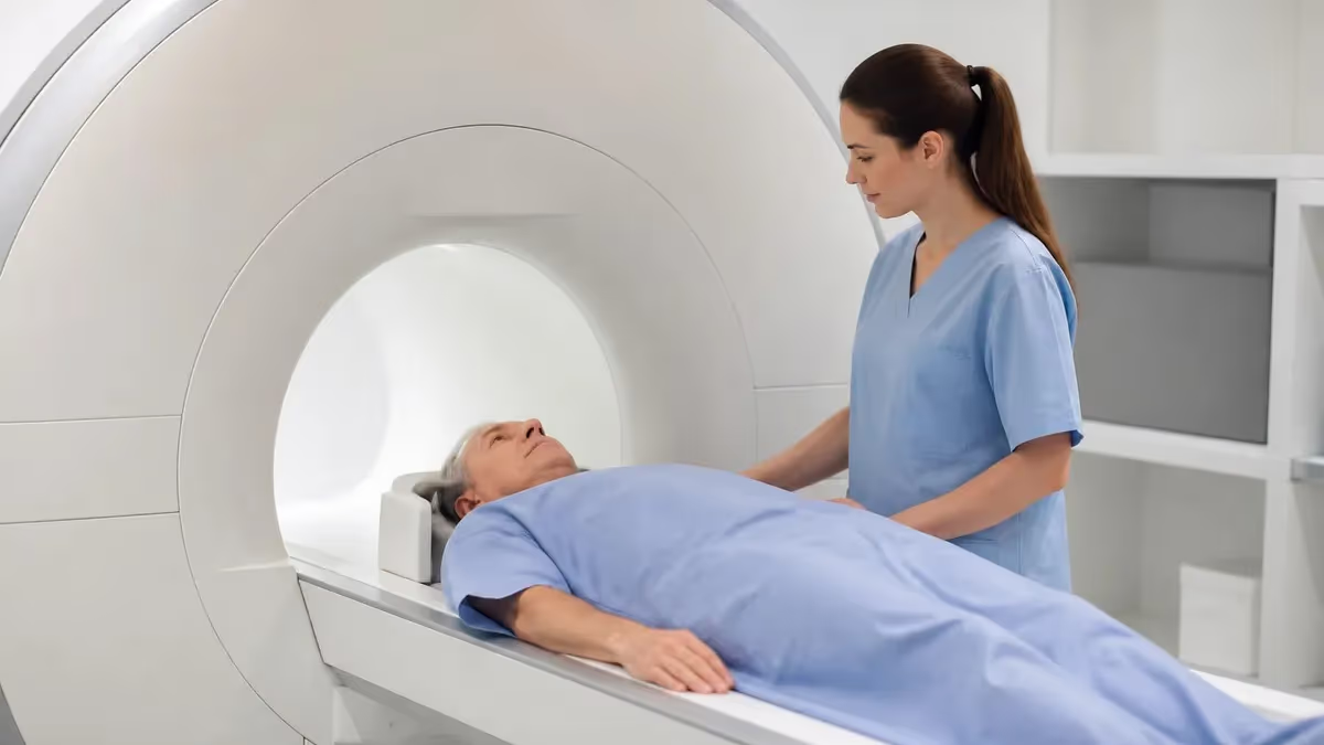
An MRI scan for brain is one of the most powerful diagnostic tools in modern medicine, offering unparalleled soft-tissue contrast that allows neurologists, radiologists, and neurosurgeons to detect abnormalities that other imaging modalities simply cannot resolve. Unlike X-rays or CT scans, brain MRI does not use ionizing radiation. Instead, it harnesses powerful magnetic fields and radiofrequency pulses to generate detailed cross-sectional images of the brain's internal structures, making it the gold standard for evaluating neurological conditions from stroke and tumor to multiple sclerosis and traumatic injury.
The procedure has become remarkably common in the United States, with more than 40 million MRI examinations performed annually across all body parts, and brain MRIs accounting for a significant proportion of that volume. Neuroimaging referrals have climbed steadily over the past decade as scanner availability has expanded into outpatient imaging centers, hospital systems, and even mobile units serving rural communities. Understanding what happens during a brain MRI — from patient positioning and coil selection to pulse sequence design and image interpretation — is essential knowledge for radiologic technologists preparing for the ARRT MRI registry exam.
A brain MRI can detect an extraordinary range of pathologies. Ischemic strokes appear as bright areas on diffusion-weighted imaging (DWI) within minutes of onset, long before they are visible on CT. Primary brain tumors, metastatic lesions, and lymphoma light up with contrast enhancement on post-gadolinium T1-weighted sequences. Demyelinating plaques characteristic of multiple sclerosis appear as hyperintense foci on T2 and FLAIR sequences, particularly in periventricular white matter. Epileptogenic foci, pituitary adenomas, cerebral aneurysms, arteriovenous malformations, and hydrocephalus all have characteristic MRI signatures that guide clinical decision-making and surgical planning.
For radiologic technology students and practicing MRI technologists, the brain is both the most frequently imaged organ and the most sequence-intensive. A standard brain protocol at most academic medical centers includes at minimum T1-weighted sagittal localizer images, axial T2, axial FLAIR, axial DWI with apparent diffusion coefficient (ADC) maps, axial T2* gradient echo or susceptibility-weighted imaging (SWI) for hemorrhage detection, and pre- and post-contrast axial and coronal T1. More advanced protocols add MR spectroscopy, perfusion imaging, or functional MRI depending on the clinical question being answered.
Patient preparation plays a critical role in image quality and safety. Screening every patient for ferromagnetic implants, pacemakers, cochlear implants, and other MRI-contraindicated devices is a non-negotiable step that technologists must perform meticulously before any brain scan. Claustrophobia affects an estimated 5–10% of patients and may necessitate open-bore scanners, anxiolytic premedication, or careful patient communication strategies. Motion artifact remains one of the most common causes of nondiagnostic brain MRI studies, so coaching patients on breath-holding techniques (when applicable) and remaining still during acquisitions directly impacts diagnostic yield.
This comprehensive guide covers everything you need to know about brain MRI — the physics behind image contrast, the anatomy visible on each sequence, common pathologies and their imaging characteristics, patient preparation best practices, and the clinical indications that drive ordering decisions. Whether you are a patient seeking to understand your upcoming scan, a student preparing for board exams, or a technologist brushing up on neuroimaging protocols, this article provides the depth and clinical context you need. You may also find it useful to review our companion resource on mri scan for brain pathology for adjacent anatomical coverage.
Beyond clinical utility, brain MRI occupies a central place in current neuroscience research. Resting-state functional MRI has revealed large-scale brain networks including the default mode network, salience network, and frontoparietal control network, deepening our understanding of consciousness, cognition, and psychiatric disorders. Diffusion tensor imaging (DTI) maps white matter tracts with a precision that was unimaginable two decades ago, enabling presurgical planning that preserves eloquent cortex and critical fiber pathways. As scanner technology advances toward ultra-high-field systems at 7 Tesla and beyond, the diagnostic ceiling for brain MRI continues to rise.
Brain MRI by the Numbers
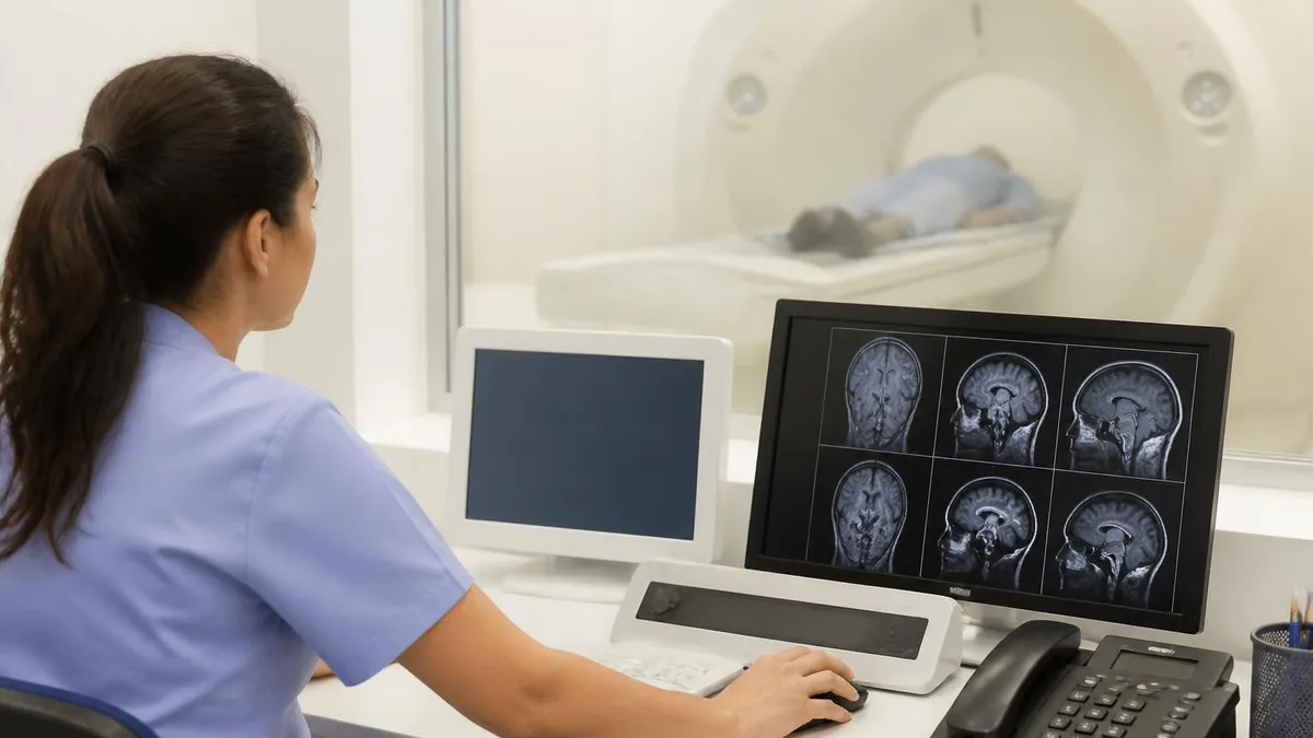
Standard Brain MRI Protocol: Step-by-Step Workflow
Patient Screening and Safety Check
Patient Positioning and Coil Selection
Localizer and Sequence Planning
Acquisition of Diagnostic Sequences
Contrast Administration (When Ordered)
Image Review and Transfer
Understanding what an MRI scan for brain can and cannot detect is fundamental for both ordering clinicians and imaging technologists. The brain MRI excels at soft-tissue characterization because of its inherently high contrast between gray matter, white matter, cerebrospinal fluid (CSF), and pathological tissue. This contrast arises from differences in T1 relaxation time, T2 relaxation time, proton density, diffusion characteristics, and magnetic susceptibility — each exploited by a different pulse sequence within the standard protocol.
Acute ischemic stroke is perhaps the most time-critical indication for brain MRI. Diffusion-weighted imaging (DWI) detects the cytotoxic edema that occurs within minutes of arterial occlusion, showing restricted diffusion as bright signal on DWI and correspondingly dark signal on ADC maps. This combination allows radiologists to distinguish acute from chronic infarction with high confidence. CT remains faster and more accessible in the hyperacute window, but DWI MRI surpasses CT for detecting small cortical, deep white matter, and posterior fossa infarcts that are frequently missed on non-contrast CT due to beam-hardening artifact near the skull base.
Neoplastic disease represents another major indication. Primary brain tumors — including glioblastoma multiforme (GBM), lower-grade gliomas, meningiomas, and acoustic neuromas — each have characteristic appearances on multiparametric MRI. GBM classically shows a thick, irregular enhancing rim surrounding a central necrotic core on post-gadolinium T1, with extensive surrounding vasogenic edema on T2/FLAIR. Meningiomas enhance avidly and homogeneously, often with a dural tail sign. MR spectroscopy adds metabolic information by measuring peaks for N-acetylaspartate (NAA), choline, creatine, and lactate — ratios that help differentiate tumor recurrence from treatment-related changes (radiation necrosis) in post-treatment patients.
Demyelinating diseases including multiple sclerosis (MS), neuromyelitis optica spectrum disorder (NMOSD), and acute disseminated encephalomyelitis (ADEM) are evaluated primarily with T2 and FLAIR sequences. Classic MS plaques are ovoid, periventricular, and oriented perpendicular to the ventricular surface — a pattern sometimes called Dawson's fingers. Active plaques show gadolinium enhancement during acute inflammation. The 2017 McDonald criteria formalized MRI's role in MS diagnosis by defining dissemination in space and time based on lesion location and enhancement characteristics, allowing earlier, more confident diagnosis and earlier treatment initiation.
Infectious and inflammatory conditions — meningitis, encephalitis, brain abscess, and neurocysticercosis — all have recognizable MRI patterns. Pyogenic brain abscess shows restricted diffusion within the cavity (distinguishing it from cystic tumor), a smooth thin ring of enhancement, and surrounding edema. Herpes simplex encephalitis characteristically involves the medial temporal lobes and insular cortex bilaterally with T2 hyperintensity and cortical swelling. Leptomeningeal carcinomatosis enhances along sulci and cisterns on post-contrast FLAIR, a sequence increasingly included in brain tumor protocols for its superior sensitivity to meningeal disease compared with standard T1 post-contrast.
Vascular abnormalities including cerebral aneurysms, arteriovenous malformations (AVMs), cavernous malformations (cavernomas), and cerebral venous sinus thrombosis each have distinct MRI signatures. SWI is exquisitely sensitive to hemosiderin from prior hemorrhage, revealing cavernomas as a characteristic popcorn-ball appearance with a hypointense hemosiderin rim. MR angiography (MRA) — using time-of-flight (TOF) or contrast-enhanced technique — delineates arterial anatomy without catheter-based angiography, making it the first-line study for screening intracranial aneurysms in patients with subarachnoid hemorrhage or family history. TOF-MRA is particularly sensitive for detecting basilar artery and middle cerebral artery bifurcation aneurysms larger than 3 mm.
White matter diseases beyond MS include small vessel ischemic disease (SVID), which appears as diffuse or patchy T2/FLAIR hyperintensity in the periventricular and subcortical white matter, particularly in elderly patients with hypertension and diabetes. The Fazekas scale grades SVID severity from 0 (absent) to 3 (confluent), and higher grades correlate with cognitive decline, gait impairment, and increased stroke risk. Neurodegeneration — including Alzheimer's disease, Parkinson's disease, and frontotemporal dementia — is increasingly characterized by structural MRI volumetrics, with hippocampal atrophy serving as a biomarker for Alzheimer's-type pathology even before clinical dementia emerges.
MRI Practice Test Questions
Prepare for the MRI - Magnetic Resonance Imaging exam with our free practice test modules. Each quiz covers key topics to help you pass on your first try.
MRI Knowledge
MRI Exam Questions covering Knowledge. Master MRI Test concepts for certification prep.
MRI Physics
Free MRI Practice Test featuring Physics. Improve your MRI Exam score with mock test prep.
MRI Anatomy and Pathology
MRI Test Prep for MRI Anatomy and Pathology. Practice MRI Quiz questions and boost your score.
MRI Anatomy and Positioning
MRI Questions and Answers on MRI Anatomy and Positioning. Free MRI practice for exam readiness.
MRI Contrast Agents
Free MRI Quiz on MRI Contrast Agents. MRI Exam prep questions with detailed explanations.
MRI Patient Care and Positioning
MRI Practice Questions for MRI Patient Care and Positioning. Build confidence for your MRI certification exam.
MRI Sequences Used in Brain Imaging
T1-weighted sequences display fat and subacute blood as bright (hyperintense) and CSF as dark (hypointense). They are the primary sequences for evaluating anatomy, detecting lipomas, dermoid cysts, and methemoglobin in subacute hemorrhage. Post-contrast T1 images exploit gadolinium's T1-shortening effect to reveal blood-brain barrier disruption in tumors, abscesses, active demyelination, and meningeal disease. Most brain protocols now use a 3D T1 MPRAGE or VIBE sequence for thin isotropic slices reformattable in any plane.
T2-weighted sequences display CSF as bright and solid tissue as intermediate-to-dark signal. Pathological processes that increase tissue water content — edema, tumor, inflammation, demyelination, infarction — all appear hyperintense on T2. The FLAIR (fluid-attenuated inversion recovery) sequence is a T2-weighted technique that nulls the CSF signal, dramatically improving the visibility of periventricular and cortical lesions that would otherwise be obscured by adjacent bright CSF. FLAIR has largely replaced conventional T2 for brain lesion detection in most clinical protocols.
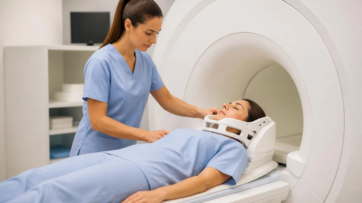
Brain MRI vs. CT Scan: Which Is Better?
- +No ionizing radiation — safe for repeated imaging and pediatric or pregnant patients (after first trimester with clinical justification)
- +Superior soft-tissue contrast compared to CT, clearly differentiating gray matter, white matter, and CSF
- +Multiplanar capability with isotropic 3D sequences reformattable in any plane without additional scanning
- +DWI detects acute ischemic stroke within minutes of onset — much earlier than CT can reveal infarction
- +Identifies posterior fossa and brainstem pathology without bone-hardening artifact that degrades CT in these regions
- +Advanced sequences (spectroscopy, perfusion, fMRI, DTI) provide functional and metabolic information beyond anatomy
- −Longer scan time (30–60 minutes vs. 5–10 minutes for CT) increases motion artifact risk and limits use in unstable or uncooperative patients
- −Contraindicated in patients with certain metallic implants, pacemakers, cochlear implants, and some aneurysm clips
- −Louder acoustic noise from gradient coils can be distressing — patients require earplugs and may need sedation
- −More expensive than CT, with brain MRI costing $1,600–$8,400 depending on contrast and facility type
- −Less available in emergency settings; CT is the first-line study for acute head trauma and suspected intracranial hemorrhage
- −Claustrophobia affects 5–10% of patients, sometimes preventing completion of the study in standard-bore scanners
Brain MRI Patient Preparation Checklist
- ✓Complete and sign the MRI safety screening questionnaire honestly, listing all implants, surgeries, and foreign body exposures.
- ✓Remove all metallic objects before entering the scan room: jewelry, hairpins, hearing aids, glasses, and body piercings.
- ✓Inform the technologist of any prior allergic reactions to gadolinium or other MRI contrast agents.
- ✓Tell the technologist if you have a cardiac pacemaker, implantable cardioverter-defibrillator (ICD), or neurostimulator device.
- ✓Notify staff if you are pregnant or suspect you may be pregnant before any contrast injection is administered.
- ✓Avoid applying hair gel, mousse, or metallic-containing cosmetics on the day of the scan to prevent susceptibility artifact.
- ✓Follow contrast prep instructions if ordered: ensure adequate renal function (eGFR ≥30 mL/min/1.73m²) before gadolinium administration.
- ✓Request anti-anxiety medication from your referring physician in advance if you experience claustrophobia in enclosed spaces.
- ✓Plan to arrive 15–20 minutes early for paperwork and to allow time for IV placement if contrast has been ordered.
- ✓Arrange transportation if you have received sedation — you will not be permitted to drive after anxiolytic premedication.
FLAIR Sequence Is the Most Important for White Matter Disease
Among all brain MRI sequences, axial FLAIR consistently provides the highest sensitivity for detecting white matter lesions, cortical infarcts, and subarachnoid hemorrhage. By nulling CSF signal, FLAIR unmasks periventricular plaques and cortical lesions that are invisible on conventional T2 — making it the single most diagnostically impactful sequence in routine neuroimaging. When reviewing brain MRIs for study or board exams, always start your systematic review on FLAIR before moving to other sequences.
Interpreting brain MRI results requires a systematic approach that prevents errors of omission — the most common source of missed diagnoses in neuroimaging. Experienced radiologists follow a structured search pattern that evaluates each anatomical region and tissue type in turn, regardless of what initially catches the eye. For ARRT MRI registry candidates, understanding this interpretive framework is as important as knowing pulse sequence physics, because exam questions frequently ask you to identify normal anatomy, recognize classic pathological patterns, and understand what clinical information each sequence provides.
The first step in systematic brain MRI review is assessing the extra-axial spaces — the subdural and epidural compartments, the subarachnoid space, and the cisterns. Subdural hematomas appear as crescent-shaped collections following the brain's convexity; their signal intensity on T1 and T2 changes predictably over time as hemoglobin evolves from oxyhemoglobin through deoxyhemoglobin, methemoglobin, and hemosiderin.
Acute subdural blood is isointense to brain on T1 and hypointense on T2 — easily overlooked on T1 — making FLAIR and SWI essential complements. Epidural hematomas, typically lenticular (biconvex) in shape, are associated with temporal bone fracture and middle meningeal artery injury and tend to be brighter on T1 due to clot retraction.
Next, evaluate the brain parenchyma systematically by lobe: frontal, parietal, temporal, occipital, and then the deep structures — basal ganglia, thalami, internal capsule, and brainstem. Signal abnormalities in the basal ganglia have a broad differential including Wilson's disease (copper deposition showing T2 hypointensity in the putamen), Huntington's disease (caudate atrophy), toxic encephalopathies from carbon monoxide or methanol, and hypoxic-ischemic injury. The thalami are particularly vulnerable to top-of-the-basilar syndrome, osmotic demyelination (central pontine and extrapontine myelinolysis), and Wernicke's encephalopathy, which classically shows T2/FLAIR hyperintensity in the mammillary bodies, periaqueductal gray, and medial thalami.
The posterior fossa deserves special attention because it is the region most degraded by CT artifact and most frequently harbors findings missed in cursory review. The cerebellum, brainstem, and fourth ventricle should be assessed on axial and sagittal T2 and FLAIR. Cerebellar infarcts in the PICA, AICA, and SCA territories have distinct distributions that correlate with the involved vessel.
Brainstem gliomas — predominantly pontine in children — present as diffuse T2 hyperintensity expanding the pons. Chiari I malformation, defined by tonsillar herniation more than 5 mm below the foramen magnum, is often an incidental finding but may require surgical decompression when associated with syringohydromyelia.
Ventricular size and CSF flow should be assessed on every brain MRI. Hydrocephalus — enlarged ventricles with periventricular T2/FLAIR halo of transependymal CSF migration — must be distinguished from cerebral atrophy, where sulci are also enlarged. Obstructive hydrocephalus implies a blockage at or near the aqueduct, fourth ventricle, or outlet foramina (Luschka and Magendie), while communicating hydrocephalus implies impaired CSF resorption at the arachnoid granulations, as occurs following subarachnoid hemorrhage or meningitis. Normal pressure hydrocephalus (NPH) presents with the clinical triad of gait ataxia, urinary incontinence, and cognitive decline, and shows disproportionately enlarged ventricles relative to sulci on MRI.
Contrast enhancement patterns provide crucial information about disease activity and blood-brain barrier integrity. Leptomeningeal enhancement (coating of cortical surface and cisterns) suggests meningitis, leptomeningeal carcinomatosis, or neurosarcoidosis. Cortical enhancement in a gyriform pattern is characteristic of subacute infarction (luxury perfusion) or encephalitis. Ring-enhancing lesions — the classic teaching point — have a differential diagnosis remembered by the mnemonic MAGIC DR: Metastasis, Abscess, Glioblastoma (high-grade), Infarct (subacute), Contusion, Demyelination (tumefactive MS), and Radiation necrosis. The distinction between these entities often requires clinical correlation, serial imaging, and sometimes advanced MRI techniques or tissue sampling.
For technologists, understanding the radiological report's structure helps in communicating effectively with the clinical team. A standard neuroradiology report includes clinical indication, technique (field strength, sequences acquired, contrast agent and dose), comparison studies, findings organized by anatomical region, and impression summarizing the most significant findings and their clinical significance. When a technologist identifies a potentially urgent finding on the console — herniation, large territorial infarct, expanding hemorrhage — prompt notification of the radiologist using the ACR's tiered communication framework is a professional and ethical obligation that can directly impact patient outcomes.
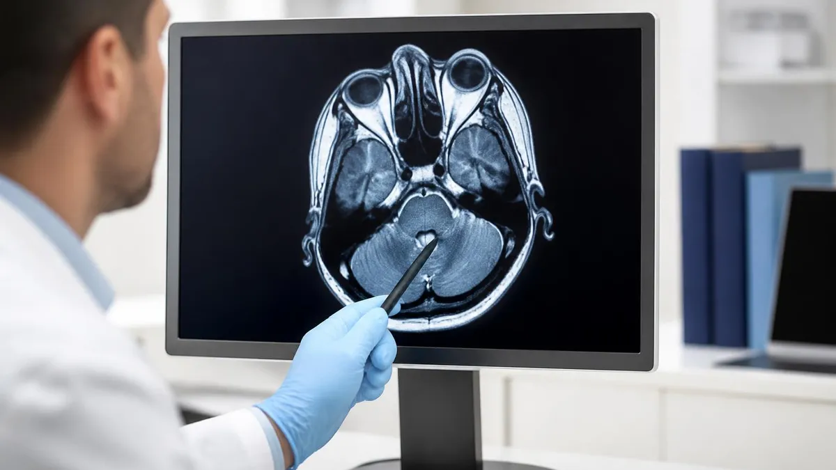
Nephrogenic systemic fibrosis (NSF) is a rare but serious complication of gadolinium-based contrast agents (GBCAs) in patients with severe renal impairment (eGFR below 30 mL/min/1.73m²). Before administering any GBCA for brain MRI, verify current renal function labs — particularly in patients older than 60, diabetics, and those with known kidney disease. The ACR Manual on Contrast Media provides updated risk-stratification guidelines; always consult institutional policy before injecting high-risk patients.
Preparing for the ARRT MRI registry exam requires a deep understanding of brain imaging protocols, normal anatomy, and pathological MRI appearances. The ARRT content specifications for the MRI examination include patient care and education (11%), safety (18%), image production (42%), and procedures (29%). Brain and neurological procedures fall within the procedures domain and encompass a substantial portion of exam content, making neuroimaging one of the highest-yield study areas for registry candidates.
Key physics concepts that frequently appear on brain MRI registry questions include the relationship between TR, TE, and image contrast (short TR/short TE = T1-weighted; long TR/long TE = T2-weighted; long TR/short TE = proton density); the role of inversion recovery sequences (FLAIR nulls CSF with an appropriate TI; STIR nulls fat); the principles of diffusion-weighted imaging and how b-values determine diffusion sensitivity; and the susceptibility effects exploited in GRE and SWI sequences. Understanding how changing flip angle affects T1 contrast in gradient echo sequences — and why this matters for 3D MPRAGE brain protocols — is another high-yield topic.
Anatomy questions on the registry exam often involve identifying structures on axial, coronal, and sagittal MRI images. The basal ganglia anatomy — caudate head, putamen, globus pallidus, and their relationships to the internal capsule (anterior limb, genu, posterior limb) — is a perennial favorite. The circle of Willis, including its anterior, middle, and posterior cerebral artery territories, is essential for stroke localization. Cranial nerve anatomy — especially the trigeminal, facial, vestibulocochlear, and optic nerves — is tested in both axial and coronal planes because these nerves are frequently evaluated in clinical brain MRI protocols.
Pathology recognition on the registry exam focuses on classic imaging presentations rather than rare entities. Candidates should be able to identify the DWI appearance of acute stroke, the ring-enhancing appearance of glioblastoma versus abscess, the FLAIR hyperintensity pattern of MS plaques, the T1 bright signal of subacute hemorrhage (methemoglobin), the SWI blooming artifact of cavernous malformation, and the enhancement pattern of a vestibular schwannoma on post-contrast T1 in the internal auditory canal. Memorizing these signal characteristics systematically — T1 signal, T2 signal, FLAIR signal, enhancement pattern, and DWI signal for each entity — is the most efficient exam preparation strategy.
Safety questions on the registry exam are weighted heavily and include MRI zones, screening procedures, implant compatibility assessment, specific absorption rate (SAR) limits, acoustic noise regulations, and emergency procedures including quench protocol. Candidates must know the four MRI zone system (Zones I–IV) defined by the ACR, which dictates access control and personnel screening requirements. Zone IV is the magnet room itself, and access must be controlled to screened individuals only. Questions about ferromagnetic detection systems, conditional versus unsafe implant classifications, and the management of a patient who experiences an adverse event inside the magnet bore are all within scope.
Study resources for brain MRI registry preparation include the ARRT content specifications document (freely available on arrt.org), the ACR Practice Parameters and Technical Standards for MRI, and standard radiology textbooks such as Brant and Helms' Fundamentals of Diagnostic Radiology. Case-based learning using platforms that provide annotated neuroimaging cases is particularly effective for pattern recognition. Practice tests remain one of the highest-yield preparation strategies — reviewing rationales for both correct and incorrect answer choices deepens conceptual understanding beyond simple fact memorization. The quiz resources on this site provide registry-caliber questions with detailed explanations for all answer choices.
For technologists who want to deepen their neuroimaging expertise beyond the registry exam, subspecialty MRI certifications and advanced practice pathways are increasingly available. The ARRT offers advanced-level examinations in MRI that test more nuanced clinical and technical knowledge. Many academic medical centers offer structured neuroimaging education programs for technologists, including didactic lectures, case conferences, and hands-on scanning experience with advanced protocols. Staying current with evolving techniques — including AI-assisted image reconstruction, ultra-high-field neuroimaging, and quantitative MRI methods — positions technologists for the rapidly changing landscape of clinical neuroimaging practice.
Practical preparation for your brain MRI scan — whether you are a patient or a student learning to optimize image quality — involves understanding the factors that most commonly compromise scan quality. Motion artifact is the leading cause of nondiagnostic brain MRI studies in clinical practice.
Even subtle head movement during a 4-minute FLAIR acquisition can smear the image sufficiently to obscure periventricular lesions or create ghosting artifacts that mimic pathology. Coaching patients effectively before and during the scan — explaining exactly when each sequence starts and ends, warning them about gradient noise levels, and encouraging relaxed breathing — significantly reduces motion-related reacquisitions.
Physiological noise from cardiac pulsation and respiratory motion affects image quality even in cooperative patients, particularly in the posterior fossa where CSF flow creates pulsation artifact. Cardiac gating or triggering synchronizes the acquisition to the R-wave of the ECG, acquiring data during the quietest phase of the cardiac cycle. This is particularly important for high-resolution posterior fossa sequences and for distinguishing true brainstem lesions from flow-related artifact. Some centers use peripheral pulse gating as an alternative when electrode placement is impractical. Understanding gating techniques is important for registry candidates and for troubleshooting artifact on clinical scans.
Parallel imaging techniques — GRAPPA, SENSE, and their variants — have transformed brain MRI by dramatically reducing acquisition time without proportional loss of SNR. Rather than acquiring every phase-encoding line sequentially, parallel imaging uses the spatial sensitivity profiles of multichannel phased-array coils to reconstruct full images from undersampled k-space data. Acceleration factors of 2–4 are routine on modern 3T scanners, cutting FLAIR acquisition time from 6 minutes to 3 minutes while maintaining diagnostic quality. Understanding the trade-off between acceleration factor and residual aliasing artifact (g-factor noise) is important for protocol optimization and appears on advanced registry examinations.
Field strength profoundly affects brain MRI quality and protocol design. Moving from 1.5T to 3T doubles SNR in theory (proportional to field strength), which can be reinvested as increased spatial resolution, faster acquisitions, or thinner slices. In practice, 3T brain MRI provides superior lesion detection for MS, improved small vessel anatomy on MRA, and better spectroscopic resolution.
However, 3T also increases susceptibility artifacts near air-tissue interfaces (frontal sinuses, skull base), increases SAR (limiting certain fast spin echo sequences), and amplifies B1 field inhomogeneity that can create shading artifacts in the temporal lobes. Protocol optimization at 3T requires understanding these trade-offs and adjusting parameters accordingly.
Gadolinium contrast agents vary in their molecular structure (linear vs. macrocyclic) and protein-binding characteristics, which affect both relaxivity (enhancement strength) and stability. The ACR and regulatory agencies have increasingly recognized that gadolinium deposition in the brain — particularly in the dentate nucleus and globus pallidus — occurs with repeated GBCA administration, with linear agents showing higher deposition rates than macrocyclic agents.
While the clinical significance of gadolinium deposition at currently observed concentrations remains under investigation, the trend in clinical practice has been toward preferring macrocyclic GBCAs for patients expected to receive multiple contrast-enhanced brain MRIs, such as those with MS or brain tumor follow-up.
Emerging brain MRI techniques are rapidly moving from research to clinical practice. Synthetic MRI acquires a single quantitative dataset from which T1, T2, PD, and FLAIR images are computationally generated, reducing total scan time while simultaneously providing tissue parameter maps useful for myelin water imaging and white matter characterization.
Compressed sensing accelerates acquisition by exploiting image sparsity in the wavelet domain, enabling high-resolution 3D brain imaging in scan times that were previously achievable only with lower resolution. Artificial intelligence image reconstruction algorithms (deep learning reconstruction, DLR) now routinely improve SNR and reduce acquisition time by factors of 2–4 on clinical scanners, and familiarity with these techniques is increasingly expected of practicing MRI technologists.
The future of brain MRI is moving toward quantitative, reproducible, and standardized imaging that can serve as a biomarker for neurological disease progression, treatment response, and even early detection of neurodegenerative changes before symptoms emerge. Initiatives like the ADNI (Alzheimer's Disease Neuroimaging Initiative) and the Human Connectome Project have demonstrated the scientific and clinical value of harmonized MRI protocols across multiple scanner platforms and field strengths.
For technologists and radiologists who master the fundamentals of brain MRI today, this evolving landscape represents an extraordinary opportunity to contribute to one of the most impactful fields in all of medicine. Investing in deep protocol knowledge, systematic interpretation skills, and continued education is the foundation for excellence in neuroimaging practice.
MRI Questions and Answers

MRI of Cervical Spine: Complete Guide to Imaging, Anatomy, Pathology, and Patient Preparation

Knee MRI Images: A Complete Guide to Reading, Understanding, and Interpreting Knee Scans
Ace Your Exam with MRI Practice Test 2026 Prep

MRI Tech School 2026 — Programs, Requirements, and What to Expect
MRI Procedure: What to Expect Before, During and After Your Scan
About the Author

Medical Laboratory Scientist & Clinical Certification Expert
Johns Hopkins UniversityDr. Sandra Kim holds a PhD in Clinical Laboratory Science from Johns Hopkins University and is certified as a Medical Technologist (MT) and Medical Laboratory Scientist (MLS) through ASCP. With 16 years of clinical laboratory experience spanning hematology, microbiology, and molecular diagnostics, she prepares candidates for ASCP board exams, MLT, MLS, and specialist certification tests.
Join the Discussion
Connect with other students preparing for this exam. Share tips, ask questions, and get advice from people who have been there.
View discussion (8 replies)