
Shoulder MRI How Long: Complete Guide to MRI Scan Duration by Body Part 2026
Shoulder MRI how long does it take? ⏳ Learn exact scan times by body part, what affects duration, and how to prepare for a faster, smoother exam.
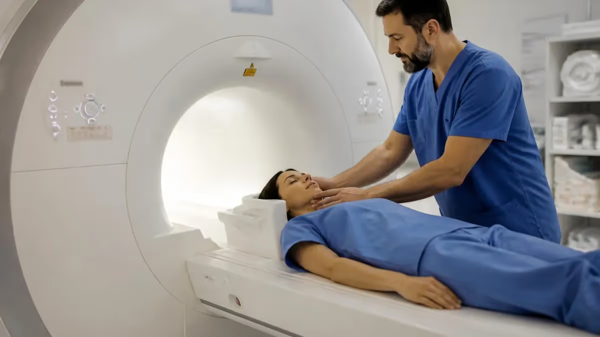
If you're scheduled for an imaging appointment and wondering about shoulder MRI how long it actually takes, you're not alone. A shoulder MRI typically runs between 45 and 60 minutes, though that window can shift based on the specific sequences your radiologist orders, whether contrast dye is used, and how well you remain still throughout the exam. Understanding what determines scan length helps you plan your day and arrive mentally prepared for the experience ahead. Practice with the free MRI - Magnetic Resonance Imaging practice test on PracticeTestGeeks (200+ questions with answer explanations).
MRI duration varies significantly across different body parts. A brain MRI without contrast often finishes in 30 to 45 minutes, while a full spine study covering cervical, thoracic, and lumbar regions can stretch to 90 minutes or more. Abdominal and pelvic MRIs typically fall in the 45- to 75-minute range because multiple sequences are needed to capture soft tissue detail. Cardiac MRI, which requires synchronization with the heartbeat, commonly takes 60 to 90 minutes. Knowing these benchmarks helps set realistic expectations before you walk through the scanner door.
Several technical factors drive how long any given MRI takes. Field strength matters enormously: a 3 Tesla (3T) magnet acquires data faster than a 1.5T unit and can complete identical protocols in roughly 20 to 30 percent less time. The number of imaging planes—axial, sagittal, coronal, and oblique—multiplies scan time because each plane requires its own acquisition run. Sequences such as fat suppression, STIR, or proton density weighting each add minutes but deliver clinically critical diagnostic information that a shorter exam would miss.
Patient-related variables are equally important. Motion artifacts are the leading cause of repeat sequences and extended scan times. Even small, involuntary movements like a swallow, a breath held too long, or an anxious shoulder twitch can blur images enough that the technologist must run the sequence again. Patients with claustrophobia sometimes need additional reassurance or, in some facilities, mild sedation, which adds preparation and recovery time on either end of the actual scan window. Larger body habitus may require adjusted coil positioning and additional sequences to maintain image quality.
Contrast-enhanced studies add a predictable block of time. After the non-contrast sequences are complete, the technologist will inject gadolinium-based contrast agent intravenously, wait several minutes for it to distribute through the target tissue, and then run a second set of sequences. The injection, waiting period, and additional imaging together typically add 15 to 25 minutes to the total appointment. Your physician orders contrast when they need to assess vascularity, rule out tumor enhancement, evaluate infection, or detect subtle post-surgical changes that non-contrast sequences cannot reliably show.
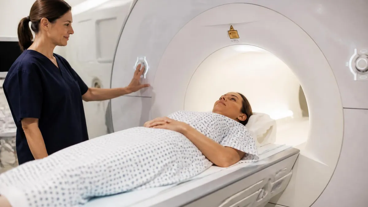
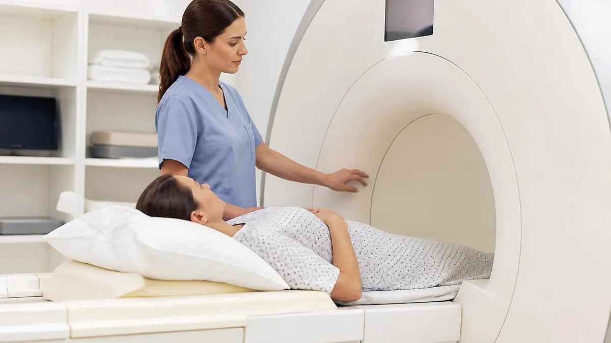
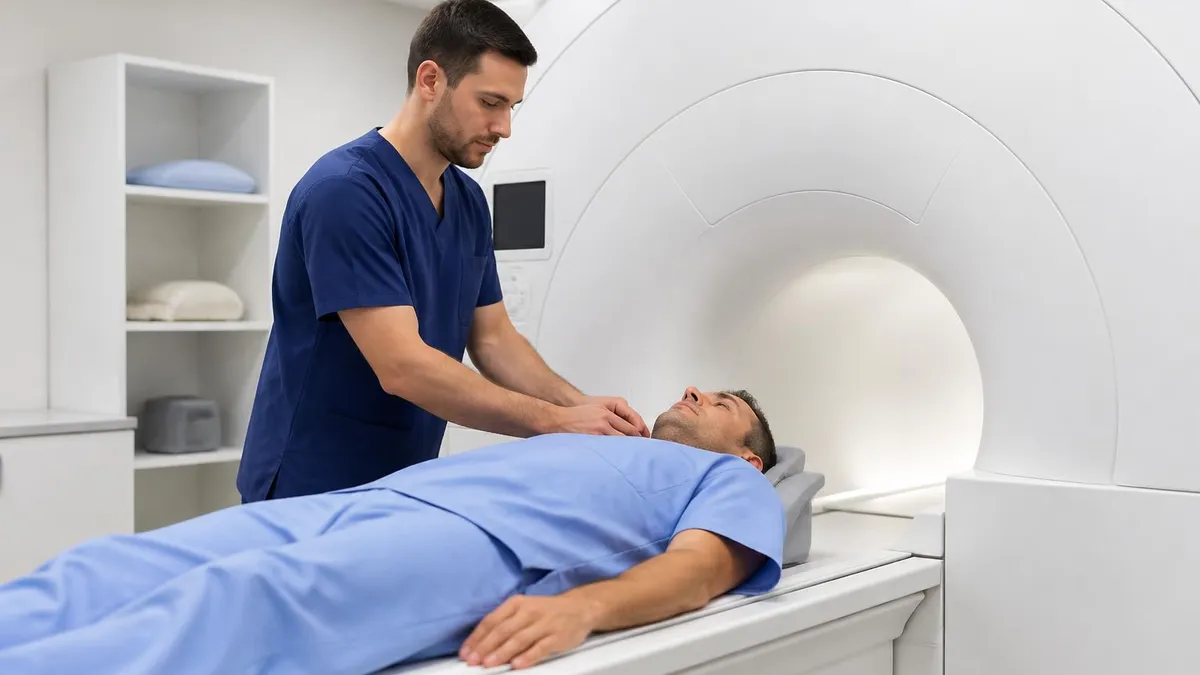
Preparation time is separate from scan time but affects your total visit length. You'll spend 10 to 20 minutes at check-in completing MRI safety screening forms, removing all metal objects, and changing into a gown. A technologist will then position you on the table, place the appropriate surface coil over the region being imaged, and run a brief localizer scan to confirm alignment before the diagnostic sequences begin. Factoring in this preparation, most shoulder MRI appointments should be budgeted at 90 minutes from arrival to departure, even if the scan itself is closer to 50 minutes.
Open MRI units and standing MRI systems—sometimes marketed to patients with severe claustrophobia or obesity—generally operate at lower field strengths (0.3T to 1.0T), which means longer scan times for equivalent image quality. A shoulder study on an open 0.7T unit might take 70 to 90 minutes compared to 45 to 60 minutes on a closed 1.5T or 3T bore. If you are researching mri scan duration across different MRI modalities and sequence types, it helps to understand that faster is not always better—the right protocol for your clinical question matters more than raw speed.
MRI: upcoming dates and official changes
- Aug 17, 2026: ARRT launched a separate entry-level Imaging Assistant, Magnetic Resonance (I.A.) credential. A reduced $75 fee applied to applications made Aug. 17–Oct. 14, 2026, for testing Oct. 15–Nov. 30, 2026. Later applicants pay $225 and can test on or after Jan. 1, 2027.[4]
- The ARRT MRI exam for postprimary-pathway candidates has 220 questions: 200 scored and 20 unscored pilot items.[1]
- The ARRT MRI exam for primary-pathway candidates has 230 questions: 200 scored and 30 unscored pilot items.[2]
- Postprimary-pathway candidates get 210 minutes for the ARRT MRI exam, inside a 230-minute (3 hours, 50 minutes) appointment.[1]
- Primary-pathway candidates get 230 minutes for the ARRT MRI exam, inside a 250-minute (4 hours, 10 minutes) appointment that also covers the tutorial, the NDA and a survey.[2]
- To pass the ARRT MRI exam you need a total scaled score of 75 on ARRT's 1–99 scale. This is not a percentage.[3]
- The ARRT MRI exam through the postprimary pathway costs $225, or $450 for candidates whose supporting credential is from NMTCB or ARDMS.[4]
- The ARRT MRI certification exam costs $225 to apply for through the primary eligibility pathway.[4]
- ARRT MRI candidates have three attempts to pass the exam, all within three years of the date their first exam window opens.[2]
- ARRT MRI certification and registration lasts 10 years, even with annual renewal. After that, holders must complete Continuing Qualifications Requirements (CQR).[5]
Numbers in brackets link to the official sources listed under Sources & last verified. Dates that have passed are removed automatically.
MRI Exam at a Glance
| Item | Detail | Source · Verified |
|---|---|---|
| Number of questions (Postprimary pathway) | 220 questions (200 scored + 20 unscored pilot)[1] | assets-us-01.kc-usercontent.com · Oct 15, 2026 |
| Number of questions (Primary pathway) | 230 questions (200 scored + 30 unscored pilot)[2] | assets-us-01.kc-usercontent.com · Oct 15, 2026 |
| Time limit (Postprimary pathway) | 210 minutes test time (230-minute appointment)[1] | assets-us-01.kc-usercontent.com · Oct 15, 2026 |
| Time limit (Primary pathway) | 230 minutes test time (250-minute appointment)[2] | assets-us-01.kc-usercontent.com · Oct 15, 2026 |
| Passing score | Scaled score of 75 (scale 1–99)[3] | The American Registry of Radiologic Technologists · Oct 15, 2026 |
| Exam fee (Postprimary pathway) | $225 application fee ($450 with an NMTCB or ARDMS supporting credential)[4] | The American Registry of Radiologic Technologists · Oct 15, 2026 |
| Exam fee (Primary pathway) | $225 application fee[4] | The American Registry of Radiologic Technologists · Oct 15, 2026 |
| Retake policy | 3 attempts within 3 years of the first exam window[2] | assets-us-01.kc-usercontent.com · Oct 15, 2026 |
| Validity | 10 years, then Continuing Qualifications Requirements (CQR)[5] | assets-us-01.kc-usercontent.com · Oct 15, 2026 |
| Renewal | Annual renewal, $65/year; 24 CE credits every 2 years[5] | assets-us-01.kc-usercontent.com · Oct 15, 2026 |
| Delivery | Computer-based at Pearson VUE test centers[2] | assets-us-01.kc-usercontent.com · Oct 15, 2026 |
| Eligibility (Postprimary pathway) | Supporting ARRT credential (or NMTCB/ARDMS) + structured education + clinical experience[1] | assets-us-01.kc-usercontent.com · Oct 15, 2026 |
| Eligibility (Primary pathway) | ARRT-recognized MRI program + associate degree or higher + ethics[2] | assets-us-01.kc-usercontent.com · Oct 15, 2026 |
| Score release | Preliminary score at the test center; official results in about 10 business days[3] | The American Registry of Radiologic Technologists · Oct 15, 2026 |
| Testing window | One-year (365-day) exam window per application[2] | assets-us-01.kc-usercontent.com · Oct 15, 2026 |
Free MRI - Magnetic Resonance Imaging Study Tools
Interactive MRI - Magnetic Resonance Imaging resources on PracticeTestGeeks, free to start:
- Free MRI Knowledge Questions and Answers — 11 questions with instant scoring and answer explanations
- MRI - Magnetic Resonance Imaging flashcards — review key terms and concepts card by card
- MRI - Magnetic Resonance Imaging study guide — topic-by-topic overview of what the exam covers
- MRI - Magnetic Resonance Imaging cheat sheet — one-page printable summary for last-minute review
MRI Scan Duration by the Numbers
MRI Appointment Timeline: What Happens and When
Arrival and Safety Screening
Gowning and Coil Placement
Localizer and Sequence Setup
Diagnostic MRI Sequences
Contrast Injection (if ordered)
Image Review and Discharge
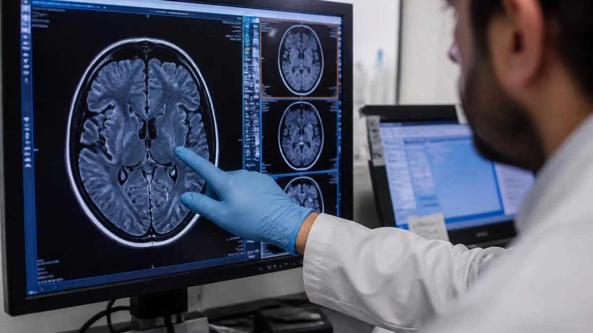
Understanding what makes an MRI shorter or longer enables patients to be active participants in their own care. The single biggest technical variable is the imaging protocol—the specific combination of sequences, planes, and parameters your radiologist selects based on the clinical question. A shoulder MRI ordered to evaluate a suspected rotator cuff tear will include sequences like proton density fat-saturated, T2 fat-saturated in multiple planes, and possibly a T1 sequence, each contributing several minutes to the total run time.
Field strength is the most influential hardware factor. Modern 3 Tesla MRI systems acquire higher signal-to-noise ratio images in less time compared to older 1.5 Tesla units. In clinical practice, a shoulder protocol that takes 55 minutes on a 1.5T scanner might complete in 38 to 42 minutes on a 3T system running the same sequences. Some academic and large hospital centers now operate 7 Tesla research scanners that acquire data even faster, though these remain rare in routine clinical settings and are not yet standard for musculoskeletal imaging.
Parallel imaging techniques have dramatically shortened MRI scan times over the past decade. Methods such as GRAPPA (Generalized Autocalibrating Partial Parallel Acquisition) and SENSE (Sensitivity Encoding) allow the scanner to use multiple coil elements simultaneously to collect data faster without sacrificing image quality. These acceleration factors can cut individual sequence times by 30 to 50 percent. Compressed sensing and simultaneous multi-slice techniques, increasingly available on newer platforms, push that acceleration even further and are beginning to reach routine clinical scanners.
Anatomy-specific challenges also extend scan time. The shoulder joint is inherently complex: it includes the glenohumeral joint, the acromioclavicular joint, the rotator cuff tendons, the biceps tendon, the labrum, the subacromial bursa, and surrounding neurovascular structures. Thoroughly evaluating all of these elements requires imaging in at least three planes—oblique coronal, oblique sagittal, and axial—and multiple sequence types. The oblique planes must be aligned to the individual patient's anatomy, which the technologist sets manually from the localizer images, adding a small amount of setup time for each acquisition block.
Patient cooperation is equally critical to scan efficiency. Technologists routinely instruct patients to breathe normally for extremity studies and to hold their breath for specific abdominal and cardiac sequences. For a shoulder scan, you'll typically be asked to lie supine with your arm at your side in a neutral position. Some protocols position the arm in slight external rotation to better visualize the posterior labrum. If you shift position between sequences, the technologist may need to re-run the localizer and adjust slice orientation, adding 5 to 10 minutes per occurrence.
Sedation and monitoring protocols add time on both ends of the scan. Patients who require conscious sedation—often children, patients with severe claustrophobia, or those with certain cognitive conditions—must be monitored before, during, and after the procedure. IV access must be established, sedation administered, and vital signs observed throughout. Recovery monitoring after the scan can add 30 to 60 minutes to the total visit length. Facilities that routinely handle sedated patients have simplified workflows, but the fundamental physiological requirements cannot be rushed.
Specialized MRI techniques also carry their own time profiles. MR arthrography—in which a radiologist injects dilute gadolinium directly into the shoulder joint under fluoroscopic or ultrasound guidance before MRI—adds 20 to 30 minutes to the pre-scan phase. This technique dramatically improves detection of subtle labral tears and is often ordered for younger, athletic patients with suspected instability. Dynamic or functional MRI sequences that capture the joint in motion add additional acquisition time but can reveal pathology invisible on static images alone.
MRI Scan Duration by Body Region
A shoulder MRI typically takes 45 to 60 minutes without contrast and 60 to 80 minutes with gadolinium enhancement. Wrist MRI runs shorter—about 30 to 45 minutes—because the field of view is smaller and fewer sequences are required. Elbow MRI falls between these two, averaging 40 to 55 minutes. All upper extremity studies benefit from dedicated surface coils that improve signal-to-noise ratio and allow higher resolution imaging of small structures like the ulnar collateral ligament or triangular fibrocartilage complex.
MR arthrography of the shoulder adds 20 to 30 minutes before the MRI itself begins, as a radiologist injects dilute gadolinium into the glenohumeral joint under image guidance. This pre-scan injection dramatically improves labral tear detection and is standard practice for evaluating instability in athletes. Total appointment time including the injection, patient repositioning, and MRI sequences typically runs 90 to 120 minutes. Patients should plan for a half-day commitment when arthrography is ordered alongside their MRI.
Is a Longer MRI Scan Better or Worse for Diagnostic Quality?
- +More sequences mean more anatomical perspectives and higher diagnostic confidence
- +Longer protocols allow thinner slices and higher resolution for detecting small tears or lesions
- +Additional time permits fat-suppressed sequences that distinguish fluid from fat precisely
- +Contrast-enhanced sequences added with extra time reveal vascularity and active inflammation
- +Multi-plane coverage catches pathology that a single-plane scan would miss entirely
- +Specialized sequences like MR neurography or spectroscopy require extra time but provide unique diagnostic value
- −Longer scans increase the risk of patient motion, which degrades image quality and may require repeats
- −Extended table time is physically uncomfortable, especially for patients with acute pain or limited mobility
- −Patients with claustrophobia find longer scans significantly more distressing than shorter protocols
- −More time in the scanner increases radiation exposure risk for patients who incorrectly believe MRI uses X-rays (it does not, but anxiety is real)
- −Longer appointments reduce scanner availability and increase healthcare costs per exam
- −Sedation risks accumulate proportionally with time for pediatric or cognitively impaired patients
Pros and Cons at a Glance
| Pros | Cons |
|---|---|
| More sequences mean more anatomical perspectives and higher diagnostic confidence | Longer scans increase the risk of patient motion, which degrades image quality and may require repeats |
| Longer protocols allow thinner slices and higher resolution for detecting small tears or lesions | Extended table time is physically uncomfortable, especially for patients with acute pain or limited mobility |
| Additional time permits fat-suppressed sequences that distinguish fluid from fat precisely | Patients with claustrophobia find longer scans significantly more distressing than shorter protocols |
| Contrast-enhanced sequences added with extra time reveal vascularity and active inflammation | More time in the scanner increases radiation exposure risk for patients who incorrectly believe MRI uses X-rays (it does not, but anxiety is real) |
| Multi-plane coverage catches pathology that a single-plane scan would miss entirely | Longer appointments reduce scanner availability and increase healthcare costs per exam |
| Specialized sequences like MR neurography or spectroscopy require extra time but provide unique diagnostic value | Sedation risks accumulate proportionally with time for pediatric or cognitively impaired patients |
Pre-MRI Preparation Checklist to Minimize Scan Time
- ✓Complete all MRI safety screening paperwork online before arriving at the facility.
- ✓Remove all jewelry, piercings, and metal accessories at home before your appointment.
- ✓Wear loose, comfortable clothing with no metal zippers, underwire, or snaps.
- ✓Notify the scheduling team of any implants, pacemakers, cochlear implants, or metal fragments.
- ✓Inform the technologist about previous surgeries, especially joint replacements or spinal hardware.
- ✓Arrive 15 minutes early to allow time for check-in, gowning, and IV placement if contrast is ordered.
- ✓Use the restroom immediately before entering the scanner to avoid discomfort during long sequences.
- ✓Discuss any anxiety or claustrophobia with your doctor in advance so a mild anxiolytic can be prescribed if needed.
- ✓Follow fasting instructions if contrast is ordered—usually nothing to eat or drink for four hours prior.
- ✓Practice controlled, even breathing and rehearse staying completely still for 5-minute intervals before your appointment.
Stay Still, Finish Faster
Even a single involuntary movement lasting less than a second can blur an entire MRI sequence, forcing the technologist to repeat it. For a shoulder MRI, one repeated sequence adds 4 to 8 minutes to your scan time. Patients who practice deliberate stillness—focusing on slow, shallow breathing and relaxing muscle tension before each sequence—consistently complete their scans 10 to 15 minutes faster than those who shift or fidget. Ask the technologist how long each sequence lasts so you know exactly how long to hold still.
Brain MRI is among the most frequently ordered scans in the United States, and its duration depends heavily on the clinical indication. A routine brain MRI without contrast—ordered to evaluate headaches, dizziness, or cognitive changes—typically completes in 30 to 45 minutes using sequences such as axial T1, axial T2, axial FLAIR, and diffusion-weighted imaging. Each sequence acquires 20 to 40 slices through the brain and takes 3 to 6 minutes to complete on a modern 1.5T or 3T scanner.
When the ordering physician adds contrast to a brain MRI, the total time increases by 15 to 20 minutes. The technologist completes the non-contrast sequences first, administers gadolinium intravenously, waits three to five minutes for the contrast to cross the blood-brain barrier and enhance any abnormal vascularity, and then runs post-contrast T1-weighted sequences in multiple planes. This protocol is standard for evaluating suspected tumor, metastatic disease, meningitis, MS plaques, and post-surgical changes near the brain parenchyma.
Comprehensive neuroimaging workups can extend a single MRI session to 90 minutes or more. Adding MR angiography of the intracranial or neck vessels takes 10 to 20 additional minutes per vascular territory. MR perfusion imaging, used to assess stroke territory and salvageable penumbra, adds another 10 to 15 minutes. MR spectroscopy—which characterizes the metabolic profile of a brain lesion—can add 20 to 30 minutes and requires precise voxel placement. These extended protocols are most common in academic medical centers and dedicated neuroimaging departments.
Spinal MRI duration varies by level and indication. A cervical spine MRI typically runs 45 to 60 minutes and includes sagittal T1, sagittal T2, axial T2, and often a STIR or fat-saturated sequence to detect marrow edema or disc inflammation. Swallowing artifacts frequently degrade cervical images and may require instructing patients to swallow before each sequence and then hold swallows, which the technologist coordinates verbally through the intercom. Thoracic spine MRI is similar in duration but less subject to swallowing motion.
Lumbar spine MRI is arguably the most ordered spinal study in the country, typically completing in 30 to 50 minutes for a non-contrast protocol. Standard sequences include sagittal T1, sagittal T2, and axial T2 through the disc levels. When the clinical question includes infection, tumor, or post-surgical evaluation, gadolinium-enhanced sagittal and axial T1 sequences are added, extending the exam by 15 to 20 minutes. The lumbar spine is mechanically stable during imaging and rarely causes motion artifact, making it one of the more reliable scans for predictable timing.
Abdominal and pelvic MRI requires a fundamentally different approach to timing because respiratory motion is unavoidable. Technologists use two strategies: breath-hold sequences, which capture data in a single suspended breath of 15 to 25 seconds, and respiratory-triggered or navigator sequences, which track the diaphragm and acquire data only during the quiet phase of breathing. Both add complexity and time. A complete abdominal MRI typically runs 45 to 75 minutes and may extend to 90 minutes when both the liver and pancreas require detailed characterization with multiple contrast phases.
Cardiac MRI represents one of the most technically demanding scan types in terms of duration. The heart never stops moving, so every sequence must be electrocardiogram-gated to acquire data during the brief diastolic pause between heartbeats. A comprehensive cardiac MRI covering function, structure, perfusion, and viability takes 60 to 90 minutes and requires close coordination between the patient, the technologist, and often a cardiac MRI specialist physician. Patients with irregular heart rhythms present particular challenges because gating becomes unreliable, potentially requiring repeated sequences or alternative acquisition strategies.
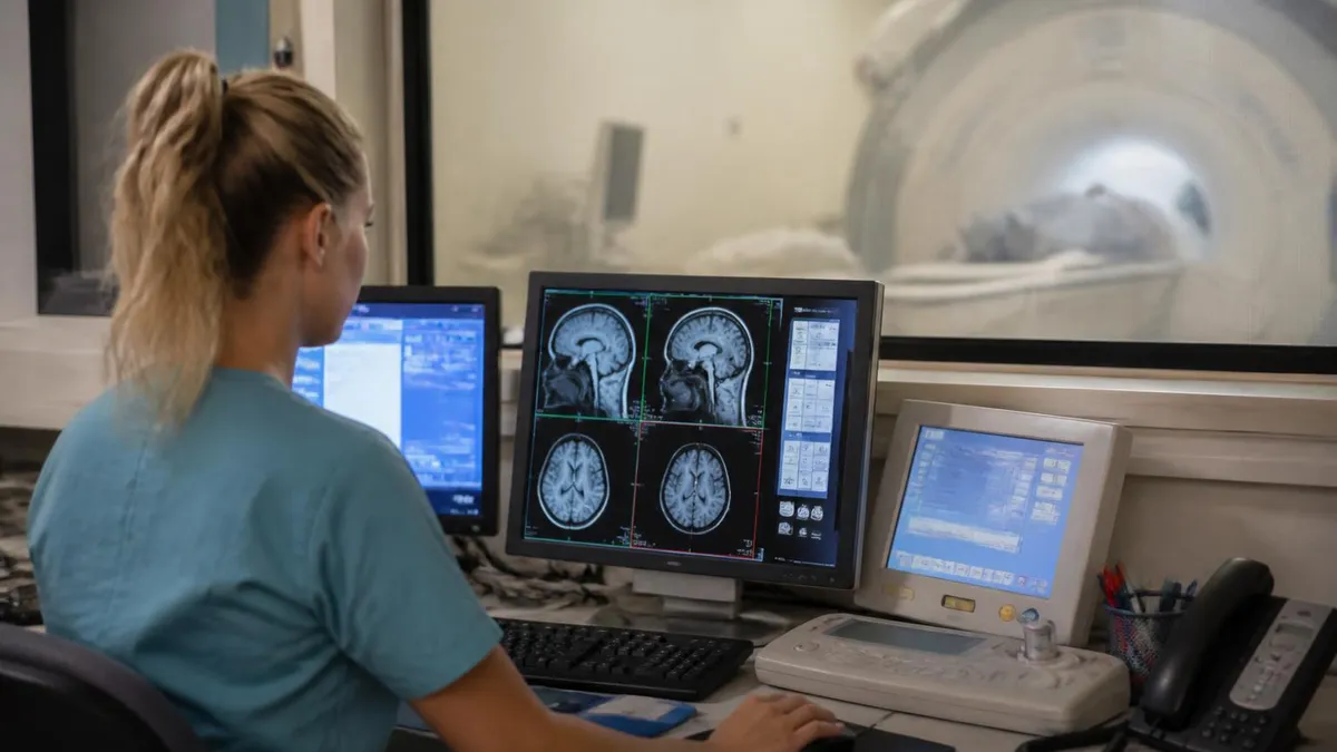
Your facility may quote a standard scan time that does not account for patient-specific variables. If you have a large body habitus, metal implants requiring safety assessment and adjusted protocols, a history of prior surgery at the imaging site, or a clinical question requiring bilateral comparison (both shoulders, for example), your actual scan time may be 20 to 40 percent longer than the standard estimate. Always ask specifically whether your order includes contrast and whether any specialized sequences have been added, and budget your appointment accordingly.
Patients who struggle with claustrophobia face a unique challenge when MRI scan duration is long. The enclosed bore of a standard closed MRI scanner is approximately 60 to 70 centimeters in diameter—wide enough for most adults, but the feeling of confinement intensifies significantly as scan time extends past 30 minutes. Head and neck studies are particularly difficult because the head coil sits close to the face, and patients often feel more enclosed than they do for extremity or abdominal studies where the head may remain outside the bore.
Several evidence-based strategies help claustrophobic patients complete their MRI without sedation. Cognitive techniques include focusing on a single calming image, counting backward from 100, or synchronized deep breathing. Facilities increasingly offer MRI-compatible headphones with music or guided meditation tracks. Some centers use virtual reality headsets that project open outdoor scenes and have demonstrated measurable reductions in anxiety scores during scans. Transparent mirror glasses that allow patients to see outside the bore rather than the tunnel wall provide a simple, low-tech solution many patients find remarkably effective.
When non-pharmacological approaches are insufficient, anxiolytic premedication is a safe and effective option. A low dose of a benzodiazepine—such as lorazepam 0.5 to 1 mg—taken orally 30 to 60 minutes before the scan reduces anxiety without requiring IV access or post-procedure monitoring for most adult patients. Patients who take oral sedation must arrange transportation, as driving is not permitted for at least 12 hours. Discussing this option with your ordering physician or the MRI facility at least 48 hours in advance ensures the prescription can be obtained and filled before your appointment day.
Open MRI systems provide an alternative for patients who cannot tolerate closed bore scanners even with anxiolytic support. Wide-bore scanners—typically 70 centimeters in diameter compared to 60 centimeters in traditional units—offer meaningfully more space without sacrificing field strength. True open MRI systems with C-arm or double-donut magnet designs have large lateral openings, which many claustrophobic patients find far more tolerable. The tradeoff is scan time: open and wide-bore scanners at 1.0T or below require longer acquisition times for equivalent image quality compared to 3T closed bore systems.
Pediatric MRI presents distinct duration challenges. Children under age seven generally cannot remain still for the 30 to 60 minutes required for most protocols, making sedation or general anesthesia necessary in most cases. Facilities that specialize in pediatric imaging have developed child-life programs that use age-appropriate preparation, mock scanner familiarization, and distraction techniques to successfully scan children as young as five or six without sedation in select cases. Anesthesia-assisted MRI adds 30 to 60 minutes of pre-scan preparation and a similar recovery period post-scan, making a routine brain MRI a 3- to 4-hour total facility visit for a sedated toddler.
MRI technology is evolving rapidly in ways that promise to substantially reduce scan times without compromising diagnostic quality. Deep learning-based image reconstruction algorithms—commercially available from multiple scanner manufacturers—can accelerate acquisition by factors of 2 to 4 while maintaining or improving image quality compared to conventional reconstruction. Compressed sensing techniques exploit the natural sparsity of MRI data to reconstruct full images from deliberately undersampled k-space data. These approaches are gradually reaching routine clinical practice and may cut standard shoulder MRI times to 20 to 30 minutes within the next few years.
Understanding MRI scan duration also matters for radiologic technologists and MRI technologists preparing for credentialing examinations. The ARRT MRI registry covers physics, patient care, imaging procedures, and safety—all of which include time-related concepts such as repetition time (TR), echo time (TE), and acquisition time formulas. Candidates who grasp how pulse sequence parameters affect both image contrast and total scan duration demonstrate deeper physics comprehension that translates directly to examination success and clinical competence. Connecting scan duration knowledge to registry preparation reinforces both the practical and theoretical dimensions of the field.
You can continue preparing with the totally open mri near me, which includes answer explanations for every question.
Practical tips for getting through your MRI efficiently begin before you even arrive at the facility. Contact the scheduling team when you book your appointment and ask specifically which sequences are planned, whether contrast is included, and whether you need any special preparation such as fasting or bowel preparation for an abdominal study. This single phone call can prevent surprises on the day of your scan and allows you to plan childcare, work schedules, or transportation arrangements accurately based on a realistic total visit time.
On the day of your scan, wear metal-free clothing from head to toe. Athletic wear made entirely of synthetic fabric with no metal grommets or zippers is ideal. Avoid wearing makeup, hairspray, or nail polish if possible, as some formulations contain metallic particles that can interfere with image quality in certain sequences, particularly facial MRI. Leave all jewelry, watches, and piercing hardware at home rather than removing them at the facility—this eliminates the risk of forgetting something in your pocket and having to pause the scan for removal after you're already positioned in the bore.
During the scan itself, the technologist is your primary resource and is monitoring you continuously from the control room. Every MRI facility is required to provide a call button or squeeze ball that allows you to alert the technologist at any time. Do not hesitate to signal if you feel uncomfortable, need a break, or experience any unusual sensation. Taking a brief 2- to 3-minute pause to stretch or reposition is far preferable to powering through discomfort that causes involuntary movement and ruins a sequence, potentially adding 10 minutes of repeat time.
Understanding the sound patterns of MRI sequences helps many patients feel more in control during the exam. The loud knocking, banging, and buzzing sounds are caused by rapid switching of gradient coils inside the magnet. They are completely normal and pose no risk. Different sequences produce distinctly different sound signatures—some are rhythmic and almost musical, others are sharper and more irregular. Knowing that a long, repetitive knocking pattern typically indicates a T2-weighted sequence lasting 4 to 6 minutes gives you a mental anchor for gauging progress and anticipating when a sequence will end.
After your MRI, the technologist will quickly review the images on the console to confirm technical adequacy before you leave the scanner. If a sequence shows motion artifact or positioning error, they will ask you to complete a repeat run while you're still on the table—it is much more efficient to repeat immediately than to discover the problem after you've dressed and left the facility. Once the images are approved, you're free to go. Contrast patients should hydrate well after the scan to help gadolinium clearance through the kidneys, as recommended by major radiology societies.
For MRI students and technologists, mastering the relationship between pulse sequence parameters and acquisition time is foundational knowledge. Acquisition time in a spin-echo sequence equals TR × number of phase-encoding steps × number of excitations (NEX), divided by the number of slices acquired per TR interval. Increasing NEX from 1 to 2 doubles the signal-to-noise ratio but also doubles scan time. Reducing the phase field of view by 50 percent cuts time in half but may introduce wrap artifact. These tradeoffs are central to protocol optimization and appear consistently on registry and board examinations.
Finally, advocating for yourself in the healthcare environment means asking your ordering physician whether the specific protocol planned for your scan is the minimum necessary to answer the clinical question. Some facilities default to comprehensive protocols that include sequences your clinical situation may not require.
If you are returning for a follow-up scan to track a known, stable finding, ask whether a shorter focused protocol—rather than the full initial workup—would be appropriate. This conversation can sometimes reduce your scan time by 20 to 30 percent while still providing all the clinical information your care team needs to make sound treatment decisions.
Sample MRI - Magnetic Resonance Imaging Practice Questions
Try these questions from our free MRI - Magnetic Resonance Imaging practice tests. The correct answer and an explanation follow each question.
For MRI of the shoulder, the patient should be positioned with the arm in which orientation to best center the glenohumeral joint?
- A. Arm elevated above the head
- B. Arm at the side in neutral or slight external rotation, shoulder centered in the coil
- C. Arm crossed over the chest
- D. Arm in full internal rotation only
Answer: B. Arm at the side in neutral or slight external rotation, shoulder centered in the coil
Positioning the shoulder with the arm at the side in neutral or slight external rotation and centering the glenohumeral joint within the dedicated shoulder coil optimizes image quality.
A technologist who holds an RT(MR)(ARRT) credential and allows it to lapse must complete which process to reinstate it?
- A. Retake the full MRI registry examination
- B. Apply for reinstatement and fulfill CE requirements
- C. Resubmit all original application materials and retake the exam
- D. Complete only a practical competency evaluation
Answer: B. Apply for reinstatement and fulfill CE requirements
ARRT allows lapsed credential holders to reinstate by applying for reinstatement and meeting outstanding CE obligations.
Which ARRT ethics requirement must be fulfilled as part of the MRI registry application process?
- A. CPR certification
- B. Pre-application ethics review if applicable
- C. Magnetic field safety officer training
- D. Advanced cardiac life support (ACLS)
Answer: B. Pre-application ethics review if applicable
Applicants with certain violations must complete a pre-application review, and all applicants must attest to meeting ARRT's Standards of Ethics.
Which SAR limit (Specific Absorption Rate) is the FDA guideline for whole-body RF energy deposition in a 15-minute period for non-controlled conditions?
- A. 1.5 W/kg
- B. 2 W/kg
- C. 3 W/kg
- D. 4 W/kg
Answer: D. 4 W/kg
The FDA guideline for whole-body average SAR under normal operating mode is 4 W/kg over any 15-minute period.
Take the full MRI - Magnetic Resonance Imaging practice test
MRI Questions and Answers
Sources & last verified
- assets-us-01.kc-usercontent.com — number of questions, time limit, eligibility (official source). “MRI Scored Items 200 Pilot Items 20 Total Items 220 Test Time 210 min” Verified Oct 15, 2026.
- assets-us-01.kc-usercontent.com — number of questions, time limit, retake policy, delivery (official source). “MRI Scored Items 200 Pilot Items 30 Total Items 230 Test Time 230 min” Verified Oct 15, 2026.
- The American Registry of Radiologic Technologists — passing score, score release (official source). “Our exam scoring scale ranges from 1 to 99, and you’ll need a total score of 75 to pass the exam.” Verified Oct 15, 2026.
- The American Registry of Radiologic Technologists — exam fee, other (official source). “Postprimary (M, CT, MR, BD, CI, VI, VS, BS, R.R.A.): $225 Postprimary (CT, MR, BD, or VS—using NMTCB as supporting category): $450 Postprimary (MR, VS,…” Verified Oct 15, 2026.
- assets-us-01.kc-usercontent.com — validity, renewal (official source). “ARRT certification and registration—even with annual renewal—is limited to 10 years.” Verified Oct 15, 2026.
How we verify: editorial & fact-checking policy. Report an error: contact us.
DWI MRI: Diffusion-Weighted Imaging Explained (2026 Guide)
Side Effects of Brain MRI: Understanding Risks, Safety, and What Every Patient Should Know
MRI Lumbar Spine Without Contrast: Complete Guide to the Scan, What It Shows, and How to Prepare
MRI History: From Nuclear Resonance to Modern Scanners

MRI of Cervical Spine: Complete Guide to Imaging, Anatomy, Pathology, and Patient Preparation
How this page was made: written with AI tools. Exam facts are checked against the official sources listed below. Not reviewed by a subject-matter expert. Editorial policy
Sources & references
Exam facts on this page (fees, format, dates, passing scores) are checked against these official sources; the date is our last check. Editorial policy
- arrt.org — Passing score, score release, fees, exam information · verified Oct 2026
- assets-us-01.kc-usercontent.com — Renewal, score validity, test delivery, eligibility, exam format, test dates, time limit · verified Oct 2026