
MRI Contraindications: A Complete Guide to Safety, Risks, and Patient Screening
MRI contraindications explained: pacemakers, implants, metal, pregnancy, and gadolinium risks. A complete safety screening guide for patients and techs. 🧠
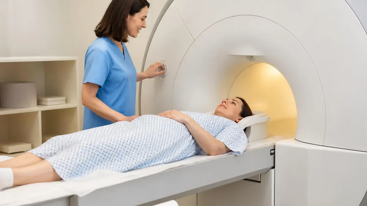
Understanding mri contraindications is one of the most critical responsibilities of every MRI technologist, radiologist, and referring physician. The powerful static magnetic field, switching gradient fields, and radiofrequency energy used in magnetic resonance imaging can interact dangerously with certain implants, metallic foreign bodies, and physiological conditions. A missed contraindication can mean device malfunction, tissue burns, projectile injuries, or even patient death. Before any scan begins, a structured screening process must rule out absolute and relative risks that could turn a routine diagnostic exam into a sentinel event. Practice with the free MRI - Magnetic Resonance Imaging practice test on PracticeTestGeeks (200+ questions with answer explanations).
MRI safety has evolved tremendously since the first clinical scanners appeared in the early 1980s. As field strengths climbed from 0.5T to 1.5T, 3T, and now research 7T systems, the list of contraindications expanded and the screening process became more nuanced. Modern implants are often labeled MR Conditional rather than MR Unsafe, meaning they can be scanned only under specific parameters such as field strength limits, SAR thresholds, or scan duration restrictions. Knowing how to interpret these labels has become a core competency.
Absolute contraindications include ferromagnetic intracranial aneurysm clips, certain older cardiac pacemakers, cochlear implants without MR-conditional labeling, and metallic foreign bodies in the eye. Relative contraindications include pregnancy in the first trimester, severe claustrophobia, renal insufficiency when gadolinium contrast is planned, and recent surgical implants within their healing window. Each of these requires careful documentation, often a phone call to the manufacturer, and sometimes additional imaging like an orbital X-ray to confirm safety before the patient ever enters Zone IV.
The American College of Radiology (ACR) publishes detailed guidance on MR safety zones, screening protocols, and contraindication management. These guidelines are the backbone of every accredited MRI department in the United States, and registry exams routinely test candidates on the specifics. Failure to follow established screening workflows is one of the leading causes of MRI-related adverse events reported to the FDA's MAUDE database each year, with projectile incidents and burns leading the statistics.
This guide walks through the full landscape of MRI contraindications, from the physics of why certain materials are dangerous to the practical workflow of screening a patient at the front desk. We will cover device-specific concerns, contrast agent risks, pregnancy considerations, and the special challenges of pediatric and unconscious patients. Whether you are studying for the ARRT MRI registry, training new staff, or simply trying to understand why your doctor canceled your scan, the information below reflects current 2026 best practices.
If you are preparing for the ARRT or ARMRIT registry, knowing the difference between absolute and relative contraindications is essential, and you should also brush up on related concepts in our guide to the MRI Medical Abbreviation overview. The terminology used in safety screening forms is dense, and many test questions hinge on understanding what acronyms like SAR, dB/dt, and MRC actually mean in clinical context.
Throughout this article you will find practice quiz links, comparison tables, and a comprehensive FAQ designed to reinforce key safety concepts. By the end, you should be able to identify the most common contraindications, explain why each one matters, describe the screening workflow, and know what to do when a patient presents with an unknown implant or uncertain medical history.
MRI: upcoming dates and official changes
- Aug 17, 2026: ARRT launched a separate entry-level Imaging Assistant, Magnetic Resonance (I.A.) credential. A reduced $75 fee applied to applications made Aug. 17–Oct. 14, 2026, for testing Oct. 15–Nov. 30, 2026. Later applicants pay $225 and can test on or after Jan. 1, 2027.[4]
- The ARRT MRI exam for postprimary-pathway candidates has 220 questions: 200 scored and 20 unscored pilot items.[1]
- The ARRT MRI exam for primary-pathway candidates has 230 questions: 200 scored and 30 unscored pilot items.[2]
- Postprimary-pathway candidates get 210 minutes for the ARRT MRI exam, inside a 230-minute (3 hours, 50 minutes) appointment.[1]
- Primary-pathway candidates get 230 minutes for the ARRT MRI exam, inside a 250-minute (4 hours, 10 minutes) appointment that also covers the tutorial, the NDA and a survey.[2]
- To pass the ARRT MRI exam you need a total scaled score of 75 on ARRT's 1–99 scale. This is not a percentage.[3]
- The ARRT MRI exam through the postprimary pathway costs $225, or $450 for candidates whose supporting credential is from NMTCB or ARDMS.[4]
- The ARRT MRI certification exam costs $225 to apply for through the primary eligibility pathway.[4]
- ARRT MRI candidates have three attempts to pass the exam, all within three years of the date their first exam window opens.[2]
- ARRT MRI certification and registration lasts 10 years, even with annual renewal. After that, holders must complete Continuing Qualifications Requirements (CQR).[5]
Numbers in brackets link to the official sources listed under Sources & last verified. Dates that have passed are removed automatically.
MRI Exam at a Glance
| Item | Detail | Source · Verified |
|---|---|---|
| Number of questions (Postprimary pathway) | 220 questions (200 scored + 20 unscored pilot)[1] | assets-us-01.kc-usercontent.com · Oct 15, 2026 |
| Number of questions (Primary pathway) | 230 questions (200 scored + 30 unscored pilot)[2] | assets-us-01.kc-usercontent.com · Oct 15, 2026 |
| Time limit (Postprimary pathway) | 210 minutes test time (230-minute appointment)[1] | assets-us-01.kc-usercontent.com · Oct 15, 2026 |
| Time limit (Primary pathway) | 230 minutes test time (250-minute appointment)[2] | assets-us-01.kc-usercontent.com · Oct 15, 2026 |
| Passing score | Scaled score of 75 (scale 1–99)[3] | The American Registry of Radiologic Technologists · Oct 15, 2026 |
| Exam fee (Postprimary pathway) | $225 application fee ($450 with an NMTCB or ARDMS supporting credential)[4] | The American Registry of Radiologic Technologists · Oct 15, 2026 |
| Exam fee (Primary pathway) | $225 application fee[4] | The American Registry of Radiologic Technologists · Oct 15, 2026 |
| Retake policy | 3 attempts within 3 years of the first exam window[2] | assets-us-01.kc-usercontent.com · Oct 15, 2026 |
| Validity | 10 years, then Continuing Qualifications Requirements (CQR)[5] | assets-us-01.kc-usercontent.com · Oct 15, 2026 |
| Renewal | Annual renewal, $65/year; 24 CE credits every 2 years[5] | assets-us-01.kc-usercontent.com · Oct 15, 2026 |
| Delivery | Computer-based at Pearson VUE test centers[2] | assets-us-01.kc-usercontent.com · Oct 15, 2026 |
| Eligibility (Postprimary pathway) | Supporting ARRT credential (or NMTCB/ARDMS) + structured education + clinical experience[1] | assets-us-01.kc-usercontent.com · Oct 15, 2026 |
| Eligibility (Primary pathway) | ARRT-recognized MRI program + associate degree or higher + ethics[2] | assets-us-01.kc-usercontent.com · Oct 15, 2026 |
| Score release | Preliminary score at the test center; official results in about 10 business days[3] | The American Registry of Radiologic Technologists · Oct 15, 2026 |
| Testing window | One-year (365-day) exam window per application[2] | assets-us-01.kc-usercontent.com · Oct 15, 2026 |
Free MRI - Magnetic Resonance Imaging Study Tools
Interactive MRI - Magnetic Resonance Imaging resources on PracticeTestGeeks, free to start:
- MRI - Magnetic Resonance Imaging practice test — 200+ questions with instant scoring and answer explanations
- MRI - Magnetic Resonance Imaging flashcards — review key terms and concepts card by card
- MRI - Magnetic Resonance Imaging study guide — topic-by-topic overview of what the exam covers
- MRI - Magnetic Resonance Imaging cheat sheet — one-page printable summary for last-minute review
MRI Safety by the Numbers
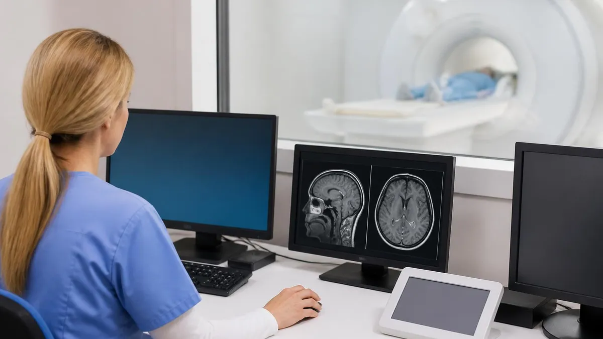
Absolute vs. Relative MRI Contraindications
Conditions that completely prohibit MRI scanning under any circumstances. Includes ferromagnetic aneurysm clips, certain non-conditional pacemakers, cochlear implants without MR labeling, and metallic intraocular foreign bodies. No imaging benefit outweighs these risks.
Conditions where MRI can proceed with caution, modified parameters, or additional precautions. Examples include first-trimester pregnancy, recent surgery, severe claustrophobia, and renal impairment when contrast is required. Each case demands physician judgment.
Implants labeled safe under specific conditions such as maximum field strength, SAR limits, gradient slew rates, and scan region. The manufacturer's IFU must be consulted and documented before scanning to verify compliance with every condition.
Devices or foreign bodies of uncertain origin require investigation before scanning. Patients may need orbital radiography, manufacturer cards, or surgical records. When in doubt, default to caution and reschedule rather than risk harm.
The most common implants encountered in MRI screening are cardiac devices, orthopedic hardware, vascular stents, neurostimulators, and drug infusion pumps. Each category has its own historical timeline of MR compatibility. Cardiac pacemakers manufactured before 2011 are almost universally non-conditional, while newer models from Medtronic, Boston Scientific, and Abbott often carry MR Conditional labeling that permits 1.5T scanning under tightly controlled parameters. Verifying the model, lead configuration, and implant date is non-negotiable before any cardiac device patient enters the magnet room.
Cochlear implants present a particularly complex situation. Older models contain internal magnets that can demagnetize, dislodge, or cause severe pain when exposed to the static field. Newer implants from Cochlear, Advanced Bionics, and MED-EL may be MR Conditional with the magnet in place at 1.5T, or may require surgical removal of the magnet before scanning. The patient's audiologist or surgeon should always be consulted, and the device's identification card carried by the patient is the gold standard reference.
Orthopedic implants such as hip and knee replacements, spinal fusion hardware, and bone plates are generally considered safe at 1.5T and 3T because most modern implants use titanium or non-ferromagnetic stainless steel alloys. However, hardware can produce significant susceptibility artifacts that obscure adjacent anatomy, and recent surgical placement may require a waiting period of 6 to 8 weeks for tissue healing before scanning. Image quality near hardware can sometimes be improved with metal artifact reduction sequences.
Vascular stents, coils, filters, and clips fall into a gray zone that depends entirely on the specific manufacturer and model. Most coronary and peripheral stents manufactured after 2007 are MR Conditional at 1.5T immediately after placement, while older devices traditionally required a 6-week waiting period for endothelialization. Inferior vena cava filters, embolization coils, and surgical clips on blood vessels all need individual verification. The MRIsafety.com database maintained by Dr. Frank Shellock is an indispensable resource for technologists.
Neurostimulators including deep brain stimulators, spinal cord stimulators, and vagus nerve stimulators have their own MR conditional protocols that often specify head-only or extremity-only scanning, restricted SAR values, and reduced gradient performance. Some devices must be programmed into a special MRI mode by a manufacturer representative before the scan and reprogrammed afterward. Drug infusion pumps such as intrathecal baclofen pumps and insulin pumps must be evaluated case by case.
Metallic foreign bodies are perhaps the most insidious contraindication because patients often do not know they have them. Anyone with a history of metalwork, welding, grinding, or military combat injury must be screened with orbital radiography to rule out intraocular metal fragments before MRI. Bullets, shrapnel, and BB pellets present case-specific risks depending on location and composition. For more on imaging alternatives when MRI cannot be performed, see our breakdown of MRI Alternatives for CT, ultrasound, and other modalities.
Tattoos and permanent makeup containing iron-oxide pigments can cause first-degree burns at the tattoo site due to local heating effects, particularly with large or recently applied tattoos. Patients should be warned to alert the technologist immediately if they feel warmth or burning during scanning, and a cold compress can be applied to high-risk tattoos as a precaution. Transdermal medication patches with metallic backing are another often-overlooked source of potential burns and must be removed before scanning.
MRI Practice Test Questions
Prepare for the MRI - Magnetic Resonance Imaging exam with our free practice test modules. Each quiz covers key topics to help you pass on your first try.
MRI Knowledge
MRI Exam Questions covering Knowledge. Master MRI Test concepts for certification prep.
MRI Physics
Free MRI Practice Test featuring Physics. Improve your MRI Exam score with mock test prep.
MRI Anatomy and Pathology
MRI Test Prep for MRI Anatomy and Pathology. Practice MRI Quiz questions and boost your score.
MRI Anatomy and Positioning
MRI Questions and Answers on MRI Anatomy and Positioning. Free MRI practice for exam readiness.
MRI Contrast Agents
Free MRI Quiz on MRI Contrast Agents. MRI Exam prep questions with detailed explanations.
MRI Patient Care and Positioning
MRI Practice Questions for MRI Patient Care and Positioning. Build confidence for your MRI certification exam.
Contrast, Pregnancy, and Special Population Risks
Gadolinium-based contrast agents (GBCAs) are generally safe but carry specific contraindications. Patients with severe acute or chronic kidney injury (eGFR below 30 mL/min/1.73m²) face an elevated risk of nephrogenic systemic fibrosis (NSF), a debilitating condition characterized by fibrotic skin and organ changes. Modern macrocyclic agents like gadobutrol and gadoterate have dramatically reduced this risk compared to older linear agents, but caution remains the standard of care.
Patients with a history of allergic reaction to gadolinium, those on dialysis, and pregnant women in the first trimester are generally not given contrast unless absolutely necessary. Recent research has also examined gadolinium retention in brain tissue after repeated contrast-enhanced scans, though clinical significance remains unclear. Always document GFR within the past 30 to 90 days depending on facility policy and verify no contrast allergy history before injection.
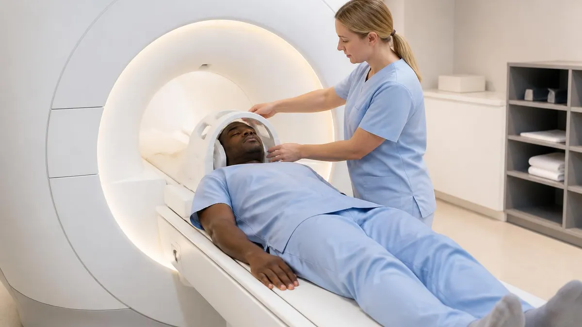
Should This Patient Be Scanned? Weighing the Decision
- +MRI provides unmatched soft-tissue contrast for neurological and musculoskeletal diagnosis
- +No ionizing radiation, making it safer than CT for repeat imaging
- +Modern MR Conditional devices allow most implant patients to be scanned safely
- +Multi-planar imaging eliminates the need for repositioning in many studies
- +Functional MRI techniques like diffusion and perfusion offer unique diagnostic information
- +Gadolinium contrast carries far lower allergic reaction rates than iodinated CT contrast
- −Strict screening protocols add time and complexity to every patient encounter
- −Ferromagnetic objects within Zone IV can become deadly projectiles
- −Claustrophobic patients may require sedation, open MRI, or alternative imaging
- −Long scan times of 30 to 60 minutes can be difficult for unstable or pediatric patients
- −Implant verification can delay urgent scans by hours or days
- −Acoustic noise up to 120 dB requires hearing protection for every scan
Pros and Cons at a Glance
| Pros | Cons |
|---|---|
| MRI provides unmatched soft-tissue contrast for neurological and musculoskeletal diagnosis | Strict screening protocols add time and complexity to every patient encounter |
| No ionizing radiation, making it safer than CT for repeat imaging | Ferromagnetic objects within Zone IV can become deadly projectiles |
| Modern MR Conditional devices allow most implant patients to be scanned safely | Claustrophobic patients may require sedation, open MRI, or alternative imaging |
| Multi-planar imaging eliminates the need for repositioning in many studies | Implant verification can delay urgent scans by hours or days |
Essential MRI Contraindications Screening Checklist
- ✓Verify patient identity with two identifiers before entering Zone III
- ✓Complete the full written MRI safety screening questionnaire with the patient
- ✓Ask specifically about pacemakers, defibrillators, and any implanted electronic devices
- ✓Document any history of metal in the eyes, with orbital X-ray when indicated
- ✓Confirm absence of cochlear implants, neurostimulators, and drug infusion pumps
- ✓Check for surgical clips, stents, coils, and orthopedic hardware with implant dates
- ✓Verify renal function with recent eGFR if gadolinium contrast is planned
- ✓Screen for pregnancy status in all women of childbearing age
- ✓Remove all external metal including jewelry, hairpins, hearing aids, and transdermal patches
- ✓Provide hearing protection and confirm patient communication device works before scanning
The Magnet Is Always On
One of the most dangerous misconceptions in MRI safety is that the magnet can be turned off between scans. The static magnetic field is always active, even during power outages, and only an emergency quench can deactivate it. This means every person entering Zone IV at any time must be screened, including cleaning staff, maintenance workers, and emergency responders. Quenching damages the magnet and costs tens of thousands of dollars to refill the cryogens.
The ACR Zone safety framework provides a structured approach to controlling access to the MRI environment. Zone I is the publicly accessible area outside the imaging suite where no MRI dangers of mri exist. Zone II is the transition area where patients are greeted, screened, and changed into MRI-safe gowns. Zone III is the controlled access area immediately surrounding the scanner room where screening is verified again, and Zone IV is the scanner room itself where the magnetic field exceeds 5 Gauss and where projectile and burn risks are highest.
Access to Zone III and IV must be physically controlled with locked doors, key cards, or constant staff supervision. Only MR-trained personnel designated as Level 2 staff should permit others to enter, and every individual entering Zone IV requires direct supervision unless they have completed equivalent safety training. This includes anesthesiologists, biomedical engineers, housekeeping staff, and visiting clinicians. Lapses in zone control are the proximate cause of most projectile incidents reported each year.
Ferromagnetic detection systems mounted at the entrance to Zone IV add a layer of automated screening but should never replace human screening. These systems can detect metal objects on or inside a person and alert staff before entry. Studies have shown they catch a significant percentage of missed items, including hair clips, pocket knives, oxygen tanks, and even ferromagnetic stretcher wheels. They are now considered standard of care at most accredited facilities and are recommended in updated ACR guidance.
The 5 Gauss line marks the boundary at which the magnetic field is considered safe for the general public, including those with pacemakers in passing exposure scenarios. This line is typically marked on the floor and walls and serves as a visual reminder of where the danger zone begins. Inside the 5 Gauss line, ferromagnetic objects experience increasing force and torque that can wrest them from a person's grip or pocket and accelerate them toward the bore at speeds that can cause fatal injury.
Acoustic safety is another often-overlooked dimension of MRI safety. Scanner noise from gradient switching can reach 110 to 130 dB during high-performance sequences, exceeding the threshold for permanent hearing damage with even brief unprotected exposure. Every patient and any companion in Zone IV during scanning must wear earplugs, MRI-compatible headphones, or both. To learn more about the acoustic environment, see our deep dive on the mri machine noise and what to expect during your scan.
Emergency procedures including code blue response, fire response, and quench procedures require specific MRI-safe protocols. Resuscitation equipment and crash carts must remain outside Zone IV, and the patient must be moved to a safe area before standard equipment can be used. Quench buttons should only be activated in true emergencies such as a person pinned against the magnet by a ferromagnetic object, as a quench releases hundreds of liters of helium gas and can asphyxiate occupants if ventilation fails.
Documentation of every screening encounter is essential for both patient safety and legal protection. Most facilities use a two-tier screening system where a written questionnaire is completed by the patient and then reviewed verbally by an MR-trained technologist. Any inconsistencies, uncertainties, or red flags must be resolved before the patient enters Zone IV, with consultation from the radiologist and referring provider as needed.
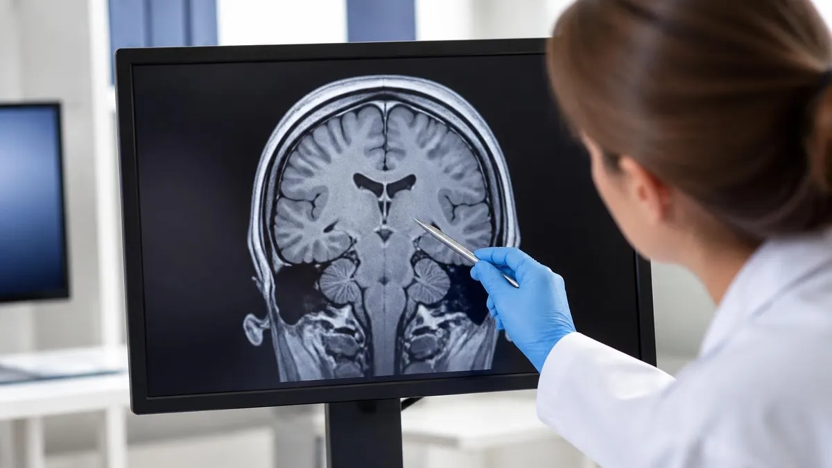
The 2001 death of 6-year-old Michael Colombini at Westchester Medical Center, struck by a ferromagnetic oxygen cylinder brought into Zone IV, remains the most cited cautionary tale in MRI safety. Despite two decades of awareness, projectile incidents continue to be reported to the FDA. Vigilance, screening, and zone control are not bureaucratic burdens. They are life-saving protocols that must be enforced without exception, every single shift.
For radiologic technologists preparing for the ARRT MRI registry or the ARMRIT certification, contraindications and patient safety questions represent one of the largest sections of the exam. Expect questions on absolute versus relative contraindications, specific device compatibility, contrast safety, pregnancy considerations, zone management, and emergency procedures. Many questions test your ability to recognize the correct action when a borderline contraindication is identified rather than simply memorizing lists.
The ARRT content specifications for MRI explicitly include screening, patient interaction, and safety as core competency areas. Successful candidates typically study the ACR Manual on MR Safety, the SMRT safety guidelines, and Dr. Frank Shellock's Reference Manual for Magnetic Resonance Safety, Implants, and Devices. These references should be supplemented with practice questions that build pattern recognition for common contraindication scenarios encountered in clinical practice.
Beyond memorizing device names and field strength limits, registry candidates should understand the underlying physics that make certain conditions dangerous. The static field B0 attracts ferromagnetic objects with force proportional to field strength squared. Switching gradient fields induce eddy currents that can stimulate peripheral nerves and produce the loud knocking noise characteristic of MRI. RF energy at the Larmor frequency causes tissue heating measured as Specific Absorption Rate or SAR, which must be limited to prevent burns.
Documentation standards have tightened significantly since the FDA began requiring more detailed reporting of MR-related adverse events. Most facilities now use electronic screening platforms that integrate with the radiology information system, flagging high-risk patients automatically and preventing scan scheduling until safety verification is complete. Familiarity with these workflows is increasingly tested in registry exams and expected on the first day of any new MR position.
Continuing education in MR safety is now mandatory in most states and is required for ARRT credential maintenance. The ACR offers a free MR safety online course, and SMRT provides advanced certification for MR safety officers. Hospitals are increasingly designating Medical Directors of MR Safety (MMRSDs) and MR Safety Officers (MRSOs) with formal training requirements. These roles ensure ongoing compliance, incident review, and policy updates as new devices and field strengths enter clinical use.
The history of MRI safety has been shaped by real patient harm, and each incident has prompted regulatory and procedural changes that protect future patients. To understand how the modality and its safety practices evolved over time, our guide to the History of MRI traces the journey from Lauterbur's first images through the development of modern safety frameworks. Learning this history reinforces why every screening question matters and why shortcuts are never acceptable.
Finally, communication with the patient is a contraindication safeguard in its own right. Patients often forget about implants placed years ago, do not realize that a metal sliver from a workshop accident counts as foreign body history, or feel embarrassed to admit claustrophobia. Skilled technologists ask open-ended questions, build trust, and create a screening environment where patients feel comfortable disclosing complete medical history. This human dimension is what ultimately separates competent MR practice from truly safe MR practice.
For practical day-to-day work in an MRI department, building habits around contraindication screening pays dividends in both safety and efficiency. Start every shift by reviewing the day's schedule for any patients flagged with implants, contrast allergies, or special handling requirements. Coordinate with nursing, anesthesia, and referring providers in advance so that information gaps are filled before the patient arrives rather than discovered at the screening desk while a busy schedule waits.
Develop a personal library of trusted resources you can access quickly during a screening encounter. Bookmark MRIsafety.com on your workstation, keep a current edition of the Shellock reference within reach, and maintain phone numbers for major device manufacturers. When uncertainty arises about a specific implant, calling the manufacturer's MR safety hotline is always preferable to guessing. Most companies respond within minutes and can email documentation directly to your facility.
Practice clear, jargon-free patient communication during screening. Many patients do not understand terms like stent, prosthesis, or pacemaker in the same way clinicians do. Use plain language and concrete examples such as asking whether they have ever had heart surgery, brain surgery, eye surgery, or any device put under their skin. Show pictures of common devices when needed, and reassure patients that disclosing implants helps you keep them safe rather than disqualifying them from imaging.
Train every staff member who enters Zone III on basic ferromagnetic awareness, including transport, housekeeping, and facilities personnel. A common pattern in adverse events is a non-MR-trained employee bringing a wheelchair, oxygen tank, or cleaning equipment into the magnet room without screening. Annual safety training, signed competency documentation, and routine walk-through audits help prevent these incidents and demonstrate compliance during accreditation surveys.
For students and new technologists, shadowing experienced screeners is invaluable. Pay attention to the questions they ask, the order in which they ask them, and the follow-up probes they use when a patient gives an uncertain answer. Notice how they verify implant cards, read manufacturer documentation, and decide when to escalate to the radiologist. These soft skills take months to develop and cannot be learned from textbooks alone.
When you do encounter a complicated case such as an MR Conditional pacemaker patient or someone with multiple stents from different manufacturers, take the time to document your decision-making process thoroughly. Record which manufacturer was contacted, which IFU was referenced, what scan parameters were modified, and which physician approved the protocol. This documentation protects the patient, the staff, and the institution in the event of an adverse outcome or regulatory review.
Above all, cultivate a culture where it is acceptable, even encouraged, to stop the line if something feels off. Many MR adverse events occurred because staff felt pressure to keep the schedule moving and ignored their instincts. A canceled or rescheduled scan due to safety concerns is always preferable to a sentinel event. The best MR departments treat every screening encounter as a high-reliability process where slowing down and asking questions is recognized as professional excellence.
Sample MRI - Magnetic Resonance Imaging Practice Questions
Try these questions from our free MRI - Magnetic Resonance Imaging practice tests. The correct answer and an explanation follow each question.
A patient reports having a cardiac pacemaker. What is the standard MRI safety classification for most older pacemakers?
- A. MR Safe
- B. MR Conditional
- C. MR Unsafe
- D. MR Compatible
Answer: C. MR Unsafe
Most older pacemakers are classified MR Unsafe because the magnetic field can interfere with device function or cause heating.
Which MRI-specific safety topic is explicitly listed under the ARRT exam content specifications within Patient Care and Education?
- A. Radiofrequency power amplifier maintenance
- B. Screening for implants and metallic foreign bodies
- C. Cryogen manufacturing standards
- D. Gradient coil winding techniques
Answer: B. Screening for implants and metallic foreign bodies
Screening patients for implants and metallic foreign bodies is a core patient safety responsibility explicitly included in the content specifications.
On the ARRT MRI registry exam, which content area covers patient care, contrast agents, and MRI safety screening?
- A. Image Production
- B. Patient Care and Education
- C. Procedures
- D. Physics and Instrumentation
Answer: B. Patient Care and Education
Patient Care and Education encompasses screening, safety, contrast administration, and patient communication topics.
Which patient condition requires consultation with the ordering physician regarding MRI safety due to potential risks during the first trimester?
- A. Known diabetes mellitus
- B. Known pregnancy, especially first trimester
- C. Mild hypertension controlled with oral medication
- D. Prior abdominal surgery more than 1 year ago
Answer: B. Known pregnancy, especially first trimester
MRI in the first trimester is performed with caution; although no confirmed harm is established, ACR guidelines recommend physician consultation and clinical justification for first-trimester MRI.
Take the full MRI - Magnetic Resonance Imaging practice test
MRI Questions and Answers
Sources & last verified
- assets-us-01.kc-usercontent.com — number of questions, time limit, eligibility (official source). “MRI Scored Items 200 Pilot Items 20 Total Items 220 Test Time 210 min” Verified Oct 15, 2026.
- assets-us-01.kc-usercontent.com — number of questions, time limit, retake policy, delivery (official source). “MRI Scored Items 200 Pilot Items 30 Total Items 230 Test Time 230 min” Verified Oct 15, 2026.
- The American Registry of Radiologic Technologists — passing score, score release (official source). “Our exam scoring scale ranges from 1 to 99, and you’ll need a total score of 75 to pass the exam.” Verified Oct 15, 2026.
- The American Registry of Radiologic Technologists — exam fee, other (official source). “Postprimary (M, CT, MR, BD, CI, VI, VS, BS, R.R.A.): $225 Postprimary (CT, MR, BD, or VS—using NMTCB as supporting category): $450 Postprimary (MR, VS,…” Verified Oct 15, 2026.
- assets-us-01.kc-usercontent.com — validity, renewal (official source). “ARRT certification and registration—even with annual renewal—is limited to 10 years.” Verified Oct 15, 2026.
How we verify: editorial & fact-checking policy. Report an error: contact us.
MRI Medical Abbreviation: What MRI Stands For and Why It Matters
The History of MRI: From Discovery to Modern Medicine

Knee MRI Images: A Complete Guide to Reading, Understanding, and Interpreting Knee Scans
Noise of MRI Machine: Why MRI Scanners Are So Loud and What to Expect
MRI Alternatives: When CT, Ultrasound, X-Ray, and PET Make More Sense Than an MRI
About the Author

Medical Laboratory Scientist & Clinical Certification Expert
Johns Hopkins UniversityDr. Sandra Kim holds a PhD in Clinical Laboratory Science from Johns Hopkins University and is certified as a Medical Technologist (MT) and Medical Laboratory Scientist (MLS) through ASCP. With 16 years of clinical laboratory experience spanning hematology, microbiology, and molecular diagnostics, she prepares candidates for ASCP board exams, MLT, MLS, and specialist certification tests.