CPR and the Heart: How Compressions Keep Blood Flowing When the Heart Stops (2026 July)
CPR heart guide: how chest compressions, AED shocks, and rescue breaths keep oxygenated blood moving when the heart stops. Save a life today. 🔎
At a glance: CPR doesn’t restart a stopped heart — chest compressions keep oxygenated blood flowing to the brain until a defibrillator fixes the underlying electrical problem. Sudden Cardiac Arrest (SCA) is an electrical malfunction; a heart attack (MI) is a plumbing blockage — the two are completely different emergencies. Adult CPR depth is 2–2.4 inches, rate is 100–120 compressions per minute, hands on the lower half of the sternum. Every minute without defibrillation drops SCA survival by roughly 10%; CPR + AED within 3 minutes lifts survival from about 7% to 49–75%. AEDs only shock V-Fib and pulseless V-Tach — they refuse to shock asystole or PEA, and that is not a malfunction.
Here’s the thing most folks get wrong about cpr heart mechanics: chest compressions don’t actually restart a stopped heart. They never have. CPR is a stall tactic — a brutally effective one — that buys minutes until something else, usually a defibrillator, fixes the underlying electrical mess. Understanding that single fact changes how you respond to a collapse.
If you’ve ever watched a TV medical drama, you’ve seen the lie. Hero pumps on a chest. Heart kicks back. Patient gasps. Roll credits. Reality is messier. The heart isn’t a stalled engine waiting for a jump start. It’s a finely tuned electrical organ, and when it goes silent, only one of two things tends to bring it back: a shock delivered at the right rhythm, or advanced drugs at the right time. CPR keeps the door open until either of those arrives.
Cardiopulmonary resuscitation — that’s the full name — literally means resuscitating both the heart (cardio) and the lungs (pulmonary). Two systems, one rescuer. You compress to push blood. You breathe to oxygenate it. Together they substitute for a circulation that has, for whatever reason, quit on its owner.
This guide walks through how the heart actually fails, what your hands do to the chest cavity during compressions, why an AED matters more than perfect form, and the survival math that should put every hesitating bystander into motion. We’ll cover sudden cardiac arrest, heart attacks (which are completely different despite the constant mix-up), pulse points, hand placement, depth, rate, and what happens after the heart restarts. There’s a lot here. Most of it could save a life you know.

CPR generates only one-third of normal cardiac output even when performed perfectly. It’s not designed to keep someone conscious — it’s designed to keep brain cells alive long enough for defibrillation or ALS to restart the heart properly.
What Actually Happens When the Heart Stops
Before you can understand why CPR works, you need a quick map of what fails. The heart is essentially a four-chamber pump driven by a built-in electrical conduction system. Sinoatrial node fires. Signal sweeps through the atria. AV node delays the pulse just enough. Bundle branches carry it into the ventricles. Ventricles squeeze. Blood flies. Roughly seventy times a minute, every minute, for eight or nine decades if you’re lucky.
When that breaks, it breaks in one of a handful of patterns. The most dangerous — and the one CPR was designed for — is Sudden Cardiac Arrest, usually shortened to SCA. The conduction system goes haywire. The ventricles either quiver uselessly (ventricular fibrillation, or V-Fib) or beat so fast they can’t fill with blood (ventricular tachycardia, V-Tach). In both cases, the pump output drops to zero. The patient collapses within seconds. No pulse. No breathing in any meaningful sense. Brain starts dying in three to four minutes.
Other arrest rhythms exist. Pulseless Electrical Activity (PEA) shows electrical signals on a monitor but produces no mechanical squeeze. Asystole — the flat line everyone associates with death — is exactly that: no electrical activity at all. These rhythms are non-shockable, meaning an AED won’t fire. CPR plus epinephrine plus reversing the underlying cause is the only option, and outcomes are grim.
Then there’s the confusion that costs lives every year: people thinking cpr heart emergencies and heart attacks are the same event. They aren’t. They’re neighbors, not twins.
How to Recognize Sudden Cardiac Arrest
- ✓Patient collapses without warning — usually within seconds
- ✓Unresponsive to shouting or shaking
- ✓No normal breathing — agonal gasps don’t count
- ✓No detectable pulse at the carotid (don’t spend >10 seconds checking)
- ✓Skin pales or turns cyanotic quickly
- ✓Begin compressions immediately — every second matters
Sudden Cardiac Arrest vs Heart Attack: The Difference Matters
This is the comparison that trips up new students every single class. Memorize it now and you’ll never confuse the two again.
A heart attack — clinically called myocardial infarction or MI — is a plumbing problem. A coronary artery, the vessels that feed blood to the heart muscle itself, gets blocked. Usually it’s a chunk of plaque that ruptured and triggered a clot. Downstream of the blockage, heart muscle starts to die from lack of oxygen.
The patient feels crushing chest pressure, shortness of breath, sweating, nausea, pain radiating into the left arm or jaw. The heart keeps beating. The person is awake, scared, often clutching their chest. They need a hospital, an angiogram, and probably a stent. They do not, in this moment, need CPR.
Sudden cardiac arrest is an electrical problem. The conduction system short-circuits. The pump quits. There’s no warning chest pain in roughly half of cases. One second the person is talking. Next second they’re on the floor, eyes rolled back, breathing maybe one or two agonal gasps before going completely still. The heart has stopped pumping. This patient absolutely needs CPR, and they need it now.
The cruel twist: a heart attack can trigger sudden cardiac arrest. Dying heart muscle gets electrically irritable. It can drop into V-Fib without warning. So someone clutching their chest at the gym today might collapse into SCA an hour later. That’s why the sudden cardiac arrest vs heart attack distinction matters operationally: same person, different emergencies, different responses, sometimes minutes apart. You treat what you see in front of you.
The mortality numbers tell the story. Roughly 805,000 Americans have heart attacks each year; the survival rate is excellent if they get to a cath lab. Roughly 350,000 Americans suffer out-of-hospital sudden cardiac arrest each year, and around 90% of them die. The difference is staggering and almost entirely explained by how fast bystander CPR and a public-access AED arrive at the scene.
Sudden Cardiac Arrest vs Heart Attack
An electrical malfunction. The heart stops pumping entirely and the patient collapses within seconds. Without immediate CPR and an AED, brain death follows in minutes.
- Underlying problem: Electrical malfunction
- What the heart does: Stops pumping entirely
- Patient consciousness: Collapses within seconds
- Pulse present: No
- Treatment: CPR + AED defibrillation
- US cases per year: ~350,000 out-of-hospital
- Survival without intervention: ~7%
- Common rhythms: V-Fib, V-Tach, PEA, Asystole
A plumbing blockage. A coronary artery is occluded by a plaque rupture and clot; heart muscle starts dying but the heart usually keeps beating and the patient remains conscious.
- Underlying problem: Blocked coronary artery
- What the heart does: Keeps pumping; muscle dying
- Patient consciousness: Awake, in pain
- Pulse present: Yes
- Treatment: Hospital, stent, clot busters
- US cases per year: ~805,000
- Survival with care: High when treated quickly
- Typical rhythm: Sinus, often with ST changes
How Chest Compressions Actually Work
The biomechanics of CPR are weirder than most people realize. When you push down on the lower half of the sternum, you’re not really “pumping the heart.” You’re changing pressure inside the entire chest cavity. Two theories try to explain why blood still moves.
The cardiac pump theory is the obvious one: your hands squeeze the heart between the breastbone in front and the spine behind. Mitral and tricuspid valves close. Blood gets shoved out through the aorta and pulmonary artery. Release. The chest recoils. Atria refill. Repeat. It’s mechanical, brute-force, and surprisingly effective.
The thoracic pump theory says the real driver is intrathoracic pressure. Compressing the chest raises pressure inside the cavity. That pressure squeezes blood out through veins and arteries. When you release, pressure drops and blood refills the chest. Reality is probably a blend of both, with the ratio depending on the patient’s chest size and stiffness.
Either way, the output is bad. Even textbook-perfect CPR generates only about one-third of normal cardiac output. That’s enough to keep the brain alive, barely, and to keep the heart muscle itself perfused so a defibrillator shock has something to work with. It is not enough to maintain consciousness, sustain organ function long-term, or do anything except buy time. Buying time is the entire job.
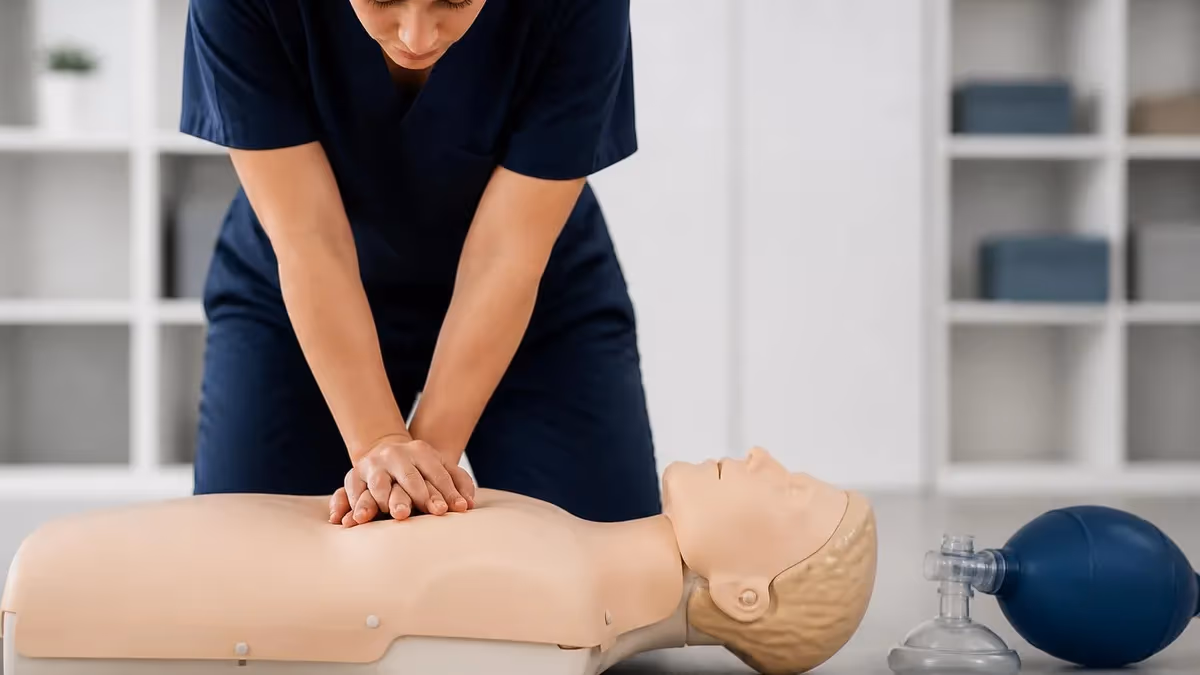
Hand placement is non-negotiable. The heel of one hand goes on the lower half of the sternum — roughly two fingers above the xiphoid process, that little pointy cartilage at the bottom. Other hand on top. Fingers interlaced and lifted off the ribs. Compress straight down. If you’re pushing on ribs you’ll break ribs without moving much blood. If you’re pushing on the xiphoid you can lacerate the liver. Centered on the lower sternum, you’re fine.
Depth and rate are the other non-negotiables. Adults: 2 to 2.4 inches deep, 100 to 120 compressions per minute. Yes, that’s the tempo of “Stayin’ Alive,” and yes, instructors really do use it. Allow full chest recoil between compressions. Incomplete recoil traps blood and kills perfusion fast. Don’t lean on the chest. Don’t pause unless you absolutely have to.
The Four Cardiac Arrest Rhythms
Ventricles quiver chaotically with no organized squeeze. Most common arrest rhythm in adults.
- ▸Shockable by AED
- ▸Triggered most often by ischemic heart disease
- ▸First shock fixes a large fraction when delivered within 3 minutes
Ventricles beat so fast they can’t fill with blood, so output drops to zero.
- ▸Shockable by AED
- ▸Often degenerates into V-Fib if not treated
- ▸Common in long QT syndrome and hereditary arrhythmias
Electrical signals show on the monitor but the heart doesn’t mechanically squeeze.
- ▸Not shockable
- ▸Treat with CPR, epinephrine, and reverse the cause
- ▸Look for the Hs and Ts (hypoxia, hypovolemia, tension pneumothorax, tamponade)
Flat line — no electrical activity at all.
- ▸Not shockable
- ▸Treat with CPR plus epinephrine
- ▸Outcomes are grim; reversing the underlying cause is critical

Why the AED Is the Real Hero
CPR keeps the patient viable. An AED — Automated External Defibrillator — actually fixes the problem. The distinction matters because too many bystanders focus on perfecting compressions while the AED on the wall ten feet away never leaves its case.
Here’s what an AED does. It reads the patient’s heart rhythm through two adhesive pads stuck to the chest. If it detects V-Fib or pulseless V-Tach, it charges and delivers a shock. That shock briefly depolarizes every cardiac cell at once. When the dust clears, the sinoatrial node — the heart’s natural pacemaker — often takes over again and produces an organized rhythm. The pump restarts.
AEDs will not shock asystole or PEA. The device is smart enough to refuse. If you hear “no shock advised, continue CPR,” it doesn’t mean the AED is broken. It means the rhythm isn’t shockable and your compressions are still doing useful work. Keep going.
The survival math is brutal and clear. Without bystander intervention, survival from out-of-hospital SCA hovers around 7%. With immediate CPR and a defibrillator shock within three minutes, survival jumps to between 49% and 75% depending on the study and the rhythm. Every minute without defibrillation drops survival by roughly 10%. That’s why public-access AEDs in airports, gyms, schools, and casinos have transformed outcomes wherever they’re deployed. If you remember nothing else from this article: when someone collapses, send a second bystander for the AED before you even start compressions. Both interventions matter. The shock matters more.
For deeper drilling on rescue mechanics, our Adult CPR step-by-step guide walks the full sequence from scene safety through handoff to EMS, including airway, breathing, and the compression-to-ventilation ratio in different rescuer scenarios.
Survival Math for Sudden Cardiac Arrest
Pulse Points: Where to Check Before You Start
Before committing to CPR, you confirm there’s no pulse. You’ve got about ten seconds. Don’t spend longer — if you can’t feel one in ten, assume there isn’t one and start compressions. Hesitation costs brain cells.
The pulse points you should know:
- Carotid — in the groove beside the trachea, just under the jaw. Primary check for adults and older children. Strong, easy to find under stress.
- Radial — thumb side of the wrist. Useful for conscious patients, but unreliable in shock or arrest because it disappears when blood pressure drops.
- Femoral — in the crease where the leg meets the torso. Used by paramedics and ER staff during arrest because it stays palpable even when peripheral pulses vanish.
- Brachial — inner upper arm, between bicep and tricep. This is the primary check for infants under one year because their short necks make carotid palpation impractical and risky.
- Pedal — top of the foot (dorsalis pedis) or behind the inner ankle (posterior tibial). Mostly used for assessing circulation in injured limbs, not arrest.
For lay rescuers, the American Heart Association now de-emphasizes pulse checking entirely. If the patient is unresponsive and not breathing normally, start compressions. The science says false negatives during pulse checks delay CPR more than false positives cause harm.
The 60-Second Collapse Response
Check the scene
Tap and shout
Call 911 and request an AED
Check breathing in ≤10 seconds
Start compressions
Attach AED the moment it arrives
Why Hearts Stop in the First Place
Different ages, different killers. Knowing the typical cause shapes your response.
Adults arrest most often from primary cardiac causes. Ventricular fibrillation triggered by ischemic heart disease tops the list. Decades of coronary plaque buildup, a sudden rupture, a clot, electrically irritable muscle, and the rhythm collapses. Long QT syndrome, hypertrophic cardiomyopathy, and arrhythmogenic right ventricular dysplasia also lurk in family histories.
Children rarely arrest from primary heart problems. Their arrests almost always start as respiratory emergencies that progress to cardiac. Drowning. Choking. Asthma attack. Severe pneumonia. Opioid overdose. The lungs fail first, oxygen levels crash, and the heart finally gives up. This is why pediatric CPR emphasizes ventilation more aggressively than adult CPR does — in adults you’re mainly fixing a pump problem, in kids you’re mainly fixing an oxygen problem.
Trauma patients can arrest from blood loss, tension pneumothorax, cardiac tamponade, or massive head injury. CPR helps less here because the underlying cause has to be fixed first — you can’t pump an empty tank.
Drug overdoses, especially opioids, kill by suppressing the respiratory drive. The patient stops breathing, blood oxygen falls, the heart eventually quits. Naloxone reverses the opioid effect within minutes if available. Rescue breaths matter enormously here.
For an absolute beginner getting up to speed on the various triggers and how field response changes, our CPR Scenarios practice cases work through realistic situations from drowning to heart attack to playground choking, with the priorities laid out side by side.
Adults arrest because their hearts give out — the rhythm fails first. Children arrest because their lungs give out — oxygen falls, and the heart finally quits last. That’s why pediatric CPR weighs ventilation more heavily and why hands-only CPR isn’t recommended for kids.
Compression-to-Breath Ratios and Rate
The ratio depends on who’s helping and who’s the patient. Memorize the standard sets and you’re ninety percent of the way there.
Single adult rescuer, any victim: 30 compressions to 2 breaths. Repeat in cycles. This is the workhorse rhythm and the one tested on most certification exams.
Two rescuers, adult victim: Still 30:2. Switch compressors every two minutes to avoid fatigue-driven shallowness.
Two rescuers, child or infant victim: 15 compressions to 2 breaths. Children need more frequent ventilation because their arrests are usually respiratory in origin.
Hands-only CPR for untrained bystanders, adult victim: Continuous compressions, 100–120 per minute, no rescue breaths. The AHA introduced this in 2008 because untrained rescuers were freezing at the mouth-to-mouth step. Compressions alone are dramatically better than nothing.
For depth: 2 to 2.4 inches in adults, about 2 inches in children, and 1.5 inches in infants. The rate stays 100–120 per minute across all ages. If you’re curious how the exact compression and breath ratios break down between certifications and patient ages, the dedicated breakdown there covers the AHA and Red Cross numbers and the small differences that crop up between providers.
Rescue breaths get a full second each. Watch for chest rise. Don’t over-ventilate — pumping the lungs too hard raises chest pressure and chokes off venous return, which kills perfusion. One second in, watch the rise, one second exhale, move on.

Adult CPR Quick Reference
- ✓Hands on lower half of sternum, two fingers above the xiphoid
- ✓Compress 2 to 2.4 inches deep
- ✓Rate of 100 to 120 compressions per minute
- ✓Allow full chest recoil between compressions
- ✓Minimize pauses — only for AED rhythm checks and rescuer swaps
- ✓Switch compressors every 2 minutes to avoid fatigue
- ✓30 compressions to 2 breaths if delivering rescue breaths
- ✓Continue until EMS arrives or patient breathes
Continue CPR until EMS takes over, the patient starts breathing, you physically cannot continue, or the scene becomes unsafe. Survival has been documented after 30+ minutes of bystander CPR. Don’t stop because you’re tired — swap compressors if you have a partner.
What Happens After the Heart Restarts
When a defibrillation shock works and an organized rhythm returns, paramedics call it ROSC — Return of Spontaneous Circulation. It’s the moment everyone’s working toward. It is also the start of an entirely new fight.
The patient’s body has been hypoxic, acidotic, and inflammatorily wrecked. Brain cells that survived the arrest are vulnerable to a cascade of injury called reperfusion damage when oxygen rushes back in. Without careful management, half of ROSC patients still die in the next 24 hours from this secondary brain injury.
Standard post-resuscitation care includes targeted temperature management — chilling the patient to around 32–36°C for 24 hours to slow brain metabolism. Mechanical ventilation gets oxygen levels exactly right, neither too high nor too low. Blood pressure is supported with fluids and vasopressors. Cardiac catheterization may follow to fix the underlying coronary blockage if one triggered the arrest.
For survivors at chronic risk — previous arrest, hereditary arrhythmia syndromes, severe heart failure — an Implantable Cardioverter-Defibrillator (ICD) often goes in before discharge. The ICD is a small device implanted under the skin near the collarbone, wired to the heart, watching every beat. When it senses V-Fib or dangerous V-Tach, it delivers an internal shock automatically. ICDs have transformed survival for high-risk patients who’d otherwise be one arrhythmia away from a repeat arrest.
If you’re studying for a paramedic or RN certification, the deeper cpr advanced cardiac life support protocols pull all of this together — airway management, drug administration, rhythm interpretation, and post-arrest care — into the ACLS algorithms tested on credentialing exams.
Common CPR Mistakes That Reduce Survival
Average bystanders spend 30 to 60 seconds checking for breathing, calling for help, finding their phone, and convincing themselves to act. Each of those seconds is brain cells. If the person is unresponsive and not breathing normally, start immediately. Don’t wait for permission, certainty, or another rescuer.
Preventing the Need for CPR in the First Place
The best CPR is the one you never have to perform. Most sudden cardiac arrests in adults trace back to coronary artery disease, and coronary artery disease is largely preventable. The risk factors are tediously familiar but they really do matter: untreated hypertension, uncontrolled diabetes, smoking, obesity, sedentary living, high LDL cholesterol. Knock those down and you knock down arrest risk by an enormous margin.
Heart-healthy living isn’t glamorous. Mediterranean-style eating, 150 minutes of moderate exercise weekly, no tobacco, moderate alcohol, sleep apnea screening if you snore, an annual physical that includes lipid panels and blood pressure. People hate hearing it. The numbers don’t care.
Family history matters enormously. If a first-degree relative dropped dead suddenly before age 50, get screened. Echocardiogram. Electrocardiogram. Stress test. Sometimes genetic testing. Inherited arrhythmia syndromes — Long QT, Brugada, Catecholaminergic Polymorphic Ventricular Tachycardia — hide quietly until they kill, and they often run in families.
Know your community’s AED locations. Map them. The PulsePoint app and similar tools crowd-source AED data and alert nearby trained bystanders when 911 dispatches a cardiac arrest call. Workplace programs, school programs, faith community programs — every additional trained responder shaves seconds off the response time. Seconds shave deaths off the annual count.
And learn CPR. Take the class. Most are four hours. Most cost less than dinner for two. The skill expires every two years for a reason — muscle memory fades — so refresh it. If you’re due for renewal, our AHA CPR recertification guide covers the online and in-person options for renewing without redoing the full course.
CPR for Pediatric and Infant Patients
The basics carry across, but the technique scales with the patient.
Children (1 year to puberty): One hand or two depending on the child’s size. Lower half of sternum. Compress about 2 inches, or one-third the depth of the chest. 100–120 per minute. Single rescuer 30:2, two rescuers 15:2.
Infants (under 1 year): Two fingers in the center of the chest, just below the nipple line, for single-rescuer CPR. Two thumbs encircling the chest for two-rescuer CPR. Compress about 1.5 inches, or one-third the depth of the chest. Same 100–120 per minute. 15:2 ratio with two rescuers.
Rescue breaths use mouth-to-mouth-and-nose for infants because their faces are small enough to cover both at once. Tiny puffs — cheek puffs, not full lung breaths — just until you see chest rise. Big breaths cause gastric distension and force vomit into the airway.
AED pediatric pads or attenuator keys reduce the shock energy for kids under 25 kg. If pediatric equipment isn’t available, adult pads are still better than no shock at all — place one on the chest and one on the back so they don’t touch each other on a small body.
Neonatal resuscitation is a different animal entirely, dominated by ventilation and warmth rather than chest compressions. Our neonatal CPR guide covers the algorithm for the first minutes of life when a newborn isn’t crying or breathing.
Infant and Child CPR Differences
- ✓Infants (under 1 year): two fingers (single rescuer) or two thumbs encircling (two rescuers)
- ✓Infant compression depth: about 1.5 inches, or one-third the chest depth
- ✓Children (1 year to puberty): one or two hands depending on size, ~2 inches deep
- ✓Pediatric ratio with two rescuers: 15 compressions to 2 breaths
- ✓Mouth-to-mouth-and-nose for infants; small cheek puffs, not full lung breaths
- ✓Check brachial pulse on inner upper arm for infants — not the carotid
- ✓Pediatric AED pads under 25 kg; if unavailable, use adult pads (one chest, one back)
- ✓Pediatric arrests are usually respiratory in origin — ventilation matters more than in adults
The Bottom Line
The heart is an electrical organ that pumps blood. When the electrics short out, the pump quits, and the brain starts dying within minutes. CPR doesn’t restart the heart — it substitutes for it, manually, until a defibrillator can reset the rhythm or advanced care arrives. Done right and done early, it’s the single highest-yield intervention any layperson can perform anywhere on the planet.
The math is clean. Bystander CPR plus an AED within three minutes lifts survival from single digits to majority odds. Every minute lost erodes the chances by ten percent. Hesitation kills. Action saves.
If you take one thing from this article, take this: when someone collapses and isn’t breathing, push hard and fast on the lower half of their sternum at the tempo of “Stayin’ Alive” until help arrives. Get the AED on as soon as anyone can hand it to you. Don’t worry about ribs. Don’t worry about looking foolish. Don’t stop. The heart you save will, statistically, be someone you love.
CPR Pros and Cons
- +CPR has a publicly available content blueprint — you know exactly what to prepare for
- +Multiple preparation pathways accommodate different schedules and budgets
- +Clear score reporting shows specific strengths and weaknesses
- +Study communities share current insights from recent test-takers
- +Retake policies allow recovery from a difficult first attempt
- −Tested content scope requires substantial preparation time
- −No single resource covers everything optimally
- −Exam-day performance can differ from practice test performance
- −Registration, prep, and retake costs accumulate significantly
- −Content changes between versions can make older materials less reliable
CPR Questions and Answers
About the Author
Registered Nurse & Healthcare Educator
Johns Hopkins University School of NursingDr. Sarah Mitchell is a board-certified registered nurse with over 15 years of clinical and academic experience. She completed her PhD in Nursing Science at Johns Hopkins University and has taught NCLEX preparation and clinical skills courses for nursing students across the United States. Her research focuses on evidence-based exam preparation strategies for healthcare certification candidates.
Join the Discussion
Connect with other students preparing for this exam. Share tips, ask questions, and get advice from people who have been there.
View discussion (5 replies)