
Can You Have MS With a Normal MRI? What the Evidence Says 2026 September
Can you have MS with a normal MRI? 🔎 Learn how MS is diagnosed, when MRI misses lesions, and what other tests matter.
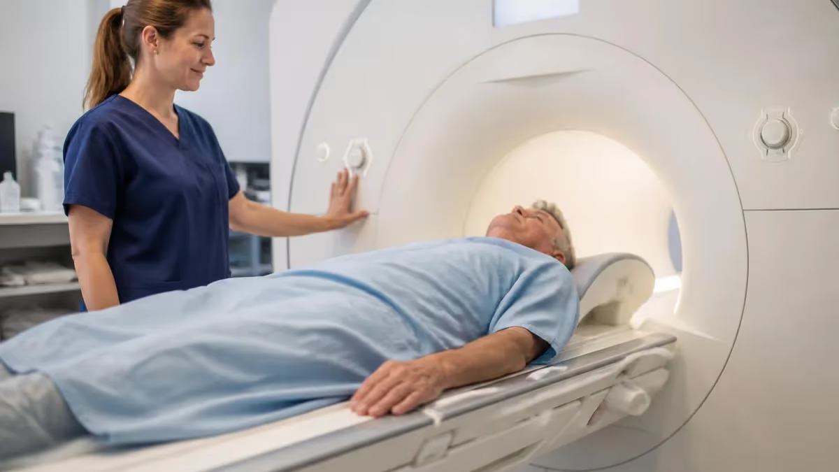
Can you have MS with a normal MRI? This is one of the most anxiety-provoking questions a neurologist hears, and the short answer is: yes, it is possible, though uncommon. Multiple sclerosis is a demyelinating disease of the central nervous system, and MRI is by far the most sensitive imaging tool available for detecting the white-matter lesions it causes.
However, MRI is not infallible. Early disease, atypical lesion locations, and technical limitations can all result in a scan that appears normal even when MS is present. Understanding why this happens requires a closer look at how MS damages the nervous system and how ms and mri technology intersects with disease biology.
Multiple sclerosis affects roughly 1 million Americans and nearly 3 million people worldwide. The disease is characterized by immune-mediated attacks on myelin, the insulating sheath around nerve fibers, leading to plaques or lesions that appear as bright spots on certain MRI sequences. The 2017 McDonald Criteria, the international diagnostic standard, rely heavily on MRI to demonstrate dissemination of lesions in space (multiple brain or spinal cord regions) and time (new lesions appearing on follow-up scans). When those criteria are met alongside a compatible clinical history, a diagnosis of MS can be made with high confidence.
The problem arises at the edges of the diagnostic picture. A patient may present with classic MS symptoms — unilateral optic neuritis, transient limb weakness, bladder dysfunction, or the striking Lhermitte sign (an electric shock sensation running down the spine on neck flexion) — yet return from imaging with a report stating no abnormality detected. For that patient, a normal MRI does not close the diagnostic door. Instead, it opens a deeper investigation that may include spinal cord imaging, cerebrospinal fluid analysis, evoked potential testing, and close clinical follow-up over months or years.
The sensitivity of brain MRI for detecting MS lesions at 1.5 Tesla is approximately 85 to 95 percent, meaning that up to 15 percent of clinically definite MS patients may have a scan that underrepresents their lesion burden at any given time. At 3 Tesla field strength, sensitivity improves substantially, and research-grade scanners at 7 Tesla reveal microstructural damage invisible on clinical machines.
The location of lesions also matters: periventricular, juxtacortical, infratentorial, and spinal cord plaques are the classic patterns, but very early or very small lesions in any of these zones may be below the resolution threshold of a standard clinical protocol.
It is equally important to understand what a normal MRI does tell you. A high-quality 3T brain and spinal cord MRI performed with standardized MS protocols, read by an experienced neuroradiologist, and showing absolutely no white-matter abnormality makes MS considerably less likely. The negative predictive value in that scenario is high. Neurologists weigh the full clinical picture: symptom character, age of onset, ethnicity, family history, and the results of supplementary tests. A normal MRI reduces the probability of MS but does not eliminate it, especially in the earliest stages of relapsing-remitting disease.
Clinically isolated syndrome (CIS) is the term used when a patient has a first clinical event suggestive of demyelination but does not yet meet full MS criteria. Studies show that roughly 60 to 80 percent of CIS patients who go on to develop MS will already have MRI abnormalities at first presentation. However, 20 to 40 percent will not.
For that subgroup, a diagnosis cannot be made on imaging alone, and follow-up protocols become essential. The risk of converting to clinically definite MS remains real even without initial MRI findings, particularly if cerebrospinal fluid shows oligoclonal bands or other inflammatory markers.
This article explores the full landscape of MS diagnosis and MRI: the specific reasons a scan can appear normal, the supplementary tests that fill the diagnostic gap, the role of spinal cord imaging, emerging MRI biomarkers, and what patients and clinicians should do when the imaging and the symptoms do not align. Whether you are a patient navigating an uncertain diagnosis or a student preparing for MRI registry examinations, understanding the nuanced relationship between MS and MRI is clinically essential.
MS and MRI by the Numbers
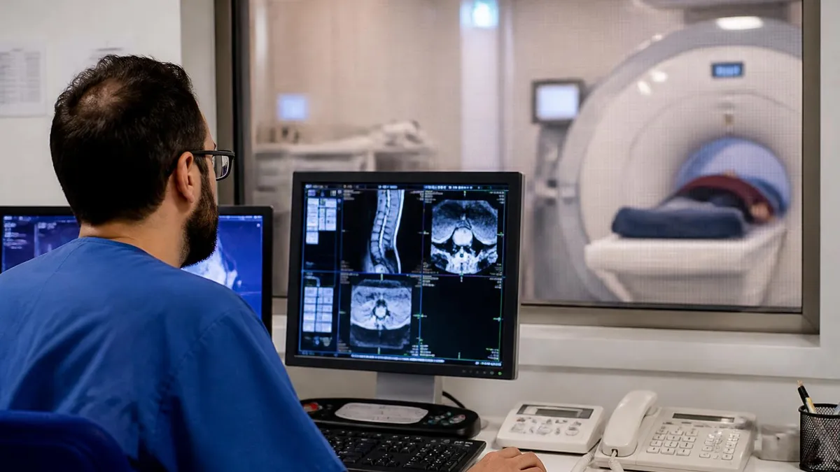
Top Reasons MRI Can Appear Normal in MS
At the time of a first clinical event, lesion burden may be minimal. The first demyelinating attack can produce symptoms from a single small plaque that falls below the detection threshold of standard 1.5T imaging protocols.
Some patients with MS have lesions confined to the spinal cord at initial presentation. A brain MRI will be entirely normal in these cases; targeted cervical and thoracic spine sequences are required to detect pathology.
MS diagnosis requires specific sequences: T2-FLAIR, T1 pre- and post-contrast, and ideally sagittal FLAIR. Scans ordered for headache or trauma may omit contrast or key planes, missing subtle periventricular lesions.
Open-bore 0.7T or older 1.0T scanners have significantly lower sensitivity. Motion artifact, incorrect slice thickness, or omitted sequences can hide lesions that would be visible on an optimized 3T protocol.
MS increasingly causes cortical gray-matter lesions invisible on standard sequences. These require ultra-high-field MRI or specialized surface-coil protocols and are systematically missed by routine clinical scanning.
When MRI results do not explain a patient's symptoms, neurologists turn to a structured set of supplementary investigations. The 2017 McDonald Criteria explicitly allow cerebrospinal fluid (CSF) analysis to substitute for the temporal dissemination requirement when paired with MRI evidence of spatial dissemination.
CSF oligoclonal bands (OCBs) — immunoglobulin G bands found in the spinal fluid but not in matched serum — are present in roughly 85 to 95 percent of patients with clinically definite MS. Their presence in a patient with a single clinical event and spatial dissemination on MRI is sufficient to fulfill McDonald criteria and make a definitive MS diagnosis, bypassing the need for a second clinical attack or new MRI lesions.
Evoked potentials provide another layer of objective evidence. Visual evoked potentials (VEPs) measure the speed of nerve signal conduction from the retina to the occipital cortex. A prolonged P100 latency indicates demyelination of the optic nerve, which is the most common site of the first MS attack. VEPs can reveal subclinical optic nerve involvement that produces no symptoms and causes no MRI-visible lesion. Similarly, brainstem auditory evoked potentials and somatosensory evoked potentials can unmask silent lesions in the posterior fossa and spinal cord. These tests are especially valuable when the imaging is borderline or negative.
Optical coherence tomography (OCT) has emerged as a sensitive biomarker of MS-related neurodegeneration. OCT measures the thickness of the retinal nerve fiber layer and the ganglion cell layer, both of which thin progressively in MS as a downstream consequence of optic neuritis and diffuse neuroinflammation. Studies show that RNFL thinning correlates with global disability scores and brain atrophy even in patients who have never had a clinically apparent episode of optic neuritis. In a patient with a normal or near-normal MRI and unexplained neurological symptoms, a thin RNFL on OCT adds meaningful biological evidence supporting a demyelinating process.
Blood-based biomarkers are reshaping the diagnostic landscape. Serum neurofilament light chain (sNfL) is released into the blood when axons are damaged, regardless of cause. In MS, sNfL levels are elevated during relapses and correlate with MRI lesion activity and disability progression. While not MS-specific, a markedly elevated sNfL in a patient with suggestive symptoms and a borderline MRI raises the clinical index of suspicion and argues for aggressive follow-up. Myelin basic protein and glial fibrillary acidic protein are additional emerging markers under active investigation in 2025 and 2026 clinical trials.
It is crucial to rule out MS mimics before settling on diagnostic uncertainty. Neuromyelitis optica spectrum disorder (NMOSD) presents with optic neuritis and transverse myelitis that can initially look identical to MS, but is caused by antibodies against aquaporin-4 or MOG rather than T-cell mediated demyelination.
NMOSD-associated optic neuritis tends to be more severe and bilateral, and spinal cord lesions are typically longer than three vertebral segments — a key distinguishing feature on MRI. Testing for AQP4-IgG and MOG-IgG antibodies is now standard practice in any patient with a demyelinating syndrome, since NMOSD and MOGAD require completely different treatments from MS.
Migraine, small-vessel cerebrovascular disease, Lyme neuroborreliosis, sarcoidosis, antiphospholipid syndrome, and vitamin B12 deficiency can all produce white-matter signal changes that mimic MS lesions, or conversely, neurological symptoms with a normal MRI. A thorough laboratory workup including ANA, ANCA, ACE level, Lyme serology, B12, folate, HTLV-1, and HIV is standard practice when the diagnosis is uncertain. In younger patients without vascular risk factors, the differential narrows substantially, but these conditions must still be excluded before a patient is labeled as probable or definite MS.
Ultimately, the diagnostic process for MS is iterative and time-sensitive. The McDonald Criteria were designed to enable early diagnosis so that disease-modifying therapy can be started before irreversible axonal damage accumulates. For patients who have a compatible clinical syndrome but a normal initial MRI, the recommended approach is to repeat brain and spinal cord MRI with gadolinium contrast at three to six months, maintain the supplementary workup described above, and refer to a specialist MS center for a second opinion. Time and serial observation remain indispensable diagnostic tools in neurology.
MS Lesion Types, MRI Sequences, and Diagnostic Protocols
MS lesions follow characteristic distributions that neuroradiologists look for systematically. Periventricular lesions — sometimes called Dawson fingers because they radiate perpendicular to the ventricles along medullary veins — are the most common finding, present in over 90 percent of MS patients. Juxtacortical lesions abut the cortex and are best seen on 3D FLAIR sequences. Infratentorial lesions occur in the cerebellum, middle cerebellar peduncles, and brainstem, often causing gait ataxia and diplopia. Spinal cord lesions, typically less than two vertebral segments in length and occupying less than half the cord cross-section, are found in 80 to 90 percent of MS patients at some point.
Cortical lesions are a fourth category increasingly recognized as important to MS pathology. These intracortical and leptomeningeal plaques are essentially invisible on standard 1.5T or even 3T MRI without specialized sequences such as double inversion recovery (DIR) or phase-sensitive inversion recovery (PSIR). Studies using 7T MRI show that cortical lesion burden correlates strongly with cognitive impairment and disability progression, suggesting that standard MRI systematically underestimates the real damage in many patients. This is one reason why a patient with extensive disability may have an MRI that appears relatively benign by conventional counting of T2 lesions.
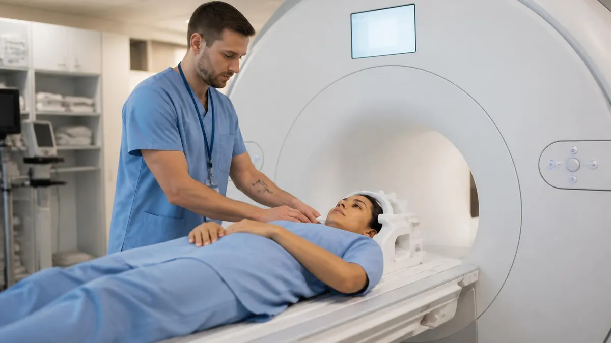
MRI for MS Diagnosis: Strengths and Limitations
- +Highest sensitivity of any imaging modality for white-matter MS lesions, detecting plaques as small as 3 mm on optimized 3T protocols
- +Gadolinium enhancement distinguishes active from chronic lesions, guiding treatment decisions and prognosis in a single scan session
- +Non-invasive and free of ionizing radiation, making it safe to repeat at 3–6 month intervals during diagnostic monitoring
- +Spinal cord MRI adds critical spatial dissemination evidence, especially when brain MRI is negative or equivocal in early disease
- +Quantitative MRI metrics such as brain volume and T2 lesion load serve as clinical trial endpoints and long-term disease-monitoring tools
- +3D FLAIR and DIR sequences can detect cortical lesions invisible on older 2D protocols, improving sensitivity in cognitively impaired patients
- −Sensitivity is imperfect: up to 15 percent of early MS patients may have a normal or near-normal MRI at initial presentation
- −Cortical and leptomeningeal lesions are systematically missed by routine clinical protocols, causing underestimation of lesion burden
- −MS mimics such as migraine, small-vessel disease, and NMOSD can produce identical-looking lesions, requiring clinical and serological correlation
- −Gadolinium-based contrast agents have raised safety questions about brain deposition with repeated exposures, though clinical significance remains debated
- −Access to 3T scanners with MS-specific protocols is uneven across the US, with rural and underserved areas relying on lower-field equipment
- −MRI lesion burden correlates only modestly with disability in individual patients — the clinico-radiological paradox limits its use as a sole outcome measure
When to Suspect MS Despite a Normal Brain MRI
- ✓Order dedicated spinal cord MRI (cervical and thoracic) with MS protocol if brain MRI is negative but clinical suspicion is high.
- ✓Request CSF analysis including oligoclonal bands and IgG index to look for intrathecal inflammation.
- ✓Perform visual evoked potentials to detect subclinical optic nerve demyelination not visible on standard imaging.
- ✓Test serum AQP4-IgG and MOG-IgG to exclude NMOSD and MOGAD before attributing symptoms to MS.
- ✓Obtain a comprehensive laboratory panel (ANA, B12, folate, Lyme titer, ACE, antiphospholipid antibodies) to rule out MS mimics.
- ✓Repeat brain and spinal cord MRI with gadolinium at 3 to 6 months if initial scan is negative and symptoms persist.
- ✓Refer to a specialized MS center for a second-opinion read of the MRI — experienced neuroradiologists detect subtle lesions others miss.
- ✓Consider 3T MRI if initial scan was performed at 1.5T or below; upgrade in field strength significantly improves lesion detection.
- ✓Check serum neurofilament light chain as a supportive biomarker of ongoing axonal injury when diagnosis remains uncertain.
- ✓Document symptom progression carefully over time — temporal dissemination of clinical events can fulfill McDonald Criteria even with limited imaging.
A Normal MRI Does Not Rule Out MS — Especially at First Presentation
Up to 40 percent of patients who ultimately develop clinically definite MS have a normal or near-normal MRI at their very first clinical event. Neurologists are trained to treat the patient, not the scan: a compatible clinical history plus positive CSF oligoclonal bands can secure an MS diagnosis under the 2017 McDonald Criteria even when MRI findings fall short of the spatial dissemination threshold. Never delay the full workup based on a single negative scan.
Advanced MRI techniques are rapidly closing the gap between what standard scanners detect and what is actually happening in MS-affected brains and spinal cords. Ultra-high-field imaging at 7 Tesla, though currently limited to research centers, offers a four-fold increase in signal-to-noise ratio compared to 3T and can resolve individual cortical lesions, perivenular lesion morphology, and leptomeningeal inflammation with unprecedented clarity.
The central vein sign — a small vein visible at the center of a periventricular MS lesion on susceptibility-weighted imaging — is a highly specific marker that distinguishes MS plaques from the non-specific white-matter lesions of aging or migraine. Several consortium studies are working to incorporate central vein sign assessment into routine diagnostic criteria.
Myelin water imaging (MWI) directly measures the fraction of total brain water trapped within the myelin bilayer, providing a quantitative index of myelination that is far more specific to the MS disease process than T2 signal changes. While conventional T2 hyperintensity reflects a mixture of edema, axonal loss, gliosis, and demyelination, MWI isolates the myelin compartment.
Early data suggest that myelin water fraction decreases even in normal-appearing white matter adjacent to lesions, implying that the disease process extends well beyond what appears on a standard scan. This has profound implications for understanding why MRI lesion counts so often underestimate patient disability.
Diffusion tensor imaging and its advanced derivatives — neurite orientation dispersion and density imaging (NODDI) and fixel-based analysis — quantify white-matter tract integrity at the microstructural level. These techniques detect axonal loss and fiber disorganization in areas that look normal on T2-FLAIR, capturing the so-called occult damage that accounts for much of the MRI–clinical paradox in MS. In clinical practice, DTI-based metrics like fractional anisotropy and mean diffusivity in the corticospinal tract predict motor disability and gait performance better than lesion counts, making them attractive candidates for inclusion in next-generation diagnostic protocols.
Spinal cord MRI deserves particular emphasis because it is where MS most directly threatens mobility and continence, and where conventional MRI performs least reliably. The spinal cord is a small, pulsatile structure surrounded by CSF and bone, making high-resolution imaging technically challenging. Standard 1.5T spine protocols have a sensitivity of only 60 to 70 percent for MS cord lesions.
Dedicated techniques — phase-array coils, reduced field-of-view sequences, and phase-sensitive inversion recovery — substantially improve detection. The MAGNIMS consensus guidelines now recommend that all patients undergoing initial MS workup receive both brain and spinal cord MRI, since cord lesions contribute independently to meeting the spatial dissemination criteria and can be the only evidence of MS in patients with normal brain MRI.
Leptomeningeal enhancement (LME) is a newly recognized MRI finding in MS associated with compartmentalized inflammation in the meningeal spaces. Detected on high-resolution post-contrast 3D FLAIR sequences, LME appears as irregular linear enhancement along the brain surface and in the sulci. Studies show that MS patients with LME have greater cortical lesion burden, faster brain atrophy, and more rapid disability progression than those without it. The presence of LME on an otherwise unremarkable brain MRI could, in the right clinical context, provide biological evidence supporting a diagnosis of progressive MS even when classic periventricular lesion criteria are not met.
PET imaging with myelin-targeting tracers such as MeDAS and Pittsburgh compound B derivatives is an emerging research tool that directly images demyelination and remyelination in vivo. Unlike MRI, PET can quantify myelin density in both lesions and normal-appearing tissue, tracking the dynamics of tissue repair that are invisible to conventional sequences. While PET is currently too expensive and isotope-limited for routine clinical use, it is already shaping clinical trial design and providing proof-of-concept data for next-generation remyelination therapies. Its eventual integration with MRI in specialized MS centers may fundamentally change how we monitor treatment response and define disease activity.
The convergence of these advanced techniques points toward a future where MS diagnosis is made earlier, with greater biological specificity, and tracked with more sensitive outcome measures than today's T2 lesion counts and clinical disability scores. For patients whose MRI is currently normal or equivocal, these tools represent real hope for a more definitive answer — and for clinicians preparing for MRI registry and registry board exams, understanding the principles behind each technique is increasingly tested in modern examination content.
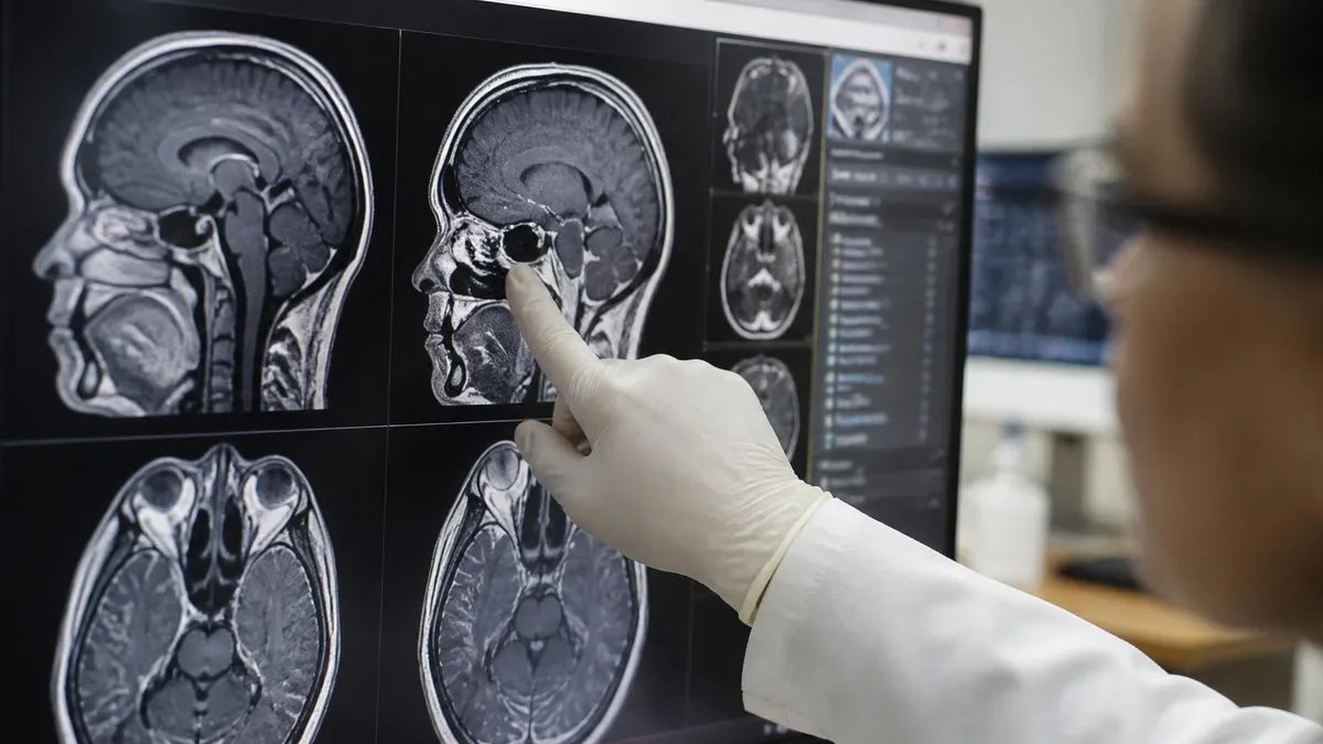
Research consistently shows that early treatment with disease-modifying therapies (DMTs) significantly reduces long-term disability in MS, and that delays of even one to two years are associated with worse outcomes. If a patient meets McDonald Criteria and has a compatible clinical syndrome, treatment should begin without waiting for additional lesions to appear on follow-up imaging. When diagnostic uncertainty genuinely remains after full workup, co-management with an MS specialist is strongly recommended before deferring therapy.
Living with diagnostic uncertainty is one of the hardest aspects of the early MS journey. Patients who have been told they may have MS — or who have been given a diagnosis of clinically isolated syndrome — often spend months or years in a limbo where their symptoms are real and sometimes disabling but their official diagnosis is provisional. This uncertainty affects employment decisions, insurance coverage, family planning, and psychological well-being in profound ways. Understanding the diagnostic process and advocating for a thorough workup is the first step in navigating this difficult period effectively.
When a patient has symptoms suggesting MS but a normal MRI, a second opinion at an accredited MS center is not merely reasonable — it is strongly advisable. MS centers concentrate expertise in both interpretation of borderline imaging and in the clinical pattern recognition that comes from seeing hundreds of MS cases each year.
Studies show that MS diagnoses made at specialized centers are both more accurate and arrived at faster than those made in general neurology practice. The National MS Society maintains a directory of certified MS care centers that can be found on their website, organized by US state and region.
Serial neurological examination is an underappreciated diagnostic tool. A careful examination at baseline and at three to six month follow-up visits can reveal subtle changes — new hyperreflexia, an afferent pupillary defect, mild coordination impairment — that were not present initially. These findings, documented over time, provide objective evidence of dissemination in time even when imaging lags behind the clinical picture. Neurologists are trained to detect these signs before patients are fully aware of them, which is why regular follow-up is so important in the early diagnostic phase.
For patients who receive a diagnosis of radiologically isolated syndrome (RIS) — incidentally discovered lesions meeting MS spatial criteria with no clinical symptoms — the risk of converting to a clinical event is approximately 34 percent within five years, and up to 60 percent within ten years. Risk factors for conversion include younger age, male sex, spinal cord lesion involvement, and infratentorial lesion location.
Recent clinical trials demonstrated that treating RIS patients with a first-line DMT significantly reduced the risk of first clinical attack, raising the question of whether asymptomatic individuals with high-risk imaging features should begin therapy — a paradigm-shifting development in MS care.
The psychological burden of diagnostic uncertainty is well documented. Anxiety, depression, and health-related quality of life impairment are common in patients awaiting an MS diagnosis, often rivaling the psychological impact of receiving a confirmed diagnosis. Mental health support — whether through a psychologist experienced with chronic illness, a structured mindfulness program, or peer support groups — should be integrated into care from the earliest stages. The MS Society of America and the National MS Society both operate peer mentor programs connecting newly diagnosed or pre-diagnosis patients with trained volunteers who have lived experience with the diagnostic process.
Diet, exercise, and sleep hygiene play a meaningful supporting role during the diagnostic period. Aerobic exercise has been shown to improve fatigue, mood, cognitive function, and even brain volume in MS patients; these benefits are likely present even in early or pre-diagnosis stages. A Mediterranean-style diet rich in anti-inflammatory foods — fatty fish, olive oil, leafy greens, and berries — is associated with lower relapse rates and slower disability progression in observational studies, though randomized trial data are still maturing.
Adequate vitamin D levels are particularly important: vitamin D insufficiency is both a risk factor for MS and a predictor of disease activity, and supplementation to achieve serum levels above 40 ng/mL is widely recommended by MS neurologists pending confirmatory trial data.
Finally, it is worth emphasizing for technologists and students preparing for registry examinations that the MRI technologist's role in MS diagnosis is more consequential than it may appear. Proper positioning for cervical and thoracic spine sequences, consistent application of contrast protocols, recognition of motion artifact, and accurate reporting of technical parameters directly affect whether subtle lesions are visible on the final images.
A technologist who understands the McDonald Criteria and the clinical importance of MS-protocol sequences will ensure that the radiologist and referring neurologist have the highest-quality data available for their diagnostic decision — a contribution that can meaningfully change a patient's trajectory.
For MRI technologists and students preparing for the ARRT MRI registry examination, the intersection of MS and MRI encompasses a broad range of testable knowledge domains. Understanding why T2-FLAIR is preferred over standard T2 for MS lesion detection — because FLAIR suppresses CSF signal and makes periventricular lesions stand out against the dark background — is fundamental physics that appears consistently on board examinations.
Knowing that gadolinium-enhancing lesions indicate active blood-brain barrier breakdown and that the enhancement window is typically four to six weeks helps explain why contrast-enhanced protocols are always requested on MS surveillance scans and why the timing of contrast administration must be standardized.
Protocol optimization questions are another high-yield area. MAGNIMS guidelines recommend sagittal 3D FLAIR for brain imaging because it captures periventricular, juxtacortical, and infratentorial lesions in a single acquisition with multiplanar reformats. Slice thickness should be 1 mm or less for 3D acquisitions, or no greater than 3 mm for 2D sequences.
In-plane resolution should target 1 x 1 mm or better. Phase-encoding direction must be chosen carefully for spinal cord imaging to avoid CSF pulsation artifact overlying the cord — an artifact that can mimic or obscure cord lesions. These are exactly the kinds of practical protocol decisions that appear in registry examination scenarios.
Safety knowledge is equally important. Gadolinium-based contrast agents are categorized by their molecular structure — linear versus macrocyclic, ionic versus nonionic — with macrocyclic agents showing significantly lower brain deposition on autopsy studies. Since MS patients may receive dozens of contrast-enhanced MRI scans over their lifetime, choosing the safest available agent has real clinical significance. Current FDA recommendations do not restrict the use of macrocyclic agents but advise practitioners to consider the smallest effective dose and to document cumulative gadolinium exposure. Understanding these regulatory nuances demonstrates the level of clinical integration expected of a competent MRI technologist.
Artifact recognition is a third high-yield domain. Susceptibility artifacts from dental hardware, cardiac pacemakers, or bone screws can obscure infratentorial lesions. Truncation or Gibbs ringing artifact along the cord can simulate or hide intramedullary pathology. Motion artifact — whether from patient movement, cardiac pulsation, or swallowing — degrades image quality in all regions relevant to MS. Technologists who can recognize these artifacts and implement corrective strategies, including cardiac gating for cervical cord imaging, contribute directly to diagnostic quality. The registry examination tests not just image reading but the problem-solving process a technologist uses when the initial acquisition is suboptimal.
Neuroanatomy relevant to MS is heavily tested, particularly the white-matter tract anatomy of the corpus callosum, cerebellum, and brainstem. The corpus callosum is a privileged MS target because it is densely myelinated and periventricular. Calloso-septal interface lesions on sagittal FLAIR — the classic Dawson fingers — are pathognomonic of MS and should be identifiable by any technologist reviewing scout images during a scan. Cerebellar peduncle involvement, particularly the middle cerebellar peduncle, produces distinctive ataxia and is a clue to infratentorial MS. The technologist who understands this anatomy can flag concerning findings for expedited radiologist review.
Differential diagnosis knowledge is increasingly tested on modern registry examinations. Candidates are expected to know that ADEM (acute disseminated encephalomyelitis) produces bilateral, often asymmetric, large confluent white-matter lesions in a monophasic pattern following infection or vaccination, and that its perilesional edema and gray-matter involvement distinguish it from MS.
Neurosarcoidosis produces leptomeningeal enhancement and cranial nerve involvement. Susac syndrome, a microangiopathy affecting the corpus callosum snowball lesions in the central fibers, is a classic MS mimic with a distinctive imaging signature. Familiarity with these entities at the level of pattern recognition prepares candidates for the clinical integration questions that comprise a growing share of registry content.
Reviewing practice questions across MRI knowledge, physics, and anatomy and pathology domains is the most effective preparation strategy for the registry examination. Spaced repetition of high-yield facts — gadolinium kinetics, sequence parameters, lesion morphology, diagnostic criteria — combined with full-length timed practice tests that simulate examination conditions produces the best outcomes. Students who integrate clinical understanding of conditions like MS with the technical knowledge of how MRI sequences detect them are better prepared not only for the examination but for the lifelong professional practice that follows it.
Many readers pair this guide with the open mri near me to check their readiness before exam day.
Many readers pair this guide with the mri abdomen and pelvis cpt code to check their readiness before exam day.
MRI Questions and Answers
DWI MRI: Diffusion-Weighted Imaging Explained (2026 Guide)
MRI Lumbar Spine Without Contrast: Complete Guide to the Scan, What It Shows, and How to Prepare
MRI History: From Nuclear Resonance to Modern Scanners

MRI of Cervical Spine: Complete Guide to Imaging, Anatomy, Pathology, and Patient Preparation
Where to Get an MRI: Facilities, Costs, and How to Choose
About the Author

Medical Laboratory Scientist & Clinical Certification Expert
Johns Hopkins UniversityDr. Sandra Kim holds a PhD in Clinical Laboratory Science from Johns Hopkins University and is certified as a Medical Technologist (MT) and Medical Laboratory Scientist (MLS) through ASCP. With 16 years of clinical laboratory experience spanning hematology, microbiology, and molecular diagnostics, she prepares candidates for ASCP board exams, MLT, MLS, and specialist certification tests.
Join the Discussion
Connect with other students preparing for this exam. Share tips, ask questions, and get advice from people who have been there.
View discussion (8 replies)