
Diazepam for MRI: What Patients Need to Know About Sedation, Dosing, and Safety
Diazepam for MRI explained: dosing, safety, alternatives & what to expect. ✅ Complete patient guide for claustrophobia and scan anxiety.
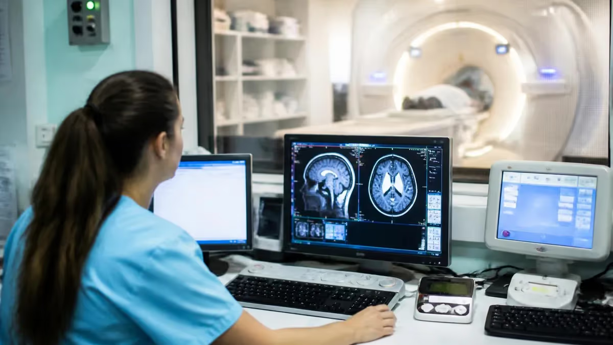
Diazepam for MRI is one of the most commonly prescribed interventions for patients who experience significant anxiety, claustrophobia, or panic when faced with the confined bore of a magnetic resonance imaging scanner. MRI machines are incredibly powerful diagnostic tools, but the enclosed tunnel, loud repetitive noise, and extended scan durations — often 30 to 90 minutes — can make the experience genuinely distressing for a substantial portion of the population. Diazepam, a benzodiazepine sedative, helps reduce that anxiety so patients can remain still long enough to produce high-quality, diagnostically useful images.
Understanding how diazepam works in the context of an MRI appointment requires a brief look at its pharmacology. Diazepam enhances the effect of gamma-aminobutyric acid (GABA), the brain's primary inhibitory neurotransmitter, producing a calming, muscle-relaxing, and mildly sedating effect within 30 to 60 minutes of oral administration. Because patients must remain motionless during each imaging sequence, even slight tremors or anxiety-driven movement can blur the resulting images, rendering the scan non-diagnostic. A single low oral dose of diazepam taken before the scan can prevent this outcome without rendering the patient unconscious.
Claustrophobia affects an estimated 2 to 15 percent of the general population, and surveys of radiology departments consistently show that between 1 and 5 percent of scheduled MRI appointments are cancelled or abandoned because a patient cannot tolerate the scanner environment. This represents both a clinical problem — delayed diagnosis — and a financial one, as scanner time is expensive and rescheduling adds administrative burden. Pre-procedural oral benzodiazepines like diazepam have been studied as a practical, cost-effective solution that allows most claustrophobic patients to complete their scan on the first attempt.
Not every anxious patient needs pharmacological sedation. Facilities increasingly offer open MRI systems with wider bores, headphones with music, scented eye masks, patient coaching, and mirror systems that allow patients to see outside the scanner. However, these accommodations do not work for everyone, and for patients with moderate to severe claustrophobia, diazepam remains one of the most reliable tools available. Radiologists and ordering physicians weigh each patient's medical history, concurrent medications, and specific anxiety level before recommending sedation.
It is important to distinguish between minimal oral sedation with diazepam and deeper procedural sedation using intravenous agents. Most MRI-related diazepam prescriptions involve a single oral tablet — typically 5 mg or 10 mg — taken at home roughly one hour before the appointment or administered in the radiology suite. This approach does not require anesthesia personnel or continuous monitoring equipment in most adult outpatient settings, though protocols vary by institution. Patients must still arrange transportation, as driving is prohibited for the remainder of the day.
For MRI technologists and radiology students preparing for certification exams, understanding patient preparation protocols — including the use of anxiolytics — is a tested competency. Being able to explain the mechanism, timing, contraindications, and monitoring requirements associated with diazepam for mri reflects the kind of integrated clinical knowledge that separates competent technologists from exceptional ones. This article covers the full picture, from pharmacology and patient selection to practical tips, safety considerations, and what happens if sedation is inadequate.
Whether you are a patient researching your upcoming appointment, a caregiver helping someone with severe anxiety, or an MRI student studying for registry boards, the following sections provide clear, evidence-based information about how diazepam is used before MRI scans, what to expect, and how to stay safe throughout the process.
MRI Sedation by the Numbers
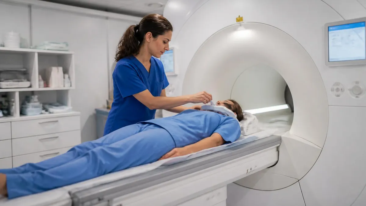
Step-by-Step: What Happens When Diazepam Is Used for an MRI
Physician Prescribes Diazepam
Pre-Scan Instructions Provided
Diazepam Taken and Onset Monitored
MRI Scan Performed
Post-Scan Monitoring and Discharge
Follow-Up and Results
Dosing diazepam appropriately for an MRI scan requires balancing adequate anxiolysis with patient safety. The goal is not to render the patient unconscious — it is to reduce anxiety enough that the patient can cooperate with positioning, tolerate the noise, and remain still during imaging sequences. Most outpatient protocols call for oral diazepam 5 mg to 10 mg administered approximately 30 to 60 minutes before the scan begins. The 5 mg dose is typically used for elderly patients, patients with low body weight, or those who have never taken a benzodiazepine before, as individual sensitivity can vary significantly.
The 10 mg dose is more commonly prescribed for adults with confirmed moderate-to-severe claustrophobia who have previously tolerated benzodiazepines without adverse effects. Some institutions allow the prescribing physician to authorize a second dose of 5 mg if the initial dose proves insufficient, though this escalation should only occur under medical supervision and is not standard practice at all facilities. Intravenous diazepam and other IV benzodiazepines such as midazolam are reserved for patients who cannot tolerate oral administration or require deeper sedation, and those situations mandate continuous monitoring and anesthesia or sedation nurse involvement.
Timing is critical. Diazepam taken too early will have partially metabolized before the scan begins, leaving the patient under-sedated during the most anxiety-provoking portions of the examination. Taken too late, it may not have reached sufficient plasma concentration to produce meaningful anxiolysis. Peak plasma levels after oral administration generally occur between 60 and 90 minutes post-ingestion, though gastric motility, body fat distribution, and concurrent food intake can shift this window. Patients should follow their facility's specific timing instructions carefully, and technologists should ask patients when they took the medication before beginning the scan.
Body composition matters more than many patients expect. Diazepam is highly lipophilic, meaning it distributes extensively into fatty tissue. In patients with high body fat percentage, the drug may exhibit a prolonged distribution phase and an extended duration of effect. Conversely, in very lean patients, peak plasma levels may be reached more quickly and may be slightly higher. Elderly patients are particularly sensitive to diazepam because they typically have reduced hepatic metabolism, decreased plasma protein binding, and altered drug distribution — all of which can increase both the intensity and the duration of sedation.
Alcohol is an absolute contraindication in the hours before and after diazepam use. Both substances are central nervous system depressants that act synergistically, and their combination dramatically increases the risk of respiratory depression, excessive sedation, and loss of protective airway reflexes. Patients should be explicitly counseled at the time of prescription to avoid alcohol for at least 24 hours before and after the scan. This instruction should appear in writing, as verbal instructions alone are frequently forgotten under the stress of a medical appointment.
Some patients ask whether they can take their usual morning medications on the day of the scan. The answer is almost always yes — with water only, and in consultation with their prescribing physician. However, certain medications warrant special attention. Opioid analgesics, other benzodiazepines, tricyclic antidepressants, antihistamines, and certain anticonvulsants all potentiate CNS depression and can turn a safe diazepam dose into an unsafe one. The MRI technologist's role includes reviewing the patient's medication list during the screening interview and escalating concerns to the supervising radiologist before the scan proceeds.
For patients taking medications that inhibit CYP3A4 or CYP2C19 — the hepatic enzymes responsible for diazepam metabolism — drug exposure may be significantly elevated. Common inhibitors include fluconazole, ketoconazole, omeprazole, fluvoxamine, and certain protease inhibitors used in HIV therapy. In these patients, a reduced diazepam dose may be appropriate, and the prescribing clinician should review the full medication list before finalizing the sedation order. Pharmacokinetic interactions are a frequently overlooked source of sedation complications in outpatient imaging settings.
Diazepam for MRI: Contraindications, Interactions, and Special Populations
Diazepam is contraindicated in patients with known hypersensitivity to benzodiazepines, severe respiratory insufficiency, sleep apnea syndrome, and myasthenia gravis. It should not be used in pregnant patients — particularly during the first trimester — because benzodiazepines cross the placental barrier and have been associated with neonatal withdrawal and sedation. Patients with severe hepatic impairment cannot adequately metabolize diazepam, leading to dangerous drug accumulation with even a single dose.
Patients with a history of substance use disorder — particularly alcohol, opioids, or benzodiazepines — present a clinical dilemma. Benzodiazepines carry addiction potential, and while a single-dose pre-procedural prescription is unlikely to trigger relapse in isolation, the prescribing physician must weigh individual risk. Alternatives such as hydroxyzine, low-dose beta-blockers, or patient coaching with nurse support may be preferred in these individuals to avoid introducing a potentially reinforcing substance.
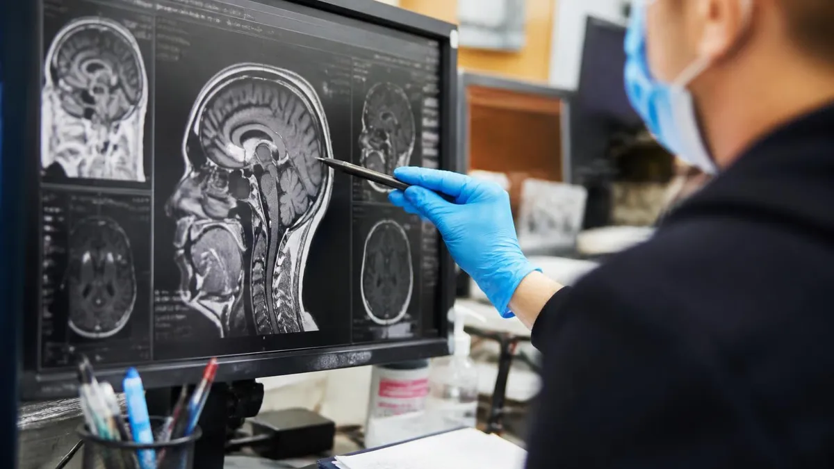
Diazepam for MRI: Benefits and Limitations
- +Highly effective at reducing moderate-to-severe claustrophobia and scan anxiety in most adults
- +Oral administration is non-invasive, inexpensive, and does not require IV access or anesthesia staff
- +Rapid onset of 30 to 60 minutes means easy timing coordination with the scheduled scan
- +Well-studied pharmacology with decades of clinical data in diverse patient populations
- +Significantly improves image quality by reducing patient motion during imaging sequences
- +Allows most claustrophobic patients to complete scans that would otherwise be impossible or require rescheduling
- −Long half-life of approximately 36 hours means residual sedation persists well into the afternoon or evening after a morning scan
- −Patients cannot drive for the rest of the day, requiring transportation arrangements that may be difficult
- −Contraindicated in pregnancy, respiratory disease, sleep apnea, myasthenia gravis, and severe liver impairment
- −Risk of paradoxical excitation — increased agitation, hostility, or disinhibition — occurs in approximately 1 to 2 percent of patients, particularly children and the elderly
- −Addiction potential makes it inappropriate for patients with active or recent substance use disorder
- −Drug interactions with opioids, alcohol, and CYP3A4 inhibitors require careful medication review before prescribing
Pros and Cons at a Glance
| Pros | Cons |
|---|---|
| Highly effective at reducing moderate-to-severe claustrophobia and scan anxiety in most adults | Patients cannot drive for the rest of the day, requiring transportation arrangements that may be difficult |
| Oral administration is non-invasive, inexpensive, and does not require IV access or anesthesia staff | Contraindicated in pregnancy, respiratory disease, sleep apnea, myasthenia gravis, and severe liver impairment |
| Well-studied pharmacology with decades of clinical data in diverse patient populations | Addiction potential makes it inappropriate for patients with active or recent substance use disorder |
Pre-MRI Diazepam Preparation Checklist
- ✓Confirm your physician has reviewed your full medication list for interactions before prescribing diazepam.
- ✓Arrange a responsible adult driver to take you home — you cannot drive after taking diazepam.
- ✓Avoid all alcohol for at least 24 hours before and after your scheduled scan.
- ✓Take the diazepam tablet 30 to 60 minutes before your appointment as instructed — not earlier or later.
- ✓Complete the MRI safety screening questionnaire honestly, disclosing all implants, devices, and prior surgeries.
- ✓Wear comfortable, metal-free clothing to avoid delays during the screening process.
- ✓Bring a list of all current medications, including over-the-counter drugs and supplements, to show facility staff.
- ✓Inform the technologist when you took your diazepam dose so scan timing can be optimized.
- ✓Request earplugs or headphones from the technologist to reduce the noise of the MRI machine during your scan.
- ✓Plan to remain at the imaging facility for 15 to 30 minutes after the scan before departure for safety monitoring.
Diazepam's Active Metabolites Extend Its Duration Significantly
Diazepam itself has a half-life of 20 to 70 hours, but its primary active metabolite — desmethyldiazepam — has a half-life of 36 to 200 hours. This means sedation and cognitive impairment can persist far longer than patients expect after even a single dose. Facilities should warn patients explicitly: the effects may still be present the morning after an afternoon scan, and patients should not assume they are safe to drive simply because they feel alert.
For patients who cannot take diazepam due to contraindications, drug interactions, or personal preference, several alternative strategies exist that span pharmacological and non-pharmacological approaches. Understanding these alternatives is important both for patients exploring their options and for MRI technologists and radiologists who counsel patients before scheduling. No single alternative works for every patient, and the optimal approach often combines techniques tailored to the individual's specific fears and clinical profile.
Hydroxyzine is a non-benzodiazepine antihistamine with anxiolytic properties that is frequently prescribed as an alternative for patients who cannot safely receive diazepam. It works through H1 histamine receptor blockade and has mild serotonin antagonist properties, producing sedation and anxiolysis without the addiction potential or respiratory risks associated with benzodiazepines. Hydroxyzine 25 mg to 50 mg taken orally one hour before the scan is a reasonable option for patients with substance use history, mild respiratory disease, or those for whom the prescribing physician prefers to avoid controlled substances.
Beta-blockers, particularly propranolol 10 mg to 20 mg taken orally one hour before scanning, can blunt the physiological symptoms of anxiety — rapid heart rate, trembling, sweating — without causing significant sedation. This approach is especially useful for patients whose primary complaint is somatic anxiety rather than psychological panic. However, beta-blockers do not address the cognitive or emotional components of claustrophobia and are contraindicated in patients with asthma, significant bradycardia, or certain cardiac conduction abnormalities.
Non-pharmacological interventions have gained considerable traction in recent years and should always be offered before or alongside medication. Wide-bore MRI scanners — those with a 70 cm or larger bore diameter compared to the standard 60 cm — significantly reduce the sense of enclosure and allow many previously intolerant patients to complete scans without sedation. Open MRI systems, while lower in field strength and generally producing lower-resolution images, can accommodate patients with extreme claustrophobia or very large body habitus. The trade-off in image quality must be weighed against the clinical need for the study.
Cognitive-behavioral techniques including guided breathing exercises, progressive muscle relaxation, and grounding strategies can reduce anxiety before and during MRI scans. Some facilities employ specially trained radiology nurses or patient navigators who spend time with anxious patients before the scan, familiarizing them with the equipment through simulation and education. Virtual reality headsets are an emerging tool that can provide an immersive distraction experience, transporting patients to a calm virtual environment while the scan proceeds around them — a particularly promising technology for pediatric patients.
Nitrous oxide, administered via nasal mask in a 50 percent oxygen mixture, has been used in some European and Australian radiology departments as a rapid-onset, rapidly-reversing anxiolytic alternative to oral benzodiazepines. Its primary advantage is its extremely short duration of action — effects resolve within five minutes of removing the mask — meaning patients can typically drive themselves home after a brief observation period. However, nitrous oxide requires specialized delivery equipment, scavenging systems, and staff training, and it is not widely available in US outpatient MRI centers as of 2026.
For patients with the most severe anxiety or those undergoing lengthy, complex protocols such as cardiac MRI, brain spectroscopy, or pediatric neuroimaging, deep procedural sedation or general anesthesia administered by an anesthesiologist may be the safest and most effective choice. These protocols require dedicated MRI-compatible monitoring equipment, anesthesia machines rated for high-field environments, and trained anesthesia personnel, all of which are available at academic medical centers and larger hospital-based imaging departments. The additional resource requirements and costs are justified when the diagnostic information is essential and lighter sedation has failed.
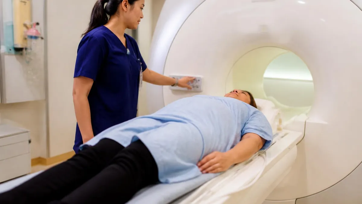
Diazepam impairs psychomotor function, reaction time, and judgment for many hours beyond the time you feel subjectively alert. Active metabolites may cause residual impairment the following morning. US law and facility policy uniformly prohibit patients from driving after receiving diazepam. Arrange transportation before your appointment — a family member, friend, rideshare service, or taxi — and do not attempt to drive regardless of how you feel when you leave the imaging center.
MRI technologists play a central role in identifying patient anxiety before it derails a scan. During the pre-scan interview, experienced technologists pick up on behavioral cues — avoidance of eye contact, hyperventilation, excessive questioning, physical trembling — that signal a patient may struggle once inside the bore. Addressing these concerns proactively, with clear explanation of what the patient will experience, how long each sequence lasts, and how to use the call button, can reduce the need for pharmacological intervention in mildly anxious patients and improve outcomes even for those who have received diazepam.
Communication during the scan is equally important. Most modern MRI systems include a two-way intercom that allows the technologist to speak with the patient between sequences. Narrating what is about to happen — "The next sequence will last about four minutes and will sound like a rapid knocking" — gives patients a sense of control and predictability that significantly reduces anxiety. Technologists should check in frequently, especially with sedated patients, monitoring for signs of excessive drowsiness, airway compromise, or paradoxical agitation that may require the scan to be paused.
For MRI registry candidates, patient care and safety is one of the primary tested domains on the ARRT MRI certification examination. Questions about pharmacological sedation protocols, monitoring requirements, diazepam pharmacology, and contraindications appear regularly. Understanding the mechanism of action, appropriate dose range, timing, duration of effect, drug interactions, and post-procedure monitoring requirements for diazepam provides not only clinical competence but also testable knowledge that separates well-prepared candidates from those who have focused exclusively on physics and pulse sequences.
The ARRT Content Specifications for MRI explicitly address patient care, which includes managing anxious and claustrophobic patients, recognizing adverse reactions to administered substances, and understanding sedation monitoring requirements. Candidates should be familiar with the concept of minimal sedation (anxiolysis) versus moderate sedation (conscious sedation) versus deep sedation, and should know that most oral diazepam protocols for outpatient MRI fall into the minimal sedation category in healthy adults — a distinction that determines monitoring requirements and who must be present during the scan.
Paradoxical reactions to diazepam deserve special emphasis because they are underrecognized and can be clinically alarming. Instead of the expected sedation and calm, approximately 1 to 2 percent of patients experience increased anxiety, agitation, combativeness, or disinhibition. This reaction is more common in elderly patients, children, patients with intellectual disabilities, and those with certain personality traits or prior trauma.
When a paradoxical reaction occurs during or before an MRI scan, the appropriate response is to stop the procedure, ensure patient safety, and consider reversal with flumazenil in cases of significant behavioral disturbance, though flumazenil use carries its own risks including seizure induction in benzodiazepine-dependent patients.
Flumazenil is a competitive benzodiazepine receptor antagonist that can rapidly reverse diazepam-induced sedation and paradoxical reactions. Its onset is approximately one to two minutes intravenously, and its duration of action — roughly 30 to 60 minutes — is substantially shorter than diazepam's. This means resedation can occur after flumazenil wears off, and patients who receive it must be monitored for an extended period afterward. Flumazenil should be available in any facility that administers benzodiazepines, and staff should be trained in its use, contraindications, and the monitoring required following its administration.
Documentation is a professional and legal requirement that is often underemphasized in discussions of MRI sedation. Technologists and nursing staff must document the patient's pre-procedure anxiety assessment, the medication administered (including dose, route, time, and lot number if applicable), vital signs where monitored, the patient's response during the scan, any adverse events, and the condition of the patient at discharge. This documentation protects the facility, provides continuity of care, and creates a record that informs future sedation decisions for the same patient. For registry exam purposes, understanding the technologist's documentation obligations is part of demonstrating professional standards knowledge.
Many readers pair this guide with the mri abdomen and pelvis cpt code to check their readiness before exam day.
Many readers pair this guide with the open mri near me to check their readiness before exam day.
Preparing for an MRI scan when you know you will be taking diazepam requires planning that begins several days before the appointment, not just the morning of the scan. The most important logistical step — arranging transportation — should be confirmed well in advance. Do not assume a family member will be available on the day. Make concrete arrangements, confirm them the day before, and have a backup plan. Rideshare apps are a reasonable alternative, but someone should ideally accompany you home rather than have you travel alone in a sedated state.
Food and hydration in the hours before the scan are generally unrestricted for patients receiving oral diazepam, unlike those undergoing general anesthesia who must follow strict NPO (nothing by mouth) guidelines. However, a very large, fatty meal immediately before taking the tablet can delay diazepam absorption. A light snack is generally fine and may actually help reduce nausea, which some patients experience with benzodiazepines on an empty stomach. Stay well hydrated in the days before the scan, as dehydration can exacerbate the sensation of confinement and dizziness that some patients notice under mild sedation.
What to wear matters more than most patients realize. Metal fasteners, underwire bras, belts with metal buckles, and jewelry all require removal before entering the MRI suite. If you take diazepam before arriving at the facility, navigating a locker room and changing into a gown while sedated — even mildly — can be disorienting. Wearing comfortable, loose-fitting, metal-free clothing (athletic wear is ideal) allows you to enter the scanner in your own clothes and avoids the need to change while your coordination may be slightly impaired.
If you have had an MRI with diazepam before, communicate that experience to your prescribing physician and the technologist. Was the previous dose adequate? Did you feel excessively drowsy? Did you experience any unusual reactions? This information allows the team to calibrate the current dose more precisely and to anticipate any issues. Patients who have had a paradoxical reaction previously should flag this immediately, as the prescribing plan should be reconsidered entirely. A documented paradoxical reaction to diazepam is a strong signal to try an alternative anxiolytic or approach for future scans.
During the scan itself, several patient behaviors significantly improve image quality independent of the diazepam effect. Breathing shallowly and regularly during breath-hold sequences, avoiding swallowing during head and neck imaging, and keeping eyes still during orbital or brain studies all reduce motion artifact. The technologist will coach you on these techniques before each sequence. Even with diazepam reducing your baseline anxiety, conscious cooperation with these instructions produces markedly better images than passive sedation alone. Think of the medication as creating the conditions for cooperation, not as a substitute for it.
After the scan, resist the temptation to assess your own fitness to drive based on how alert you feel. Diazepam and its metabolites impair reaction time and complex cognitive processing at blood levels that feel subjectively normal — this is the insidious nature of benzodiazepine impairment and one reason they are so commonly involved in vehicle accidents when patients self-assess as unimpaired.
The pharmacokinetic data is clear: meaningful impairment persists for at least 12 hours after a 10 mg oral dose in most adults, and longer in elderly patients or those with liver disease. Follow the facility's discharge instructions without exception.
Patients who find the MRI experience deeply traumatic even with diazepam should discuss this openly with their ordering physician after the scan. There are options worth exploring for future imaging needs: open MRI, wide-bore systems, alternative imaging modalities such as CT or ultrasound where clinically appropriate, or a referral to a clinical psychologist for brief cognitive-behavioral therapy specifically targeting medical procedure anxiety. Claustrophobia is a treatable condition, and several structured CBT protocols have demonstrated lasting improvement in MRI tolerance that eliminates the need for pharmacological sedation in subsequent scans.
Sample MRI - Magnetic Resonance Imaging Practice Questions
Try these questions from our free MRI - Magnetic Resonance Imaging practice tests. The correct answer and an explanation follow each question.
What is the term for the MRI safety classification that indicates a device has been tested and found safe under specified conditions?
- A. MR Safe
- B. MR Conditional
- C. MR Unsafe
- D. MR Compatible
Answer: B. MR Conditional
MR Conditional means the device poses no known hazards when used within the specific conditions (field strength, SAR, gradient limits) stated by the manufacturer.
In MRI safety, what is the specific absorption rate (SAR)?
- A. The rate of RF energy absorbed per unit mass of tissue, measured in W/kg
- B. The rate of gradient switching measured in T/m/s
- C. The static field strength absorbed by implants
- D. The rate of acoustic energy deposited in tissue
Answer: A. The rate of RF energy absorbed per unit mass of tissue, measured in W/kg
SAR measures RF power deposition in tissue; regulatory limits exist to prevent tissue heating, especially important at higher field strengths and with certain sequences.
A patient reports having a cardiac pacemaker. What is the standard MRI safety classification for most older pacemakers?
- A. MR Safe
- B. MR Conditional
- C. MR Unsafe
- D. MR Compatible
Answer: C. MR Unsafe
Most older pacemakers are classified MR Unsafe because the magnetic field can interfere with device function or cause heating.
Which MRI artifact is caused by periodic patient motion such as cardiac or respiratory movement?
- A. Chemical shift artifact
- B. Gibbs ringing artifact
- C. Ghosting (motion) artifact
- D. Magnetic susceptibility artifact
Answer: C. Ghosting (motion) artifact
Ghosting artifacts appear as repeated copies of moving structures along the phase-encoding direction due to periodic physiologic motion.
Take the full MRI - Magnetic Resonance Imaging practice test
⏱️ How long before an MRI should I take diazepam?
Take it exactly when your prescriber tells you, often 30 to 60 minutes before the scan. The FDA label for Valium (diazepam) states that oral diazepam reaches peak blood levels in about 1 to 1.5 hours on average, with a range of 15 minutes to 2.5 hours.
START MRI KNOWLEDGE PRACTICE TEST🍽️ Does eating food change how fast diazepam works before an MRI?
Yes. According to the FDA label, absorption is delayed and decreased when diazepam is taken with a moderate-fat meal, with a mean lag time of about 45 minutes. A heavy meal right before your tablet can push the calming effect later than your scan slot.
START MRI KNOWLEDGE PRACTICE TEST🚗 Can I drive home after taking diazepam for an MRI?
No. Diazepam causes drowsiness and slows reaction time, and MedlinePlus advises not to drive until you know how it affects you. RadiologyInfo.org (ACR/RSNA) tells MRI patients who take relaxation medication to make sure they have a ride home.
START MRI KNOWLEDGE PRACTICE TEST⏳ How long does diazepam stay in your system after an MRI?
A long time. The FDA label gives diazepam a terminal elimination half-life of up to 48 hours, and its active metabolite N-desmethyldiazepam up to 100 hours. That is why drowsiness can linger into the next day after a single pre-scan dose.
START MRI KNOWLEDGE PRACTICE TEST⚠️ Who should not take diazepam before an MRI?
The FDA label lists contraindications including allergy to diazepam, myasthenia gravis, severe respiratory insufficiency, severe liver insufficiency, sleep apnea syndrome, acute narrow-angle glaucoma, and infants under 6 months. Tell your doctor about pregnancy, lung disease, or past substance misuse before it is prescribed.
START MRI KNOWLEDGE PRACTICE TEST🍷 Is it safe to mix diazepam with alcohol or opioid pain medicine?
No. The FDA boxed warning states that combining benzodiazepines such as diazepam with opioids can cause profound sedation, respiratory depression, coma, and death. Alcohol adds to the sedation too, so tell your doctor about every pain or cough medicine you take before your scan.
START MRI KNOWLEDGE PRACTICE TEST🧠 How does diazepam calm MRI claustrophobia?
Diazepam is a benzodiazepine that enhances the effect of GABA, the brain's main inhibitory neurotransmitter. This slows brain activity and reduces anxiety, which helps a claustrophobic patient lie still inside the scanner bore so the images are not blurred by motion.
START MRI KNOWLEDGE PRACTICE TEST👵 Do older adults get a lower diazepam dose for an MRI?
Usually yes. For elderly or debilitated patients the FDA label recommends starting at 2 mg to 2.5 mg once or twice daily to avoid oversedation and unsteadiness. The label also notes paradoxical reactions such as agitation are more likely in children and the elderly.
START MRI KNOWLEDGE PRACTICE TEST🔒 Is diazepam a controlled substance?
Yes. Diazepam is a Schedule IV controlled substance in the United States. In September 2020 the FDA required an updated boxed warning for all benzodiazepines covering the risks of abuse, addiction, physical dependence, and withdrawal reactions.
START MRI KNOWLEDGE PRACTICE TEST🩻 Can I ask for a sedative if I am claustrophobic during an MRI?
Yes. RadiologyInfo.org advises patients with claustrophobia or anxiety to ask their doctor for a mild sedative before the exam. Ask when you book the appointment, because the prescription, timing, and a driver all have to be arranged in advance.
START MRI KNOWLEDGE PRACTICE TESTMRI Questions and Answers
Official Resources
- DailyMed (U.S. National Library of Medicine): Valium (diazepam) prescribing information — the FDA-approved label with absorption, half-life, contraindications, and boxed warnings.
- MedlinePlus: Diazepam drug information — the NIH's plain-language guide to how diazepam is taken, its side effects, and safety precautions.
- RadiologyInfo.org (ACR and RSNA): MRI of the Body — the radiology societies' patient guide to MRI preparation, including sedation for claustrophobia and arranging a ride home.
DWI MRI: Diffusion-Weighted Imaging Explained (2026 Guide)
MRI Lumbar Spine Without Contrast: Complete Guide to the Scan, What It Shows, and How to Prepare
MRI History: From Nuclear Resonance to Modern Scanners

MRI of Cervical Spine: Complete Guide to Imaging, Anatomy, Pathology, and Patient Preparation
Where to Get an MRI: Facilities, Costs, and How to Choose
About the Author

Medical Laboratory Scientist & Clinical Certification Expert
Johns Hopkins UniversityDr. Sandra Kim holds a PhD in Clinical Laboratory Science from Johns Hopkins University and is certified as a Medical Technologist (MT) and Medical Laboratory Scientist (MLS) through ASCP. With 16 years of clinical laboratory experience spanning hematology, microbiology, and molecular diagnostics, she prepares candidates for ASCP board exams, MLT, MLS, and specialist certification tests.
Join the Discussion
Connect with other students preparing for this exam. Share tips, ask questions, and get advice from people who have been there.
View discussion (8 replies)