MRI vs PET Scan: Differences, Uses, Safety, and Which Test You Need
MRI vs PET scan compared: how each works, what they detect, safety, cost, prep, and when doctors order one over the other or both together.
The mri vs pet scan question comes up constantly in oncology clinics, neurology offices, and cardiology consultations because the two tests look superficially similar but answer completely different clinical questions. Magnetic resonance imaging produces extraordinarily detailed anatomical pictures using strong magnetic fields and radio waves, while positron emission tomography reveals metabolic activity using a small dose of radioactive tracer. One shows what tissue looks like; the other shows what tissue is doing. Understanding that distinction is the first step toward knowing which scan your physician will recommend.
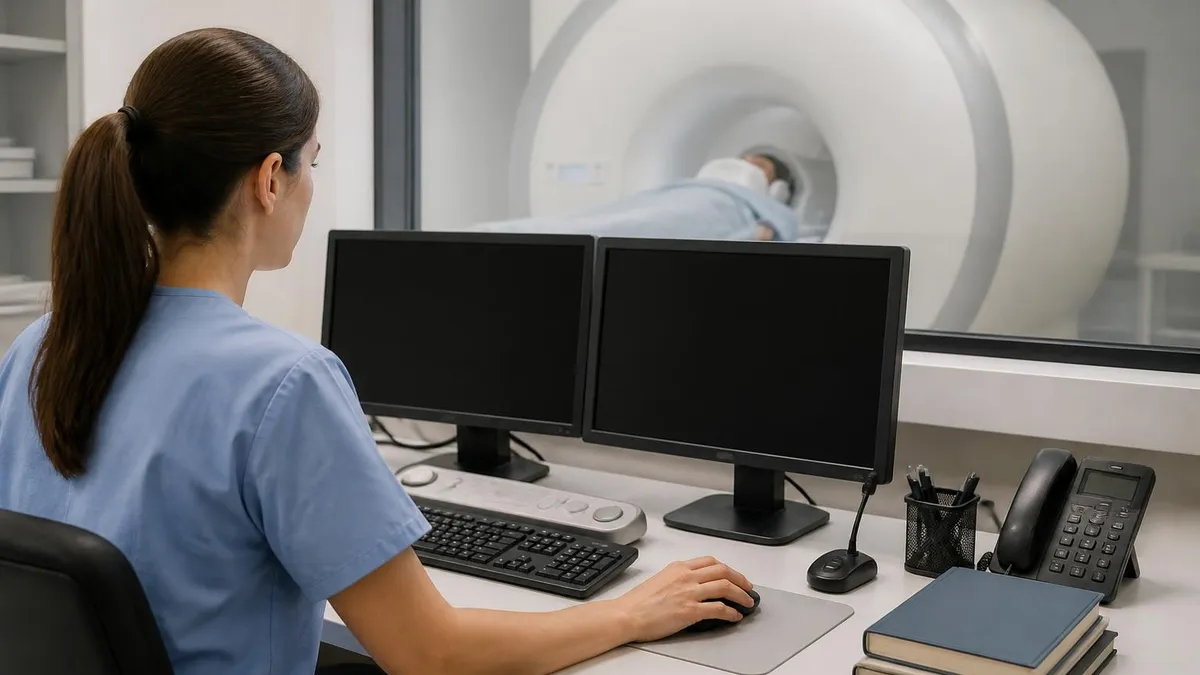
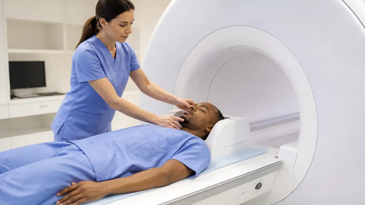
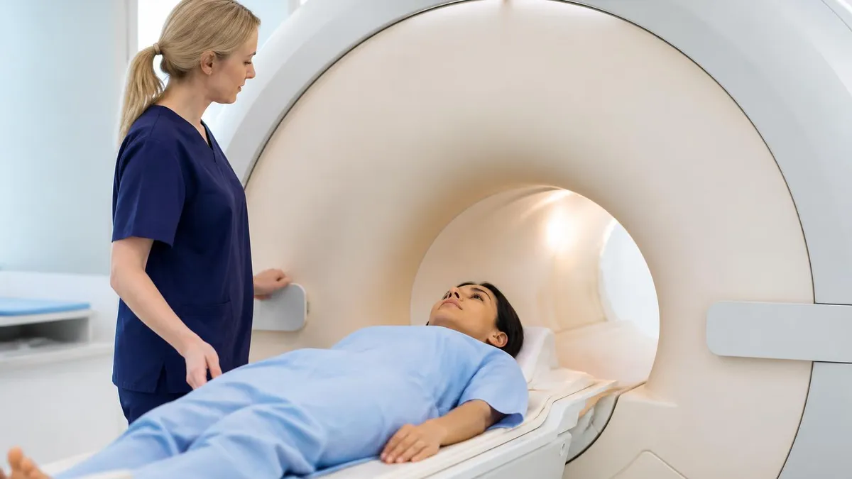
Patients often arrive at imaging departments confused because both scans involve lying still inside a large machine for thirty to ninety minutes. The hardware resembles each other in photographs, but the physics underneath is radically different. MRI generates no ionizing radiation whatsoever, relying instead on hydrogen protons aligning within a 1.5T or 3T magnetic field. PET, by contrast, depends on a cyclotron-produced radiopharmaceutical such as fluorodeoxyglucose that emits positrons as it decays inside the body. The detection methods, image appearance, and diagnostic strengths follow from that fundamental difference.
For staging cancer, evaluating treatment response, or hunting for distant metastases, PET typically wins because malignant cells consume glucose at elevated rates and light up the scan. For pinpointing a brain tumor's exact borders, assessing a torn meniscus, or evaluating spinal cord compression, MRI dominates because soft-tissue contrast resolution is unmatched. Many modern cancer workups actually use both, and hybrid PET/MRI scanners now combine the two modalities in a single session at major academic centers.
Cost, availability, and insurance coverage also factor into the decision. A standard MRI in the United States runs anywhere from $400 to $3,500 depending on body part and contrast use, while a whole-body PET/CT can range from $1,200 to over $9,000. Authorization requirements differ too — PET scans almost always need pre-approval tied to a specific diagnosis code, whereas MRI authorization depends more on the body region and clinical indication. Wait times for PET can stretch days because the tracer must be ordered fresh.
Radiologists who interpret these studies undergo years of specialized training, and the technologists operating each machine hold different certifications. MRI technologists pass ARRT or ARMRIT registry exams that test magnetic safety, pulse sequences, and anatomy, while nuclear medicine technologists earn NMTCB or ARRT(N) credentials covering radiopharmaceutical handling and detector physics. Reviewing the history of MRI alongside the parallel evolution of nuclear imaging helps explain why hospitals run separate departments for each.
This guide walks through every meaningful difference between MRI and PET scanning, from the underlying physics and clinical indications to patient prep, contraindications, image interpretation, and the growing role of hybrid imaging. Whether you are a patient trying to understand why your oncologist ordered one over the other, a student preparing for registry exams, or a clinician sharpening referral patterns, the following sections cover the practical knowledge you need to navigate these two foundational imaging modalities with confidence.
By the end you will know not only when each scan is preferred but also how to read a report, what questions to ask before your appointment, how safety precautions differ, and what emerging technologies like PET/MRI fusion and amyloid imaging mean for the future of diagnostic radiology. The goal is clarity, not jargon — both tools are remarkable, and choosing well between them often changes outcomes.
MRI vs PET Scan by the Numbers
How Each Scanner Works
Uses a superconducting magnet (typically 1.5T or 3T) plus radiofrequency pulses to flip hydrogen protons. As protons relax, they emit signals that gradient coils localize into voxels, producing T1, T2, FLAIR, and diffusion-weighted images.
Injects a positron-emitting radiotracer such as F-18 FDG. Emitted positrons annihilate with electrons, producing paired 511 keV gamma photons detected at 180 degrees. Coincidence detection reconstructs metabolic activity maps.
MRI delivers high-resolution anatomy with sub-millimeter detail of soft tissue, cartilage, white matter, and ligaments. PET delivers functional maps showing glucose, oxygen, or receptor activity, usually fused with CT or MRI for anatomical reference.
Modern PET/CT remains the workhorse for oncology staging. PET/MRI scanners combine functional and exquisite soft-tissue imaging in one session, reducing radiation and improving registration for brain, pelvic, and pediatric cases.
Clinical use cases for MRI and PET diverge sharply once you move past the surface similarity. MRI is the first-line modality for evaluating the brain when seizures, stroke, multiple sclerosis, or tumors are suspected because it resolves gray-white matter boundaries, edema, and demyelinating plaques that CT and PET simply cannot show with comparable detail. Orthopedic surgeons rely on MRI to assess rotator cuff tears, meniscal injuries, ACL ruptures, labral pathology, and bone marrow edema before deciding whether arthroscopy is warranted.
PET earns its keep in oncology. When a lung nodule appears suspicious on CT, an FDG-PET scan helps distinguish benign granulomas from active malignancy by measuring glucose uptake quantified as standardized uptake value, or SUV. Lymphoma staging, restaging after chemotherapy, and surveillance for recurrence all lean on PET because whole-body coverage reveals distant disease that targeted MRI would miss. Solitary pulmonary nodules with SUV above 2.5 raise concern for malignancy and typically trigger biopsy.
Cardiology uses both modalities differently. Cardiac MRI assesses myocardial viability, infarct scar, hypertrophic cardiomyopathy, and pericardial disease with late gadolinium enhancement sequences that no other test matches. Cardiac PET with rubidium-82 or N-13 ammonia quantifies myocardial blood flow and detects microvascular dysfunction invisible on standard stress tests. PET also evaluates cardiac sarcoidosis where inflammatory FDG uptake guides immunosuppression decisions.
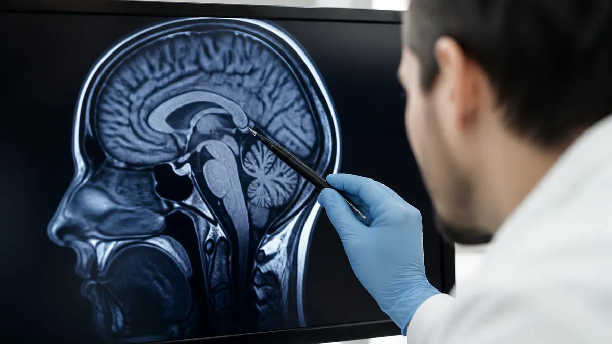
Neurology overlaps the two modalities most interestingly. MRI shows structural lesions, atrophy patterns, and white matter changes, while PET with amyloid or tau tracers reveals Alzheimer pathology years before MRI atrophy appears. Epilepsy workups often pair high-resolution 3T MRI for focal cortical dysplasia detection with interictal FDG-PET to localize the seizure focus when MRI is negative. Reading common MRI findings in the brain alongside a PET report gives neurologists complementary information.
Musculoskeletal imaging belongs almost entirely to MRI. The contrast between tendon, ligament, cartilage, muscle, and fluid is what MRI does best, and PET rarely contributes outside of rare cases involving bone tumors or osteomyelitis where PET/CT can clarify infection extent. Sports medicine physicians ordering knee or shoulder imaging will almost never request PET because the metabolic signature of musculoskeletal injury is nonspecific.
Gastrointestinal and pelvic imaging splits along similar lines. Rectal cancer staging uses MRI for local extent and circumferential resection margin assessment, while PET handles distant metastatic survey. Liver lesions get characterized by multiphase MRI with hepatobiliary contrast such as gadoxetate, but PET tracks treatment response in metastatic colorectal disease. Gynecologic malignancies typically follow the same pattern: MRI for local staging, PET for whole-body assessment.
Pediatric imaging strongly favors MRI when possible because of radiation concerns. Children with lymphoma, brain tumors, or congenital abnormalities benefit from MRI's zero ionizing radiation, though sedation may be required for younger patients unable to remain still. PET is reserved for cases where functional information clearly changes management, and pediatric PET/MRI hybrid scans are growing in popularity at children's hospitals precisely because they cut radiation dose roughly in half compared with PET/CT.
MRI vs PET Scan in Cancer, Brain, and Heart Imaging
PET/CT dominates oncologic staging for most solid tumors and lymphomas because whole-body FDG uptake reveals nodal and distant metastatic disease in a single sitting. Lung, colorectal, esophageal, head and neck, melanoma, and lymphoma all have established PET indications validated by NCCN guidelines, with SUVmax values guiding treatment response assessment via PERCIST or Deauville criteria.
MRI shines for local tumor characterization. Rectal cancer, prostate cancer, primary brain tumors, hepatocellular carcinoma, and soft-tissue sarcoma all rely on MRI for surgical planning because depth of invasion, neurovascular involvement, and tumor margin definition exceed what PET can show. Multiparametric prostate MRI with PI-RADS scoring has largely replaced random biopsy as the diagnostic standard before tissue sampling.
MRI vs PET Scan: Side-by-Side Pros and Cons
- +MRI delivers unmatched soft-tissue contrast for brain, spine, joints, and pelvis
- +No ionizing radiation, making MRI safer for repeated imaging and pediatrics
- +Multiparametric capability with T1, T2, DWI, perfusion, and spectroscopy in one session
- +Functional brain mapping and cardiac viability assessment without radiation
- +Widely available in community hospitals and outpatient imaging centers
- +Gadolinium contrast generally safer than iodinated CT contrast for kidneys
- −PET requires a fresh radiopharmaceutical with short half-life, limiting scheduling
- −PET carries 7–25 mSv ionizing radiation when combined with CT
- −PET is expensive and almost always requires insurance prior authorization
- −MRI is loud, lengthy, and intolerable for patients with severe claustrophobia
- −MRI contraindicated with many pacemakers, cochlear implants, and metallic foreign bodies
- −PET specificity limited by physiologic FDG uptake in brain, heart, and inflammation
Patient Prep Checklist for MRI and PET Scans
- ✓Confirm with your physician which scan is ordered and the clinical question it answers
- ✓Tell the technologist about any implants, pacemakers, or metal fragments before MRI
- ✓Remove all jewelry, hearing aids, dentures, and metal-containing clothing for MRI
- ✓Fast 4–6 hours before PET with FDG and avoid sugars or caffeine that morning
- ✓Hydrate well before PET to help your body clear the tracer faster after imaging
- ✓Wear loose comfortable clothing without zippers, snaps, or metallic threads
- ✓Disclose pregnancy or possible pregnancy to staff before either scan begins
- ✓Bring a list of current medications, particularly diabetes drugs that affect PET
- ✓Request open MRI or oral anxiolytic if claustrophobia is a known issue
- ✓Plan for a driver if sedation, anxiolytics, or contrast premedication is given
Different questions need different scans
MRI answers 'what does this tissue look like?' while PET answers 'what is this tissue doing?' If your physician needs both anatomical detail and metabolic activity, hybrid PET/MRI or sequential PET/CT plus dedicated MRI may be ordered together rather than choosing one over the other.
Cost differences between MRI and PET scans can shock patients seeing out-of-pocket bills for the first time. A non-contrast brain MRI at a community outpatient center might run $400 to $1,200, while the same scan at a hospital outpatient department often bills $1,500 to $3,500. PET/CT for cancer staging routinely reaches $5,000 to $9,000 because the radiopharmaceutical alone costs hundreds of dollars and must be produced fresh in a cyclotron facility. Insurance coverage hinges on diagnosis codes, prior imaging, and whether the indication appears in NCCN or AUC criteria.
Safety profiles differ in fundamental ways. MRI uses no ionizing radiation, so the cumulative dose concern that drives ALARA principles in CT and nuclear medicine simply does not apply. However, MRI carries unique safety hazards: the static magnetic field never turns off, projectile injuries from ferrous objects can be lethal, RF heating can burn skin around tattoos and certain implants, and gradient noise can damage hearing without proper protection. Quench events from helium boil-off are rare but require emergency response protocols.
PET safety centers on radiation exposure and tracer handling. A typical FDG-PET/CT delivers 7 to 25 millisieverts, roughly two to seven years of background radiation in a single exam. Pregnant patients generally avoid PET unless benefits clearly outweigh risks, and nursing mothers should pump and discard milk for several hours after FDG injection. The tracer itself is biologically inert at imaging doses, and the radiation dose decays rapidly so patients pose minimal risk to family members after a few hours.
Contraindications also differ. MRI cannot be performed safely on patients with non-MR-conditional pacemakers, cochlear implants, certain aneurysm clips, intraocular metal foreign bodies, or implanted nerve stimulators not cleared for MRI. The American College of Radiology maintains detailed safety guidelines, and every patient completes a screening questionnaire before entering Zone IV. Gadolinium contrast is contraindicated in advanced kidney disease with eGFR below 30 due to nephrogenic systemic fibrosis risk, though newer macrocyclic agents have largely eliminated this concern.
PET contraindications are fewer but significant. Uncontrolled hyperglycemia above 200 mg/dL competes with FDG uptake and degrades image quality, so diabetic patients receive specific preparation instructions. Pregnancy is a relative contraindication weighed against clinical urgency. Recent chemotherapy, radiation therapy, surgery, or infection can produce false-positive FDG uptake that confounds interpretation, so timing scans appropriately after these interventions matters for diagnostic accuracy.
Image interpretation requires different expertise. MRI reads emphasize signal intensity on multiple sequences, enhancement patterns, restricted diffusion, and anatomic relationships. PET reads quantify SUV values, compare to physiologic background, and correlate with fused CT or MRI anatomy. Reading MRI with and without contrast reports alongside PET reports gives clinicians a complete picture when staging or treatment monitoring requires both functional and structural data.
Turnaround times also vary. MRI reports often come back within 24 hours at outpatient centers and same-day at hospitals, while PET interpretation can take 48 to 72 hours because metabolic quantification and prior study comparison demand careful review. Tumor boards routinely discuss complex PET findings before treatment decisions, and discrepancies between MRI and PET sometimes trigger biopsy or short-interval follow-up imaging to resolve uncertainty.
Never enter the MRI suite with metallic implants, pacemakers, or ferrous objects without explicit clearance. Even small items like hairpins, credit cards, or oxygen tanks can become deadly projectiles in the 1.5T or 3T field. Always complete the full safety screening and disclose any prior surgeries involving metal.
Choosing between MRI and PET starts with the clinical question, not the technology. If your physician needs to evaluate the structure of a specific organ, joint, or anatomic region with maximum soft-tissue detail, MRI is almost always the correct answer. If the question involves metabolic activity, whole-body cancer surveillance, or functional assessment of disease processes invisible on structural imaging, PET earns its place. Many sophisticated workups use both, sequentially or simultaneously via hybrid scanners.
For staging newly diagnosed cancer, expect PET/CT as the workhorse. Lymphoma, lung cancer, head and neck cancer, esophageal cancer, melanoma, and many gastrointestinal malignancies have established PET indications. Dedicated MRI then characterizes the primary tumor for surgical or radiation planning. Breast cancer is an outlier where PET plays a limited role, and dedicated breast MRI dominates high-risk screening and extent-of-disease assessment.
For neurologic complaints, MRI is the default. Headache with red flags, suspected stroke, multiple sclerosis workup, seizure evaluation, suspected tumor, cognitive decline, and demyelinating disease all start with MRI. PET enters the picture for amyloid imaging in early Alzheimer evaluation, FDG patterns in dementia subtyping, and seizure focus localization when high-resolution MRI is non-diagnostic. Knowing the MRI medical abbreviation conventions on reports helps patients understand exactly what was imaged.
For musculoskeletal pain, injuries, and arthritic conditions, MRI is essentially the only answer. PET adds value only in rare bone tumor cases, suspected osteomyelitis where conventional imaging is equivocal, or prosthetic joint infection workups using specialized tracers. Sports medicine, orthopedic surgery, and rheumatology rely almost exclusively on MRI plus radiographs and ultrasound.
Cardiology splits more evenly. Cardiac MRI handles structural heart disease, cardiomyopathy characterization, viability, and pericardial disease. Cardiac PET handles quantitative perfusion, microvascular dysfunction, and inflammatory conditions like cardiac sarcoidosis. Choosing between them depends on whether the clinical question is structural or functional, and many advanced cardiology programs use both for complex patients.
Cost and access influence real-world decisions. PET requires a cyclotron-supplied tracer, so rural areas often have limited access and longer waits. MRI is more universally available but specific protocols like cardiac MRI or multiparametric prostate MRI require centers with trained technologists and radiologists. Insurance authorization for PET is more restrictive, and self-pay patients face significantly higher out-of-pocket costs for PET than for MRI.
Hybrid imaging is the future for complex cases. PET/MRI scanners combine simultaneous metabolic and anatomic imaging in a single appointment, reducing total radiation dose and improving registration for brain tumors, pelvic malignancies, pediatric cancers, and cardiac sarcoid workups. Academic medical centers increasingly offer this option, and clinical evidence supporting PET/MRI continues to grow for indications where the fused datasets change management decisions.
Practical preparation for either scan starts with reading the appointment instructions carefully and calling the imaging center if anything is unclear. MRI prep is usually minimal — wear loose comfortable clothing without metal, remove jewelry, and arrive a few minutes early to complete the safety screening questionnaire. If contrast is ordered, you may need recent kidney function labs, and patients with known claustrophobia should request oral anxiolytic premedication days before the appointment so the prescription is ready.
PET prep is more involved. Patients fast 4 to 6 hours before FDG injection, avoid sugars and caffeine that morning, and stay well hydrated with plain water. Diabetic patients receive specific instructions about insulin and oral medications because blood glucose must be reasonably controlled for accurate FDG uptake. Strenuous exercise in the 24 hours before PET is discouraged because muscle uptake can mimic disease. After injection, you rest quietly in a dimmed room for about 60 minutes while the tracer distributes.
During MRI, expect loud knocking, beeping, and buzzing from gradient coils — earplugs and headphones with music help. You must remain still because even small motion blurs the image and may require repeat sequences. The technologist communicates through an intercom and provides a squeeze ball for emergencies. Some sequences require breath-holds, particularly for abdominal or cardiac imaging. Total scan time depends on body part and protocol complexity, ranging from 15 minutes for a quick brain scan to 90 minutes for a comprehensive cardiac study.
During PET, you lie on a narrow table that moves slowly through a ring-shaped detector. The scan itself is silent and lasts 20 to 40 minutes for whole-body coverage, longer for specialized brain or cardiac protocols. The acquisition is quieter than MRI but the room can feel cold to maintain detector performance. Stay still and breathe normally unless instructed otherwise. You can usually resume normal activities immediately after, drinking extra fluids to help your body clear residual tracer faster.
After your scan, the radiologist or nuclear medicine physician interprets the images and dictates a report that goes to your ordering physician. Turnaround times vary by facility and complexity, typically 24 to 72 hours. Ask your physician when to expect results and how they will be communicated. If findings are urgent — acute stroke, large tumor, critical infection — the radiologist will call the ordering provider directly before the formal report is finalized, ensuring time-sensitive findings reach you quickly.
Follow-up imaging is common in oncology and chronic disease management. PET responses are quantified using PERCIST or Deauville criteria depending on tumor type, while MRI follow-ups track lesion size, enhancement, and diffusion changes. Bringing prior imaging on disc or arranging electronic transfer ensures the interpreting radiologist can compare directly, which is essential for treatment response assessment and surveillance. Always keep copies of your imaging studies for future reference, especially if you move or change healthcare systems.
Finally, do not hesitate to ask questions. Patients who understand why a scan was ordered, what it shows, and how results will guide treatment make better decisions and feel less anxious throughout the process. Imaging is a tool that supports clinical care; it does not replace the relationship with your physician. The clearest reports come from radiologists who receive thorough clinical histories, so giving your ordering physician complete symptom details and prior imaging information directly improves the diagnostic value of every MRI or PET scan you undergo.
MRI Questions and Answers
About the Author
Attorney & Bar Exam Preparation Specialist
Yale Law SchoolJames R. Hargrove is a practicing attorney and legal educator with a Juris Doctor from Yale Law School and an LLM in Constitutional Law. With over a decade of experience coaching bar exam candidates across multiple jurisdictions, he specializes in MBE strategy, state-specific essay preparation, and multistate performance test techniques.