
MRI Terminology for Dummies: T1, T2, TR, TE, Hyperintense and Key Terms Explained
MRI terminology explained plainly — T1, T2, TR, TE, hyperintense, flow void, STAT, isocenter, high intensity zone and more. ⏳
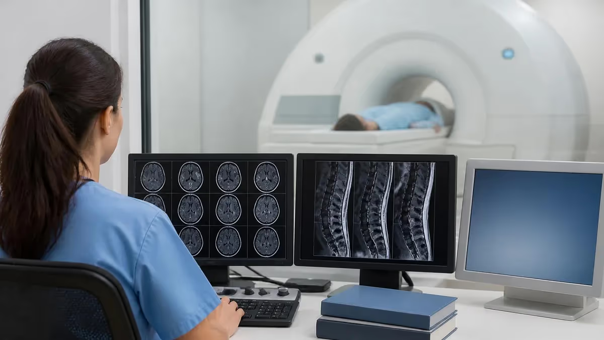
What Is MRI Terminology?
If you've ever stared at an MRI report and felt completely lost, you're not alone. Phrases like hyperintense on T2, low TR/TE, or flow void can sound like a foreign language — even to patients who've had dozens of scans. Learning the core vocabulary doesn't require a medical degree, though. It just takes a clear map of what the terms actually mean.
MRI terminology covers two broad areas: image weighting (how the scanner is tuned) and signal intensity (how bright or dark tissues appear on the resulting image). Once you understand those two pillars, almost everything else falls into place. This guide walks you through mri terminology for dummies style — plain language, real examples, no unnecessary jargon.
Whether you're a radiology student, a patient reviewing your own report, or a clinician brushing up before boards, knowing when MRI scans are used is only half the picture. The other half is understanding what the scan is actually showing you.
One thing that surprises many newcomers: MRI doesn't use radiation. Unlike X-ray or CT, it works entirely with magnetic fields and radio waves. The scanner creates a powerful magnetic field that aligns the hydrogen protons in your body. A radio wave pulse then knocks them out of alignment, and the signal they produce as they snap back into place is what creates the image. Different tissues snap back at different rates — and those differences in rate are exactly what T1 and T2 terminology describes.
This guide covers the terms you'll actually encounter in radiology reports and board exams: signal intensity words (hyperintense, hypointense, isointense), timing parameters (TR, TE), sequence types (T1, T2, FLAIR, DWI), and specialized findings (flow void, high intensity zone, STAT, isocenter). Each section builds on the previous one, so reading straight through will give you the most complete picture.
One more thing worth knowing upfront: MRI terminology is always relative. Hyperintense doesn't mean intrinsically bright — it means brighter than something else. That something else is called the reference tissue, and its identity changes depending on what you're evaluating. For brain MRI, gray matter is often the reference. For musculoskeletal MRI, it's often muscle. For abdominal MRI, it's often the liver. Keeping that relativity in mind prevents a lot of confusion when you see terms like iso or hypo used without explicit reference structures.
Signal Intensity, TR, and TE
Hyperintense means a tissue appears bright relative to a reference structure on a given MRI sequence. On T2-weighted images, fluid is hyperintense — it glows white. When radiologists say a lesion is "high T2 signal" or "hyperintense on T2," they mean it looks brighter than surrounding tissue. This often suggests fluid, edema, inflammation, or certain tumors.
Hypointense is the opposite — the tissue appears dark. Cortical bone, air, and rapidly flowing blood are hypointense on most sequences. Dense fibrous tissue is also typically hypointense. A hypointense lesion on T2 can indicate fibrosis, calcification, or blood products (depending on age).
Isointense (or iso on MRI) means a tissue has the same signal intensity as the reference tissue — usually muscle or gray matter. Isointense lesions can be hard to spot because they don't stand out from background. This is clinically significant: an isointense mass may be nearly invisible on one sequence but clearly visible on another, which is why radiologists use multiple sequences.
T1 vs T2 Weighting: What Tissues Look Like and Why It Matters
The most fundamental concept in mri terminology is the difference between T1 and T2 weighting. They're not two different scanners — they're two different ways of programming the same MRI machine to highlight different tissue properties.
T1 Weighting
T1 relaxation refers to how quickly protons in tissue realign with the main magnetic field after an RF pulse. Tissues with short T1 values (fat, proteinaceous fluid, subacute blood) realign quickly — and with a short TR, they've already recovered most of their magnetization by the time the next pulse fires. That gives them a strong signal and a bright appearance on T1 images.
Water and most pathological fluids have long T1 values — they're still recovering when the next pulse arrives, so they appear dark on T1. This is why cerebrospinal fluid is black on T1-weighted brain scans.
T1 is the go-to sequence for anatomy. It shows the layers of the brain, the boundaries of organs, and the distribution of fat — all with crisp contrast. When gadolinium contrast is used, areas with a disrupted blood-brain barrier (tumors, active inflammation) enhance brightly on T1 because the contrast agent shortens T1 relaxation time.
T2 Weighting
T2 relaxation describes how quickly the transverse magnetization of protons decays due to interactions with neighboring protons. Free water has a very long T2 — its protons interact minimally, so the signal persists and the tissue appears bright on T2 images. Solid tissues and those with restricted molecular motion have short T2 values — they lose signal quickly and appear dark.
The clinical implication is powerful: almost any pathology increases the water content of tissue (edema, inflammation, necrosis, tumor infiltration). That means almost any disease process produces high T2 signal. A high T2 signal on MRI is non-specific but sensitive — it tells you something is wrong here, even if it doesn't tell you exactly what.
What is a high T1 and T2 signal MRI finding? That combination — bright on both T1 and T2 — usually points to fat (lipoma, dermoid) or subacute blood (methemoglobin). It narrows the differential considerably.
T1 vs T2 Quick Reference
- ✓T1: short TR (~400–600 ms) + short TE (~10–30 ms) — anatomy, fat bright, fluid dark
- ✓T2: long TR (~2,000+ ms) + long TE (~80–120 ms) — fluid bright, pathology visible
- ✓High T2 signal = fluid, edema, inflammation, or tumor infiltration
- ✓High T1 signal = fat, subacute blood (methemoglobin), or proteinaceous fluid
- ✓Gadolinium shortens T1 — enhancing tissues appear bright on post-contrast T1
- ✓Proton density: long TR + short TE — tissue mass visible, minimal T1/T2 bias
- ✓FLAIR: T2 variant with CSF suppressed — best for periventricular lesions
- ✓DWI + ADC: detects restricted diffusion — acute stroke, abscess, hypercellular tumor
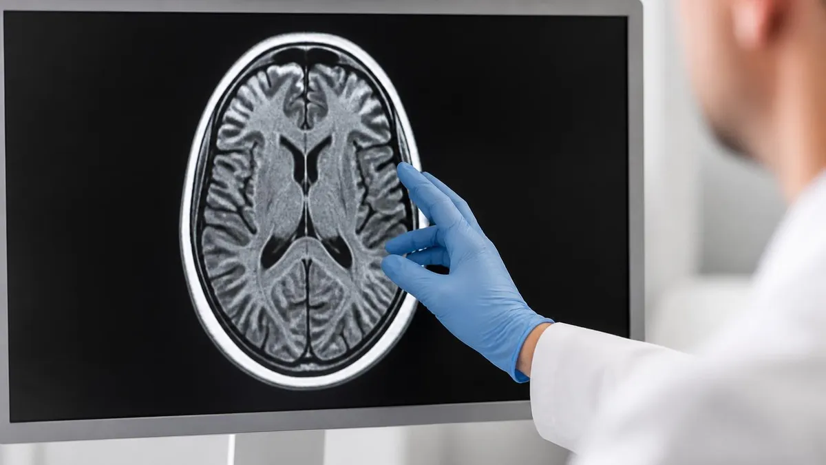
How TR and TE Create the Weighting
Understanding tr and te in mri means understanding that you're trading off between two types of tissue contrast with every sequence. For T1 weighting, you use short TR (roughly 400–600 ms) and short TE (10–30 ms). The short TR ensures T1 differences between tissues are fully expressed; the short TE minimizes T2 decay before you read the signal.
For T2 weighting, you use long TR (2,000–6,000 ms) and long TE (80–120 ms). The long TR nullifies T1 differences; the long TE lets T2 decay develop so the fast-decaying tissues go dark and the slow-decaying fluid stays bright. In tr te mri practice, radiologists don't usually pick these values themselves — they're embedded in standardized sequences. But knowing the logic helps you understand why the same patient can look completely different on two images taken in the same scanner session.
To see how these principles apply in practice, it helps to understand how MRI machines work at a hardware level — the gradient coils, the RF transmitter, and the receiver array all play roles in determining image quality and the effectiveness of each sequence.
Contrast Enhancement and Signal Behavior
When gadolinium-based contrast agents are injected, they shorten T1 relaxation time in tissues they reach. Areas where contrast accumulates — because the blood-brain barrier is broken, because a tumor has abnormal vessels, or because an infection is active — become bright on T1-weighted post-contrast images. This is called T1 shortening or T1 enhancement. It doesn't change T2 behavior directly, which is why both pre-contrast and post-contrast T1 images are acquired. Comparing them side-by-side reveals where the contrast went and gives radiologists a decisive diagnostic edge when anatomy alone isn't enough.
Key MRI Terms at a Glance
Appears bright on a given MRI sequence relative to a reference tissue. High T2 signal = fluid, edema, or pathology. High T1 signal = fat or subacute blood.
Appears dark relative to reference tissue. Common causes include cortical bone, air, rapidly flowing blood, calcification, or chronic blood products.
Time between RF pulses in milliseconds. Short TR (~400–600 ms) produces T1 weighting. Long TR (~2,000+ ms) is used for T2 and proton density sequences.
Time between the RF pulse and signal readout. Short TE (~10–30 ms) minimizes T2 contrast. Long TE (~80–120 ms) maximizes T2 differences between tissues.
Dark (signal-void) area on spin echo sequences caused by rapidly moving blood or CSF. The moving blood exits the slice before the echo is collected, producing no signal.
Bright T2 signal in the posterior annulus fibrosus of an intervertebral disc. Associated with annular tears and disc degeneration, clinically linked to discogenic pain.
STAT means immediately — from Latin statim. A STAT MRI is ordered for urgent situations: acute stroke, spinal cord compression, or suspected subarachnoid hemorrhage.
The geometric center of the MRI magnetic field, where field homogeneity is highest. Patient positioning at isocenter maximizes image quality and signal-to-noise ratio.
STAT MRI, Flow Void, High Intensity Zone, Isocenter, and Isointense: Clinical Context
STAT MRI: When Minutes Matter
Stat mri meaning comes straight from Latin: statim, meaning immediately. When a physician orders a STAT MRI, it goes to the front of the queue — the patient is scanned within minutes to hours rather than days. STAT orders are typically placed for acute neurological emergencies: sudden-onset severe headache (subarachnoid hemorrhage until proven otherwise), focal neurological deficit (stroke), vision loss, suspected spinal cord compression from metastatic disease, or cauda equina syndrome with bowel/bladder involvement.
In practice, STAT doesn't mean the radiologist drops everything — it means the order is flagged as high priority and the reading is expedited. If you see STAT on your order, it's not a reason to panic, but it does mean your doctor needs results quickly for a time-sensitive clinical decision.
How quickly does a STAT MRI actually happen? In most hospitals, a STAT order for a truly emergent indication gets the patient onto the scanner within 30–60 minutes around the clock. Preliminary reads are often available within minutes of scan completion. A STAT order placed for an urgent-but-not-emergency reason might mean same-day rather than next-week scheduling.
It's also worth knowing that STAT applies to the entire workflow — scheduling, scanning, and interpretation. A STAT order that isn't clearly communicated to the MRI technologist, the reading radiologist, and the ordering physician can fail at any link in that chain.
In well-run radiology departments, STAT orders trigger a cascade: the scanner is cleared, the tech is notified, and the radiologist is paged as soon as images are uploaded. Understanding stat mri meaning isn't just trivia — if you're ever the ordering provider, knowing what happens downstream helps you use the designation appropriately rather than overusing it and diluting its urgency signal.
What Is a Flow Void on MRI?
What is a flow void on mri? It's a dark area on spin echo sequences where you'd normally expect to see a blood vessel. Rapidly flowing blood moves out of the imaging slice between the time the RF pulse is applied and the time the echo is collected. Because those protons are no longer in position to contribute signal, the vessel appears black. Flow voids are normal findings in the carotid arteries, the basilar artery, the jugular veins, and within the cardiac chambers on brain and chest MRI.
Flow voids become clinically important when they're absent where you'd expect them (suggesting slow flow, thrombosis, or occlusion) or when they appear where you don't expect them (suggesting an arteriovenous malformation). On abnormal MRI findings, a missing flow void in the internal carotid can be one of the first signs of dissection or occlusion.
MRI Signal: What to Expect on Each Sequence
- +T1: fat and subacute blood are bright — good for anatomy and post-contrast enhancement
- +T2: fluid and edema are bright — detects most pathology by water content increase
- +FLAIR: suppresses CSF while keeping pathological fluid bright — best for MS plaques
- +DWI: restricted diffusion is bright — stroke and abscess detection within minutes
- +Gadolinium contrast on T1: enhancing lesions become unmistakably bright
- −T2 hyperintensity is non-specific — fluid, edema, tumor, and infarct all look similar
- −Isointense lesions on T1/T2 can be invisible without contrast or DWI
- −Flow voids on spin echo disappear on gradient echo — sequence context matters
- −High intensity zones in discs are present in asymptomatic patients too
- −Off-isocenter positioning degrades image quality and can distort anatomy
High Intensity Zone: The Disc's Distress Signal
What is a high intensity zone on MRI? The HIZ is a focal bright T2 signal seen in the posterior annulus fibrosus of an intervertebral disc — the tough outer ring that contains the disc. It represents a tear through the annular fibers that has filled with vascularized granulation tissue or inflammatory fluid. That fluid-filled tear lights up on T2.
Studies link HIZ findings to discogenic pain — pain coming from the disc itself rather than nerve compression. It's particularly relevant in back pain workups. If you're reviewing a lumbar spine report and see mention of a posterior HIZ at L4-L5 or L5-S1, it's a marker of annular disruption at that level. For patients with back MRI findings, HIZ is one of the more specific T2 findings for internal disc disruption.
Not everyone with an HIZ has pain. Asymptomatic individuals can have posterior annular bright zones without clinical symptoms. That's part of why discogenic pain diagnosis is challenging — the imaging finding alone doesn't make the diagnosis. Correlation with the patient's history, symptom distribution, and clinical exam remains important. The HIZ is a supportive finding, not a standalone diagnostic criterion. Discography — an invasive procedure injecting dye into the disc — is sometimes used to confirm the disc as the pain source, but MRI HIZ detection is the usual first-line imaging marker.
MRI Signal Intensity Quick Facts
Isocenter: Getting the Best Signal
Isocenter mri refers to the geometric sweet spot of the magnetic field — the point where field homogeneity is highest and gradient performance is optimal. Modern MRI machines are engineered so that the center of the bore coincides with isocenter. Radiographers aim to place the anatomical region of interest at or near isocenter — a knee scanned at the edge of the bore will have more field inhomogeneity and worse image quality than one positioned centrally.
The gradients that create spatial localization work optimally at isocenter. Off-center scanning introduces geometric distortion — anatomical structures end up misrepresented in size or shape. For surgical planning and radiation therapy, that distortion can have real consequences. Isocenter positioning isn't just about image quality; it's about diagnostic and treatment accuracy. Dedicated extremity MRI units, which scan fingers, wrists, and ankles, are designed so that the small joint sits precisely at isocenter even when the patient sits beside the machine rather than lying inside it.
Iso Signal: The Invisible Lesion Problem
Iso mri — or isointense signal — is one of the trickier concepts in MRI reporting. A lesion that is isointense on T1 and T2 is essentially the same shade of gray as surrounding brain or soft tissue. It doesn't stand out. This doesn't mean there's no pathology; it means the sequence you're looking at isn't the right tool to detect it. Recognizing isointensity is just as diagnostically important as recognizing hyperintensity, because it tells you which additional sequences to run.
Meningiomas are a classic example: they're often isointense to gray matter on both T1 and T2, making them almost invisible on non-contrast sequences. Add gadolinium contrast, and they enhance dramatically. Isointense lesions on unenhanced MRI are one reason contrast administration is so important in oncology and neurology workups. Hepatocellular carcinoma on unenhanced liver MRI, prostate cancer on T2 of the peripheral zone, and certain lymph node metastases can all be isointense — detectable only with contrast, DWI, or specialized sequences.
Understanding iso signal also helps explain why radiologists often describe lesions in terms of multiple sequences: "isointense on T1, hyperintense on T2, restricted on DWI." No single sequence tells the whole story. That's the real point of mri terminology for dummies — each term is a piece of a larger diagnostic puzzle, and the art of radiology is fitting those pieces together into a coherent picture.
After working through all of this mri terminology, a pattern emerges. MRI is fundamentally a language of contrasts. T1 vs T2 is a contrast. Hyperintense vs hypointense is a contrast. Flow void vs flow-related enhancement is a contrast. Learning this vocabulary means learning to see those contrasts clearly, and that's what separates a confident reader from someone who just sees shades of gray. The terms aren't ends in themselves — they're the building blocks of radiological reasoning, and once they're automatic, reading MRI reports becomes a lot less intimidating.
MRI Questions and Answers
About the Author

Medical Laboratory Scientist & Clinical Certification Expert
Johns Hopkins UniversityDr. Sandra Kim holds a PhD in Clinical Laboratory Science from Johns Hopkins University and is certified as a Medical Technologist (MT) and Medical Laboratory Scientist (MLS) through ASCP. With 16 years of clinical laboratory experience spanning hematology, microbiology, and molecular diagnostics, she prepares candidates for ASCP board exams, MLT, MLS, and specialist certification tests.
Join the Discussion
Connect with other students preparing for this exam. Share tips, ask questions, and get advice from people who have been there.
View discussion (8 replies)