Chest MRI: Complete Guide to Thoracic Magnetic Resonance Imaging 2026 July
Chest MRI explained: 📗 indications, protocols, sequences, contrast use, patient prep, and how it compares to CT for thoracic imaging.
A chest MRI is a powerful, non-ionizing imaging examination that uses strong magnetic fields and radiofrequency pulses to generate detailed cross-sectional images of the thoracic cavity. While computed tomography remains the workhorse for most thoracic indications, chest MRI has become indispensable for evaluating mediastinal masses, cardiac and vascular pathology, brachial plexus disorders, and selected lung diseases where soft tissue contrast or radiation avoidance matters most for diagnosis and follow-up care.
Radiologic technologists and physicians performing thoracic MRI must understand the unique physics challenges of the chest. The thorax is in constant motion from breathing and cardiac pulsation, and air-filled lung parenchyma produces low proton density and severe susceptibility artifacts. These factors historically limited MRI of the chest, but modern sequences with respiratory triggering, ECG gating, and ultrashort echo time techniques have dramatically expanded its clinical utility over the past decade.
Indications for thoracic MRI include characterizing anterior mediastinal masses such as thymoma, lymphoma, and germ cell tumors. It excels at evaluating Pancoast tumors at the lung apex because of its multiplanar capability and superior visualization of the brachial plexus, subclavian vessels, and chest wall invasion. Cardiac MRI is a subspecialty in its own right, assessing myocardial viability, congenital heart disease, cardiomyopathies, pericardial pathology, and valvular function with quantitative precision unmatched by other modalities.
Patients presenting for a chest MRI typically receive intravenous gadolinium-based contrast unless contraindicated by renal function or prior allergic reaction. Pre-contrast and post-contrast T1-weighted sequences with fat suppression are essential for tissue characterization. Diffusion-weighted imaging adds functional information about cellularity, particularly useful in oncologic staging and distinguishing benign from malignant pleural disease in patients with mesothelioma or metastatic effusions.
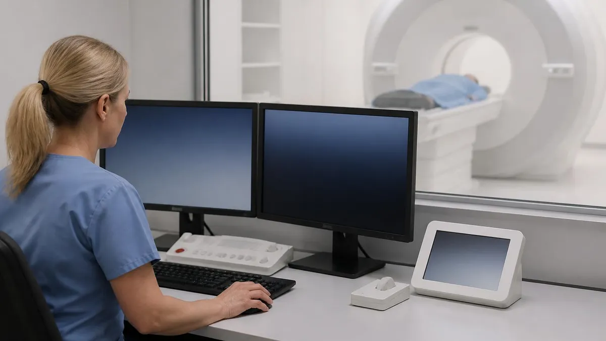
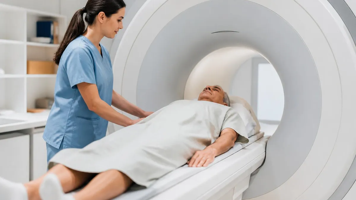
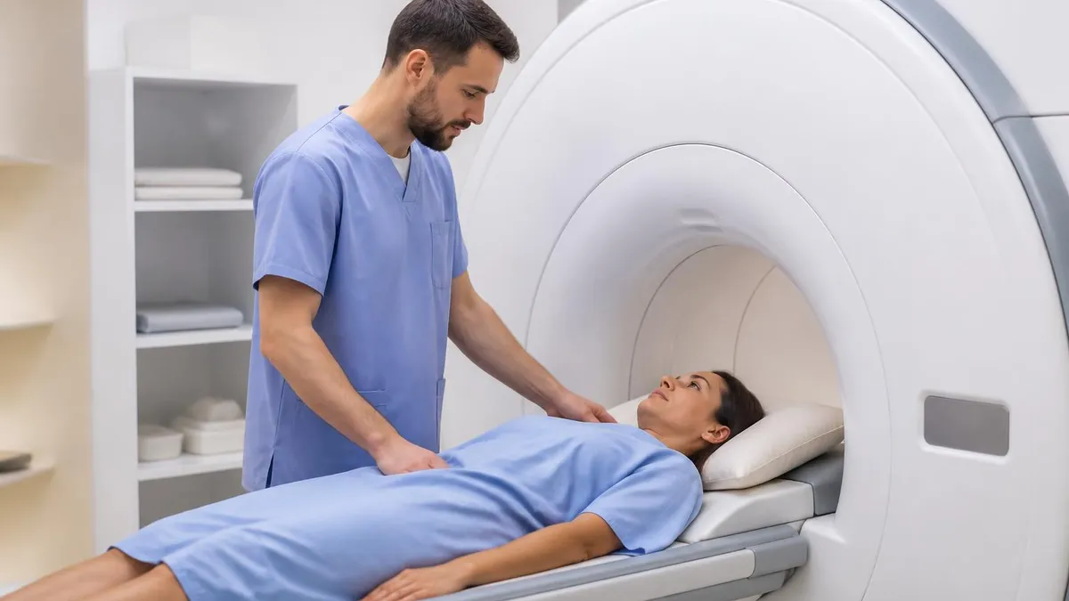
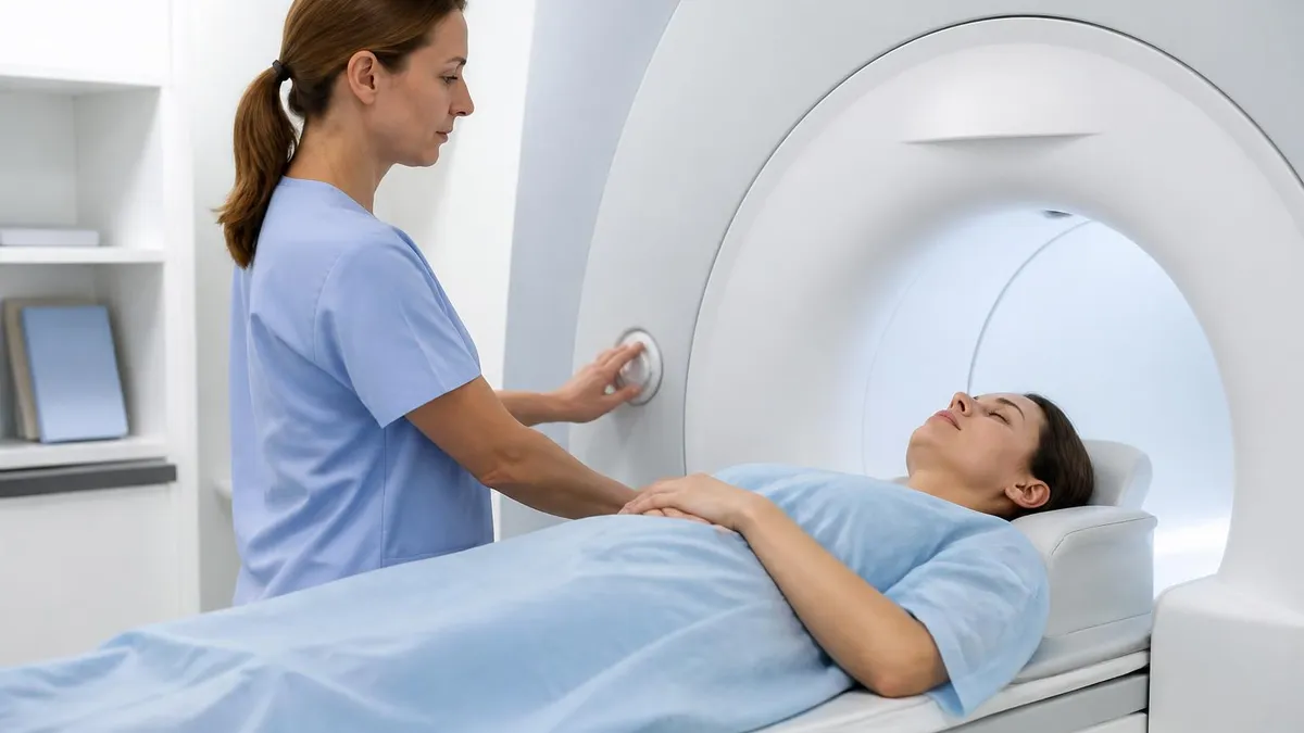
Patient preparation begins with rigorous safety screening for ferromagnetic implants, cardiac devices, and aneurysm clips. Many modern pacemakers and ICDs are now MR-conditional and can be scanned safely with appropriate programming, but each device must be verified against manufacturer guidelines. The patient lies supine with the chest centered in a phased-array torso coil, and ECG leads are applied for cardiac-gated sequences. Breath-hold instructions are rehearsed before scanning begins.
Scan times for a complete thoracic MRI typically range from 30 to 60 minutes depending on the clinical question. Cardiac protocols tend to run longer than mediastinal surveys because they include cine imaging, perfusion, late gadolinium enhancement, and sometimes flow quantification. Modern scanners with parallel imaging, compressed sensing, and motion-correction algorithms have significantly reduced acquisition times while improving image quality, making chest MRI tolerable even for dyspneic or claustrophobic patients who previously could not complete the exam.
This comprehensive guide walks through every clinical and technical aspect of chest MRI, from anatomic considerations and sequence selection to artifact troubleshooting and contrast administration. Whether you are a registry candidate preparing for board examinations, a working technologist refining your protocol, or a referring clinician trying to understand when MRI is the right test, this resource will provide the depth and specificity needed to make confident decisions about thoracic magnetic resonance imaging in real-world practice.
Chest MRI by the Numbers
Common Indications for Chest MRI
Characterization of anterior, middle, and posterior mediastinal masses including thymoma, lymphoma, germ cell tumors, neurogenic tumors, and bronchogenic cysts when CT findings are indeterminate or radiation must be avoided.
Cardiac MRI evaluates myocardial viability, cardiomyopathies, congenital heart disease, pericardial disease, and aortic pathology including dissection, aneurysm, and coarctation with quantitative flow measurements.
Superior sulcus tumors require MRI to assess brachial plexus involvement, subclavian vessel invasion, and chest wall extension before surgical planning or radiation therapy delivery.
Differentiating benign pleural plaques from malignant mesothelioma, characterizing complex effusions, and staging pleural malignancy with diffusion-weighted imaging and dynamic contrast enhancement.
Children with congenital lung malformations, vascular rings, or recurrent imaging needs benefit from MRI because it eliminates cumulative radiation exposure during follow-up surveillance over many years.
Building an effective chest MRI protocol requires matching pulse sequences to the clinical question while accounting for cardiac and respiratory motion. Most institutions begin with localizers in three planes followed by axial half-Fourier single-shot turbo spin echo, often called HASTE or SSFSE, which acquires a complete slice in under a second and provides excellent T2-weighted contrast without motion blurring. This sequence is the backbone of mediastinal and pleural imaging because it tolerates free breathing.
T1-weighted imaging is essential for tissue characterization, particularly for detecting fat within thymolipomas, teratomas, and lipomatous tumors, as well as identifying hemorrhagic components or proteinaceous fluid. Dual-echo gradient echo sequences with in-phase and opposed-phase imaging can confirm microscopic fat suggestive of thymic hyperplasia, an important distinction from neoplastic thymic enlargement in patients with myasthenia gravis or other autoimmune conditions affecting the anterior mediastinum.
Short tau inversion recovery sequences provide robust fat suppression across the inhomogeneous thoracic field, highlighting edema, inflammation, and tumor against suppressed mediastinal and chest wall fat. STIR is particularly valuable for brachial plexus imaging and chest wall pathology where uniform spectral fat saturation often fails due to magnetic field distortions near air-tissue interfaces and at the apex of the thorax where coverage drops off in conventional torso coil setups.
Diffusion-weighted imaging with apparent diffusion coefficient mapping adds quantitative functional information to anatomic sequences. Highly cellular tumors restrict water diffusion and appear bright on high b-value images with corresponding low ADC values. This characteristic helps differentiate lymphoma from benign thymic tissue, identify peritumoral invasion, and detect distant metastases. For more on principles of magnetic resonance physics, see our guide on how does an MRI work to understand the underlying mechanisms.
Cardiac protocols add ECG-gated steady state free precession cine imaging in standard short-axis and long-axis planes to assess ventricular function and wall motion. Late gadolinium enhancement using phase-sensitive inversion recovery sequences performed 10 to 15 minutes after mri contrast injection identifies myocardial scar, infiltrative disease, and inflammation. T1 and T2 mapping have emerged as quantitative biomarkers for myocardial fibrosis, edema, and iron overload in conditions such as hemochromatosis and thalassemia.
Contrast-enhanced MR angiography of the thoracic aorta and pulmonary arteries uses a time-resolved or single-phase 3D gradient echo acquisition during peak arterial enhancement. Test bolus or bolus tracking techniques ensure proper timing. For patients who cannot receive gadolinium, non-contrast techniques such as balanced steady state free precession with respiratory navigation or arterial spin labeling provide diagnostic vascular imaging without the small but documented risks of nephrogenic systemic fibrosis or gadolinium deposition.
Newer sequences continue to expand what chest MRI can show. Ultrashort echo time and zero echo time imaging capture signal from lung parenchyma with echo times below one millisecond, allowing CT-like visualization of nodules and interstitial disease without radiation. Compressed sensing accelerates free-breathing 3D acquisitions to under five minutes. These advances are gradually closing the historical gap between chest MRI and CT, particularly for follow-up imaging in young patients with cystic fibrosis, lymphoma, or other chronic conditions requiring repeated thoracic evaluation.
MRI Practice Test Questions
Prepare for the MRI - Magnetic Resonance Imaging exam with our free practice test modules. Each quiz covers key topics to help you pass on your first try.
MRI Knowledge
MRI Exam Questions covering Knowledge. Master MRI Test concepts for certification prep.
MRI Physics
Free MRI Practice Test featuring Physics. Improve your MRI Exam score with mock test prep.
MRI Anatomy and Pathology
MRI Test Prep for MRI Anatomy and Pathology. Practice MRI Quiz questions and boost your score.
MRI Anatomy and Positioning
MRI Questions and Answers on MRI Anatomy and Positioning. Free MRI practice for exam readiness.
MRI Contrast Agents
Free MRI Quiz on MRI Contrast Agents. MRI Exam prep questions with detailed explanations.
MRI Patient Care and Positioning
MRI Practice Questions for MRI Patient Care and Positioning. Build confidence for your MRI certification exam.
Chest MRI by Anatomic Region
The mediastinum is the central thoracic compartment bounded by the pleura laterally, the sternum anteriorly, the spine posteriorly, and the diaphragm inferiorly. It is traditionally divided into anterior, middle, and posterior compartments using the Felson method or the newer ITMIG cross-sectional classification, which provides more reproducible localization for surgeons and oncologists planning biopsy or resection of mediastinal lesions encountered on imaging.
MRI excels at characterizing mediastinal masses because of superior soft tissue contrast. Thymoma typically shows intermediate T1 and T2 signal with avid enhancement and possible capsular invasion. Lymphoma demonstrates marked diffusion restriction. Germ cell tumors often contain fat or calcification, while cystic lesions such as bronchogenic and pericardial cysts show fluid signal without enhancement, allowing confident diagnosis without biopsy when imaging features are characteristic.
Chest MRI vs. Chest CT: Strengths and Limitations
- +No ionizing radiation, ideal for pregnant patients and children requiring serial imaging
- +Superior soft tissue contrast for characterizing mediastinal masses and chest wall invasion
- +Multiplanar imaging without reformatting, useful for Pancoast tumors and complex anatomy
- +Functional information including diffusion, perfusion, and flow quantification
- +Excellent for cardiac and vascular evaluation with cine, late enhancement, and mapping techniques
- +Tissue characterization including fat, hemorrhage, and fluid without contrast in many cases
- −Longer scan times of 30 to 60 minutes versus seconds for CT
- −Lower spatial resolution and reduced sensitivity for small pulmonary nodules under 4 mm
- −Susceptibility artifacts at air-tissue interfaces degrade lung parenchyma evaluation
- −Cardiac and respiratory motion require gating, navigation, or breath-holding cooperation
- −Contraindicated in patients with non-conditional pacemakers, ICDs, and certain implants
- −Higher cost and limited availability compared to CT in many community hospital settings
Patient Preparation Checklist for Chest MRI
- ✓Complete MRI safety screening including all surgical implants, devices, and prior procedures
- ✓Verify renal function with recent eGFR if gadolinium contrast is planned for the examination
- ✓Ask patients about pregnancy status and breastfeeding before scheduling and again at check-in
- ✓Remove all external metallic objects including jewelry, hairpins, dentures, and hearing aids
- ✓Provide a gown or scrubs free of metallic snaps, zippers, and antimicrobial silver threading
- ✓Place IV access in an antecubital or forearm vein appropriate for power injection of contrast
- ✓Apply ECG leads on prepped skin for any cardiac-gated or cine imaging sequences planned
- ✓Rehearse breath-hold instructions and confirm the patient can hold their breath 15-20 seconds
- ✓Position the patient supine with arms at sides or above head depending on coil and protocol
- ✓Provide hearing protection and the squeeze bulb for communication during the entire exam
Always confirm cardiac device MR conditionality before scanning
Modern pacemakers and ICDs labeled as MR-conditional can be safely scanned with proper programming, monitoring, and field strength compliance. However, abandoned leads, legacy devices, and certain combinations remain absolute contraindications. Always verify the model number against the manufacturer's MRI safety database and coordinate with cardiology before bringing the patient into the scan room. Document the device interrogation and post-scan check in the medical record.
Gadolinium-based contrast agents are central to many chest MRI examinations because they improve detection and characterization of mediastinal masses, pleural disease, cardiac scar, and vascular pathology. Standard dosing is 0.1 millimoles per kilogram of body weight for macrocyclic agents such as gadobutrol, gadoterate meglumine, and gadoteridol, which have favorable safety profiles and are preferred over linear agents in current ACR Manual on Contrast Media guidelines for routine clinical use.
Renal screening is mandatory before contrast administration. Patients with estimated glomerular filtration rate below 30 milliliters per minute per 1.73 square meters have historically been at highest risk for nephrogenic systemic fibrosis, a rare but devastating fibrosing condition associated with linear gadolinium agents in severe renal insufficiency. With modern macrocyclic agents, the risk is considered exceedingly low, but most institutions still require shared decision-making and informed consent in this population to balance benefits against perceived risk.
Allergic-like reactions to gadolinium are uncommon, occurring in less than 0.1 percent of administrations, but they can range from mild urticaria to anaphylactoid shock. Departments must have emergency response protocols, trained personnel, and immediately accessible medications including epinephrine, antihistamines, and bronchodilators. Patients with prior moderate or severe reactions typically receive pre-medication with corticosteroids and antihistamines or are offered an alternative agent or a non-contrast protocol depending on the clinical indication.
Power injection is standard for dynamic contrast mri-enhanced sequences such as MR angiography of the thoracic aorta and pulmonary arteries. Typical injection rates are 2 to 3 milliliters per second followed by a 20 to 30 milliliter saline flush to push the contrast bolus into the central circulation. Lower flow rates of 1.5 to 2 milliliters per second are appropriate for cardiac perfusion and pediatric patients to maintain venous access integrity and avoid extravasation injuries.
Bolus timing for thoracic MRA can be achieved with a test bolus of 1 to 2 milliliters of contrast or with real-time bolus tracking software that detects arrival of contrast in a region of interest placed over the target vessel. Time-resolved MRA sequences with high temporal resolution eliminate the need for precise timing by acquiring multiple sequential 3D datasets over 30 to 60 seconds, capturing arterial and venous phases without operator-dependent triggering decisions during the scan.
For patients who cannot receive gadolinium, non-contrast techniques have expanded considerably. Balanced steady state free precession with respiratory navigation produces high-quality coronary and thoracic vascular images. Time-of-flight angiography works for slower-flow venous evaluation. Arterial spin labeling labels endogenous blood water magnetically and follows it into the tissue of interest, providing perfusion information without exogenous tracers. These options are particularly valuable for serial imaging in cystic fibrosis, vasculitis, and post-transplant surveillance.
Understanding when contrast adds diagnostic value is essential for protocol selection. A simple cyst characterization or assessment of mediastinal lymphadenopathy may not require contrast. Conversely, tumor staging, post-treatment surveillance, cardiac viability assessment, and suspected dissection or pulmonary embolism nearly always benefit from contrast enhancement. For a detailed comparison of when contrast is necessary versus optional, see our discussion of MRI with or without contrast across various clinical scenarios.
While MRI itself is considered safe in pregnancy, gadolinium-based contrast agents cross the placenta and are detected in amniotic fluid. The ACR recommends avoiding gadolinium in pregnancy unless absolutely essential, with documented informed consent. Diagnostic MRI without contrast is preferred whenever possible. Always confirm pregnancy status before contrast administration in women of reproductive age, and consult with the radiologist about risk-benefit balance for the specific clinical question.
Artifacts on chest MRI fall into several predictable categories, and recognizing them is critical for both technologists optimizing acquisitions and radiologists interpreting studies. Respiratory motion creates ghosting along the phase-encode direction, blurring mediastinal structures and creating false lesions. Solutions include breath-held acquisitions for cooperative patients, respiratory triggering using bellows or navigator echoes, and motion-insensitive sequences such as HASTE or radial k-space sampling that distribute motion artifacts more diffusely.
Cardiac motion blurs structures adjacent to the heart, particularly the left lower lobe, left atrium, and aortic root. ECG gating synchronizes acquisition to a specific cardiac phase, typically mid-diastole when motion is minimal. Vector ECG with three or four leads improves trigger reliability in the magnetic field where conventional ECG can show magnetohydrodynamic distortion of the T-wave that confuses standard triggers. Peripheral pulse gating is a backup when ECG signal is poor.
Susceptibility artifacts arise at air-tissue interfaces throughout the lungs, around the diaphragm, and near surgical clips or metallic implants. These appear as signal voids with surrounding distortion on gradient echo sequences and are reduced by using spin echo or turbo spin echo sequences, shortening echo time, decreasing voxel size, and increasing receiver bandwidth. Metal artifact reduction sequences specifically designed for orthopedic implants can also be applied to chest hardware when warranted.
Chemical shift artifact appears as bright or dark bands at fat-water interfaces along the frequency-encode direction. It can mimic disease at the pleural surface or around fat-containing mediastinal masses. Increasing receiver bandwidth reduces chemical shift in pixels but increases noise, requiring a balance. Spectral fat saturation and Dixon-based techniques separate fat and water signal mathematically, providing robust fat suppression that is less sensitive to field inhomogeneity than spectrally selective saturation pulses.
Aliasing or wrap-around artifact occurs when the field of view is smaller than the anatomy being imaged, causing tissue outside the FOV to appear superimposed on the opposite side. In the chest, this typically wraps arms onto mediastinum. Solutions include enlarging the FOV, using no-phase wrap settings that oversample without increasing scan time when used with parallel imaging, or positioning arms above the head file fafsaer coverage is not clinically required.
Flow-related artifacts in the thoracic aorta and pulmonary arteries can mimic dissection flaps or thrombus. Velocity compensation, saturation bands, and ECG gating reduce these confounders. On bright-blood sequences, intraluminal signal variations are normal; on dark-blood sequences, residual flow signal can suggest pathology that is not present. Reviewing multiple sequences and planes is the best safeguard against misinterpreting flow phenomena as anatomic abnormalities requiring further workup or intervention.
Understanding the differences between MRI and other modalities is essential for appropriate test selection in thoracic imaging. For a side-by-side comparison covering radiation, scan time, soft tissue characterization, and indications, review our analysis of whats difference between mri and ct scan for a complete overview of when each modality is the right choice for specific clinical scenarios.
Practical tips for high-quality chest MRI begin with thoughtful patient positioning and coil selection. The phased-array body or torso coil should be centered over the area of interest, typically the mid-sternum for mediastinal studies or the cardiac apex for cardiac protocols. Coverage extends from the thoracic inlet to the diaphragm for most indications, with extended coverage including the upper abdomen when adrenal metastases or hepatic involvement is suspected in oncology staging examinations.
Breath-hold rehearsal cannot be overstated. Many patients who appear robust struggle to hold their breath for 18 to 25 seconds required for high-resolution acquisitions. Practice runs before placing the patient in the bore allow technologists to coach breathing depth and timing. Supplemental oxygen via nasal cannula at 2 to 4 liters per minute extends breath-hold capacity in dyspneic patients. Hyperventilation immediately before breath-hold is no longer recommended due to risk of syncope and dysrhythmia.
Sequence ordering matters for workflow efficiency and image quality. Begin with localizers, then proceed to non-contrast anatomic sequences while the IV is being placed and mri with contrast prepared. Reserve contrast-enhanced sequences for after the patient has acclimated to the scan environment and breath-hold technique. End with delayed post-contrast sequences such as late gadolinium enhancement, allowing equilibration time between bolus injection and image acquisition for optimal scar visualization in cardiac applications.
Optimizing parallel imaging factors is essential at 3T where signal-to-noise abundance allows aggressive acceleration. Acceleration factors of 2 to 4 are typical, with higher factors used for time-resolved MRA and free-breathing 3D acquisitions. Compressed sensing combines random k-space undersampling with iterative reconstruction to achieve acceleration factors of 6 to 10 with preserved image quality. These techniques have transformed cardiac and free-breathing thoracic imaging into clinically routine procedures.
Working with claustrophobic patients requires patience and creativity. Wide-bore 70 cm scanners reduce closed-in feeling. Prone positioning, although uncomfortable for many, places the face outside the bore opening. Mirrors built into the head coil allow visual contact with the exit. Sedation with oral lorazepam 30 to 60 minutes before scanning is appropriate for moderate anxiety; deeper sedation or anesthesia is reserved for severe cases or when complete cooperation is impossible due to age or cognitive limitations.
Quality assurance for chest MRI includes daily phantom scans, weekly signal-to-noise checks, and periodic coil testing. Document any artifacts or non-diagnostic studies for review with the radiologist and quality officer. Track contrast extravasation rates, breath-hold completion rates, and exam duration as continuous quality improvement metrics. Regular protocol review with subspecialty radiologists ensures sequences remain optimized as scanner software, contrast agents, and clinical indications continue to evolve in thoracic magnetic resonance imaging.
For registry candidates and technologists building competency in chest MRI, structured study combining didactic learning, hands-on protocol practice, and review of edge cases yields the strongest foundation. Familiarity with the ACR Practice Parameter for the Performance of Cardiac Magnetic Resonance Imaging and the Society for Cardiovascular Magnetic Resonance protocols is highly recommended. Pair these with frequent self-assessment using high-quality practice questions to consolidate knowledge and identify gaps before encountering them in real clinical scenarios or on certification examinations.
MRI Questions and Answers
MRI MARS Protocol: Complete Guide to Metal Artifact Reduction Sequences for Orthopedic Imaging
How Does an MRI Work: Magnetic Resonance Imaging Explained
MRI With or Without Contrast: What to Expect

The History of MRI: From Discovery to Modern Medicine
What's the Difference Between MRI and CT Scan? A Complete Comparison Guide
About the Author
Medical Laboratory Scientist & Clinical Certification Expert
Johns Hopkins UniversityDr. Sandra Kim holds a PhD in Clinical Laboratory Science from Johns Hopkins University and is certified as a Medical Technologist (MT) and Medical Laboratory Scientist (MLS) through ASCP. With 16 years of clinical laboratory experience spanning hematology, microbiology, and molecular diagnostics, she prepares candidates for ASCP board exams, MLT, MLS, and specialist certification tests.
Join the Discussion
Connect with other students preparing for this exam. Share tips, ask questions, and get advice from people who have been there.
View discussion (6 replies)