
Can You Exercise Before an MRI? Prep Rules, Pelvic MRI CPT Codes, and Safety Guide
🎯 Can you exercise before an MRI? Learn safe prep rules, pelvic MRI CPT codes 72195–72197, open MRI options, and safety screening tips for US patients.
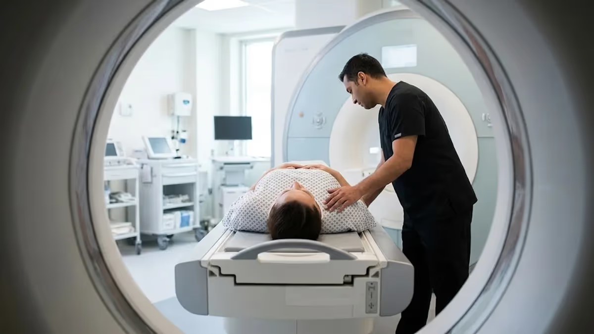
Can you exercise before an MRI? In most cases, yes. Light activity such as a walk or gentle stretching is fine, but vigorous workouts right before your scan can cause problems. Hard exercise raises your heart rate, breathing rate, and core temperature, and it leaves muscles twitchy or sore, which makes lying perfectly still harder. Because image quality depends on stillness, a rushed gym session can blur pictures and force repeated sequences. This guide explains timing, safety rules, and what to expect.
The answer changes with the type of scan. A brain or knee MRI tolerates a morning jog better than an abdominal or pelvic study, where bowel motion and bladder filling influence image clarity. Some exams also require fasting, which makes a hard workout unwise because low blood sugar can leave you shaky or lightheaded. Cardiac stress MRI is a special case, since exercise or medication is used deliberately. Always follow the written instructions from your imaging facility first.
Preparation matters because an MRI scanner uses a strong static magnet, rapidly switching gradient fields, and radiofrequency pulses. Your body absorbs a small amount of that energy as heat, measured as the specific absorption rate. Scanners keep exposure within limits, normally about 2 watts per kilogram for whole-body normal operating mode, but a person who is already overheated and sweating has less margin for comfort. Arriving calm, hydrated, and cool helps the technologist finish on schedule and avoid rescheduling.
Safety screening is the other half of preparation. You will complete a questionnaire about implants, pacemakers, aneurysm clips, shrapnel, tattoos, prior surgeries, kidney function, and pregnancy. Gym gear adds extra risks, because some athletic clothing contains metallic threads, and fitness trackers, patches, and wireless earbuds must come off. Reading about mri preparation and safety before appointment day lets you ask informed questions and avoid surprises at the front desk. It also explains why technologists repeat their screening questions so carefully.
Billing questions come up just as often, especially for women's imaging and pelvic problems. The pelvic mri cpt code your provider selects, 72195, 72196, or 72197, determines what your insurer reviews for authorization. Knowing the difference between scans without contrast, with contrast, and both helps you confirm that your referral, your prior authorization, and your scheduled study all match. Mismatches are a common reason for delayed appointments and unexpected bills, and they are easy to prevent with one phone call.
Location and equipment also shape your experience. If you feel anxious in tight spaces or have a larger body size, searching for the closest open MRI to my location may be a smart first step. Open systems have wider openings and less claustrophobia, though many run at lower field strengths and can need longer scan times. Whether an open or closed scanner suits you depends on the body part, the clinical question, and your comfort. We cover those trade-offs below.
This article walks through each step in order: safe exercise timing, pelvic MRI coding, preparation checklists, open MRI selection, implant and heating safety, and practical tips for scan day. Every section is written for patients in the United States and for students studying for MRI registry exams. Use it as a reference before you book, before you arrive, and before you ask your imaging team questions. Informed patients tend to move more calmly through the scanner, and calm patients produce better images.
Pelvic MRI Prep and Coding by the Numbers
How to Prepare for Your MRI Step by Step
Confirm Your Order and Code
Get Your Instructions in Writing
Plan Light Activity Only
Dress Metal-Free and Gather Records
Arrive Early and Complete Screening
Current Procedural Terminology codes are how US providers describe and bill imaging. For pelvic imaging, three codes cover nearly every study. The pelvic mri cpt code 72195 means magnetic resonance imaging of the pelvis without contrast material. Code 72196 means the study was performed with contrast, and code 72197 means images were acquired without contrast and then repeated with contrast. The American Medical Association maintains these codes, and facilities report them alongside ICD-10 diagnosis codes that justify the scan medically.
The distinction matters because contrast adds time, cost, and screening requirements. A scan without contrast, 72195, often takes 30 to 45 minutes and needs no IV. Codes 72196 and 72197 require an intravenous gadolinium-based agent, a kidney function review for some patients, and typically 45 to 60 minutes on the table. Because the combined study adds a second set of images, 72197 usually carries a higher allowed amount than 72195, though actual payment depends on your plan, site of service, and negotiated rate.
Gynecologic indications often lead to the code searched as mri pelvis w wo gyn cpt, which corresponds to 72197. Providers choose it for suspected endometriosis, adenomyosis, fibroid mapping, complex adnexal masses, cervical or endometrial cancer staging, and congenital uterine anomalies. Radiologists compare pre-contrast and post-contrast images to see which tissue enhances, helping separate solid tumors from blood products or fat. If your order says pelvis with and without contrast, expect an IV to be placed before you enter the scanner.
Related codes can appear on the same claim. MRI of the abdomen is reported with 74181, 74182, or 74183, and a combined abdomen and pelvis study may be billed with both families. Hip imaging uses lower extremity joint codes such as 73721 rather than the pelvic family. Always ask the scheduler for the exact code on your order so the facility can verify coverage with your insurer and so that your referral, your protocol, and your claim all describe the same study.
Prior authorization is where coding errors cause real trouble. Many commercial plans and Medicare Advantage plans require approval before elective MRI, and approval is tied to a specific code. If your doctor ordered 72195 but the radiologist decides contrast is needed, the facility may have to request a change, which can delay the appointment. Call both your insurer and the imaging center a few days ahead, confirm the code, and write down the authorization number and the name of the person you spoke with.
Costs vary widely across the country. Hospital outpatient departments commonly charge several times more than freestanding imaging centers for the same code. Cash prices at independent centers for a pelvic MRI often fall roughly between 400 and 1,500 dollars, while hospital billed charges can run to several thousand. Federal price transparency rules require hospitals to post prices, so you can compare. Asking for a self-pay quote, the radiologist's reading fee, and any separate contrast charge helps you avoid surprise bills.
Understanding the code also helps you prepare physically. Pelvic protocols commonly ask for fasting for four to six hours to reduce bowel motion, and some centers give an anti-spasmodic medication such as glucagon. A moderately full bladder may be requested, though some protocols want it empty. Because instructions differ, never assume. If your order is 72197 for a gynecologic problem, ask whether your scan needs vaginal gel, a rectal preparation, or specific timing within your menstrual cycle, and whether you should avoid exercise that morning.
Pelvic MRI CPT Code Breakdown: 72195, 72196, and MRI Pelvis W Wo GYN CPT 72197
CPT 72195 describes MRI of the pelvis performed without any contrast agent. It is often the first study for pelvic pain, suspected fibroids, hip or bone concerns, bladder abnormalities, and follow-up of known cysts. Because no needle is needed, patients with poor kidney function or contrast concerns can usually undergo this scan. T1 and T2 weighted sequences show anatomy and fluid, and many uterine and ovarian findings are evident without enhancement.
Expect about 30 to 45 minutes on the table, with a coil placed over the lower abdomen and pelvis. You will hear loud knocking sounds, and the technologist will give you headphones or earplugs. Fasting may still be requested to calm the bowel, and an anti-spasmodic injection is sometimes given. This code usually has the lowest cost and the simplest authorization path among the three pelvic MRI codes, which is why many referrals begin here.
Open MRI vs Closed MRI: Is an Open Scanner Right for You?
- +A wider, less enclosed space reduces claustrophobia and anxiety
- +Often accommodates larger body sizes and higher weight limits
- +Lets a parent or support person stay close to a child during the scan
- +Can be easier to tolerate without sedation for nervous patients
- +Some open units allow weight-bearing or positional imaging
- +Often feels less loud and less confining than a narrow tube
- −Many open units run at lower field strength, about 0.3 to 1.2 tesla, with less signal
- −Longer scan times raise the chance of motion blur
- −Small lesions may be harder to see than on 1.5T or 3T systems
- −Fewer centers offer open systems, so travel may be longer
- −Some advanced sequences, such as certain diffusion protocols, may be limited
- −A radiologist may still require a high-field repeat for cancer staging
Pre-Scan Checklist When Searching for the Closest Open MRI to My Location
- ✓Confirm your exact CPT code and authorization number with your insurer.
- ✓Ask the facility about bore size, field strength, and open or wide-bore options.
- ✓Ask whether fasting is required and for how many hours.
- ✓Limit scan-day exercise to a gentle walk or light stretching.
- ✓Avoid intense workouts for several hours before your appointment.
- ✓Follow bladder and drinking instructions exactly as given.
- ✓Wear loose, metal-free clothing without zippers or metallic fabric.
- ✓Leave jewelry, watches, hearing aids, and fitness trackers at home.
- ✓Bring implant cards, prior images, and a current medication list.
- ✓Arrive 15 to 30 minutes early for screening and IV placement.
Keep it light, keep it cool, keep it metal-free
A calm walk before your scan is fine. Intense exercise, heavy sweating, and fasting do not mix well, and an overheated, shaky body moves more and tolerates the scanner less. When in doubt, call your imaging center and ask what they recommend for your specific exam.
Many patients type closest open mri to my location into a search engine the night before a scan, often after a previous claustrophobic experience. Start with your referring provider, because an open scanner is only useful if it can answer the clinical question. Ask whether your exam can be performed on an open system, a wide-bore system, or a standard closed bore. Then use insurer directories and facility websites to compare drive time, price, and appointment availability.
Open MRI is not one thing. Traditional open units use a C-shaped or dual-pillar magnet with field strengths near 0.3 to 1.2 tesla, while wide-bore closed scanners have openings around 70 centimeters at 1.5 or 3 tesla. Many people who fear tight tubes do very well in a wide-bore scanner with a short tunnel, ceiling lighting, and a mirror. Ask the facility for the bore diameter and field strength rather than relying on a marketing label alone.
For pelvic imaging in particular, field strength matters. Gynecologic and prostate protocols rely on thin slices, diffusion-weighted imaging, and dynamic contrast enhancement, and high-field systems deliver these with better signal. A low-field open scanner may be acceptable for general pelvic pain or hip issues but less ideal for cancer staging or small lesion detection. If the order is for 72197, call ahead and confirm the center regularly performs that exact protocol and has radiologists who read pelvic studies.
Check credentials and accreditation. Look for American College of Radiology accreditation, which reviews equipment, personnel qualifications, and image quality. Ask whether technologists are ARRT-certified in MRI and whether a board-certified radiologist, ideally with body or women's imaging experience, interprets the study. These details affect diagnostic confidence far more than the bore shape. A well-run closed scanner with an experienced team can outperform a poorly maintained open unit, even for a patient who feels nervous.
Compare logistics next. Freestanding centers often schedule within days and cost less than hospital departments, while hospital sites may be needed for patients requiring sedation, anesthesia, or complex monitoring. Ask about weekend and evening slots, parking, whether prior images must be sent in advance, and how quickly results go to your doctor. If your doctor prescribes antianxiety medication, bring a driver. Our guide to choosing an imaging facility offers a deeper comparison checklist for patients weighing several centers.
Anxiety strategies can make a standard scanner manageable. Request a prescription for a mild antianxiety medication from your doctor if appropriate, practice slow breathing, and ask for a blanket, a cushion under your knees, and a cloth over your eyes. Most scanners have a call button, and technologists can speak with you between sequences. Many centers offer a brief tour or a mock scanner. Knowing that you can stop the scan at any time restores a sense of control.
If you do choose an open scanner, set expectations. Scan times may be longer, sometimes by 50 percent or more, and staying still becomes even more important because motion blur has more time to accumulate. A radiologist may still recommend repeating the study on a high-field system if images are not diagnostic. Discussing that possibility ahead of time prevents frustration, and it helps you avoid paying for two studies when one well-chosen scanner would have answered the question.
The MRI magnet is always on, and undisclosed metal, implants, or medication patches can cause burns, device malfunction, or serious projectile injuries. Report every surgery, implant, tattoo, and fragment of metal, even if it seems minor or happened years ago.
MRI is safe for most people because it uses no ionizing radiation, but the magnet is always on, even when no one is being scanned. That is why safety screening is strict. Ferromagnetic objects can be pulled toward the bore at high speed, and this projectile effect has caused serious injuries and deaths. Screening covers every item in your body and every item you carry, and the process continues from the scheduling call through the moment you enter the scanner room.
Implanted devices are the biggest concern. Many modern pacemakers, defibrillators, neurostimulators, and joint replacements are labeled MR Conditional, meaning they can be scanned under specific field strength, gradient, and programming conditions. Older leads, cochlear implants, some aneurysm clips, and retained metal fragments in the eye may rule out the scan entirely. Bring your device card, model number, and implant date. Your imaging team may need to contact the manufacturer or cardiology clinic to arrange safe programming first.
Heating and burns are the second major risk and connect directly to the exercise question. Radiofrequency energy can warm the skin, particularly where skin touches skin, cables, or conductive material. Sweat, damp clothing, and a high body temperature increase discomfort, and loops formed by crossed arms or touching thighs can concentrate current. Technologists use padding and positioning to prevent loops. Tell them right away if you feel warmth, tingling, or burning, and never wait until the sequence ends.
Transdermal patches, some medicated plasters, and even certain cosmetics contain metal particles that can heat. Tattoos with iron-oxide pigments may feel warm, and permanent eyeliner has caused rare irritation. Some compression garments, athletic shirts with silver threads, and sports bras with underwire or metal clasps are also unsafe. Changing into a facility gown eliminates most of these risks. Remove hair accessories, dentures, and piercings, and keep credit cards away, since magnets can wipe magnetic strips and damage phones.
Contrast safety is another screening topic. Gadolinium-based agents are generally well tolerated, with allergic-like reactions occurring in a small fraction of a percent of injections. Patients with severely reduced kidney function face a rare condition called nephrogenic systemic fibrosis, mostly linked to older linear agents. Facilities may check kidney function for older patients and those with kidney disease, diabetes, or hypertension. Breastfeeding can usually continue after contrast because the infant absorbs very little, but discuss any concerns with your physician.
Pregnancy needs a careful conversation. MRI without contrast is considered acceptable in any trimester when clinically necessary, but gadolinium is generally avoided during pregnancy unless it will significantly change management. Tell the technologist if you might be pregnant, and let your physician explain the benefits. Pelvic imaging for suspected appendicitis, placental problems, or masses during pregnancy often uses noncontrast protocols for this reason. Never hide a possible pregnancy to avoid rescheduling, because decisions are made with your safety in mind.
Noise and hearing protection also matter. Scanner sounds can exceed 100 decibels, so foam earplugs or headphones are required. Children, older adults, and patients with hearing devices need individualized plans. Real-world accident reports, including burns and projectile events, are explored in our guide to MRI safety and accidents. Most incidents involve a missed screening step rather than equipment failure, which is why honest, complete answers on your form are your strongest protection against harm.
The week before your scan, handle paperwork first. Confirm the appointment time, address, and CPT code, and ask whether you need to arrive early for registration. Request any prior images and reports from other facilities so the radiologist can compare. Review your medication list and ask the ordering provider about diabetes medication, blood thinners, or anxiety medicine. Write down your questions in advance. A short checklist now avoids long delays and canceled studies on the day of your appointment.
Plan your exercise around the scan. Regular workouts earlier in the week are fine and may even help you relax. On scan day, a 20 to 30 minute walk or gentle yoga is reasonable if you are not fasting and feel well. Skip heavy lifting, long runs, hot yoga, and high-intensity classes for at least several hours beforehand, and ideally the full morning. Hard exercise raises body temperature and leaves muscles shaky, which makes holding still more difficult.
Food and drink instructions vary. For many abdominal and pelvic exams, you will be asked to avoid solid food for four to six hours, though water is typically allowed unless you are told otherwise. Avoid caffeine if your center says so, since it can stimulate the bowel and increase heart rate. Diabetic patients should plan fasting around insulin or medication with their physician. If your protocol asks for a full bladder, drink the recommended amount and avoid emptying it before the scan.
Dress for comfort and safety. Choose loose cotton clothing without zippers, metal buttons, or reflective fabrics, or plan to change into a gown. Leave jewelry, watches, hairpins, and smartwatches at home. Wear socks, since scan rooms are cool, and ask for a blanket. Bring a list of your medications and your implant card. Leave valuables behind or use a locker, because phones, keys, wallets, and credit cards cannot enter the scanner room.
During the scan, your main job is to breathe normally and relax. Muscles that tense during stress or discomfort cause motion. Ask for a pillow under your knees, and tell staff if the table is uncomfortable before the scan starts. When you hear breath-hold instructions, practice them in advance. Focus on slow breathing, count your breaths, or imagine a calm scene. Each sequence lasts from a few seconds to several minutes, and the technologist announces the length.
After the scan, you can usually resume normal activity immediately, including exercise, unless you received sedation. If you had contrast, drink fluids and report any rash, swelling, or pain at the IV site. The radiologist typically sends a report to your ordering provider within one to three business days, sometimes faster for urgent studies. Ask your provider how results will be shared. Patient portals often release reports quickly, so be ready to discuss findings with your doctor rather than interpreting them alone.
If you study MRI technology, the same habits apply to your exam preparation. Learn the safety zones, the difference between MR Safe, MR Conditional, and MR Unsafe, and how SAR, gradient fields, and contrast affect patients. Practice questions help you retain these details, and quizzes reveal weak spots early. Review CPT basics, patient screening workflows, and physics together, because registry questions often combine them. Consistent short sessions beat last-minute cramming for most learners.
MRI Questions and Answers

MRI Center: How to Choose the Right Imaging Facility

Brachial Plexus MRI: Complete Guide to Imaging, Anatomy, Pathology, and Clinical Applications
Chiari Malformation MRI: Complete Patient Guide

MRI Coil: Complete Guide to Types, Functions, and Clinical Applications

MRI Safety and Accidents: Real Cases, Risks, and Screening Failures
How this page was made: written with AI tools and published by Nathan Beja, founder of Practice Test Geeks, who is responsible for its content. Exam facts are checked against the official sources listed below. Not reviewed by a subject-matter expert. Editorial policy
Sources & references
Exam facts on this page (fees, format, dates, passing scores) are checked against these official sources; the date is our last check. Editorial policy
- arrt.org — Passing score, score release, fees, exam information · verified Oct 2026
- assets-us-01.kc-usercontent.com — Renewal, score validity, test delivery, eligibility, exam format, test dates, time limit · verified Oct 2026