
MRI Safety and Accidents: Real Cases, Risks, and Screening Failures
MRI accidents: 💯 ferromagnetic projectile cases, burns, the man sucked into MRI machine, FDA MAUDE reports, pacemakers, and what every patient should check.
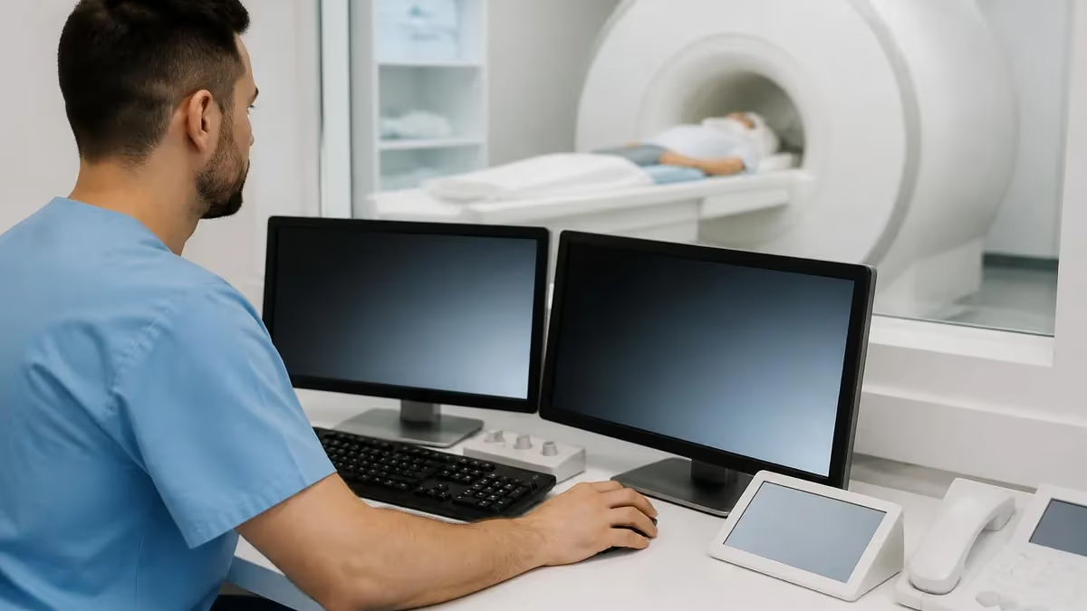
MRI accidents are rare, but the ones that happen are unforgettable — a 6-pound oxygen tank flying across a room at 40 mph, a metal floor buffer pinned to the scanner bore, a man sucked into MRI machine while delivering a chair to a patient. The American College of Radiology estimates fewer than one serious adverse event per 100,000 scans, but the FDA's MAUDE database catalogs them in detail, and the patterns repeat.
Roughly half of reported MRI accidents involve ferromagnetic objects entering Zone IV. About a third involve burns from coils, leads, or jewelry that the screener missed. The rest are split between contrast reactions, implant displacement, and acoustic injuries.
What makes MRI accidents distinctive among medical errors is that the underlying physics doesn't care about good intentions. A 1.5 Tesla scanner generates a static field roughly 30,000 times stronger than the Earth's magnetic field, and it's always on — even when no scan is running. Walk into Zone IV with a steel wrench in your back pocket and the wrench will pull you across the room, not the other way around. This is the failure mode that screening protocols exist to prevent, and it's also the failure mode that occasionally slips through.
The most widely cited single fatal MRI accident in U.S. history happened in 2001 at Westchester Medical Center, when a six-year-old boy named Michael Colombini was struck by a ferromagnetic oxygen tank that an anesthesiologist had brought into the scan room. The tank was pulled from the anesthesiologist's hands and accelerated into the scanner, striking the patient's head. Michael died two days later. The case reshaped MRI safety protocols nationwide — Zone IV access controls, mandatory ferromagnetic screening with handheld detectors at major centers, and ACR's tiered safety officer roles all trace back partly to this incident.
More recently, in 2018, a man at a Mumbai hospital was killed when he carried an oxygen cylinder into Zone IV after a relative directed him to bring it — a screening breakdown rather than an equipment failure. The case demonstrated that even well-established safety protocols fail when staff don't enforce them at the door.
Burns are the second-largest category of MRI accidents, and they're almost always preventable. The scanner's radiofrequency pulses can heat conductive loops — a patient's hand resting on the opposite arm forming a closed loop, an EKG lead coiled instead of straight, a transdermal medication patch with metallic backing, a tattoo with iron-oxide pigment. The FDA's database has hundreds of reports of second-degree burns from these mechanisms. The fix is screening: pre-scan questions about patches, metal jewelry, recent tattoos, and conductive medical devices, plus visual inspection by the technologist before the patient enters the bore.
This guide walks through the major MRI accident categories with specific cases from the FDA MAUDE database and published case reports, the screening failures that allowed each one, the safety zones and protocols that exist to prevent them, and the questions every patient should ask before a scan. It's intended for both patients trying to understand what they're walking into and for healthcare staff (technologists, nurses, ordering physicians) who need a practical refresher on the failure modes that matter.
MRI Safety: 2026 Statistics and Facts
- Reported serious adverse events: Roughly 1 per 100,000 scans (ACR estimate). Over 30 million MRI exams annually in the U.S.
- Most common accident type: Ferromagnetic projectile incidents (~50% of major reports)
- Burns: ~30% of reports — usually preventable with proper coil positioning and screening
- Implant complications: ~15% of reports — pacemakers, neurostimulators, cochlear implants, certain aneurysm clips
- Static field strength: 1.5T scanner ≈ 30,000x Earth's magnetic field; 3T ≈ 60,000x
- Field is always on: Unlike CT/X-ray, MRI magnetic field doesn't shut off between patients — Zone IV access controls must be active 24/7
The man sucked into MRI machine cases get the most public attention, and there are more of them than most people realize. The 2018 Mumbai case mentioned above is the most widely reported fatal incident in recent years. But the FDA MAUDE database documents non-fatal cases regularly — janitors pulled across scan rooms by floor buffers, patients pulled by hairpins or jewelry, contractors with toolbelts pinned to scanners during construction.
The 2014 case at the AIIMS hospital in Delhi involved a patient who entered Zone IV with an oxygen cylinder; the cylinder embedded itself in the scanner bore but the patient survived. The 2020 case at a New Zealand hospital involved a 60-year-old patient pinned against the scanner by her own clothing buttons that turned out to be ferromagnetic.
What ties these cases together is screening failure — not equipment failure. The magnetic field works exactly as designed; the breakdown is at the door. Modern Zone IV access protocols require a trained MRI technologist (the Level II safety person in ACR terminology) to physically clear every individual entering the scan room. Handheld ferromagnetic detectors, used at most modern centers, can identify metal objects that the patient doesn't know about — including some surgical implants the patient forgot to mention. But detectors are tools, not policies. The center has to actually use them.
The cases where screening fails tend to share patterns: emergency situations where staff cut corners (the Westchester case involved an emergency oxygen need), facility staff who don't normally work in MRI but enter for delivery or maintenance, contractors who haven't been trained on the safety zones, and patient interactions where the language barrier or cognitive state made screening questions unreliable. Most accident root-cause analyses identify policy, not technology, as the failure point.
Burns deserve more attention than they typically get, because they're the most frequent MRI injury and almost always preventable. RF heating happens when the scanner's radio pulses induce currents in conductive loops. A patient's hand resting on the opposite shoulder forms a loop. An EKG lead coiled into a circle forms a loop. A transdermal pain patch with metallic backing forms a small loop. Tattoo ink with iron-oxide pigment can heat enough to cause first-degree burns. The technology and the physics are well-understood; the variability is in patient positioning and screening rigor.
Practical examples from the FDA database: a patient with a Lidocaine patch developed second-degree burns under the patch during a 45-minute spine MRI. A patient with a fentanyl patch suffered similar burns. A patient with a heart monitor (Holter monitor) that wasn't properly removed before the scan suffered both burns and equipment damage. A patient who positioned her ankles touching during a pelvis MRI had bilateral lower-leg burns from the closed loop. These cases all happened despite written screening protocols — the protocols failed at the bedside.
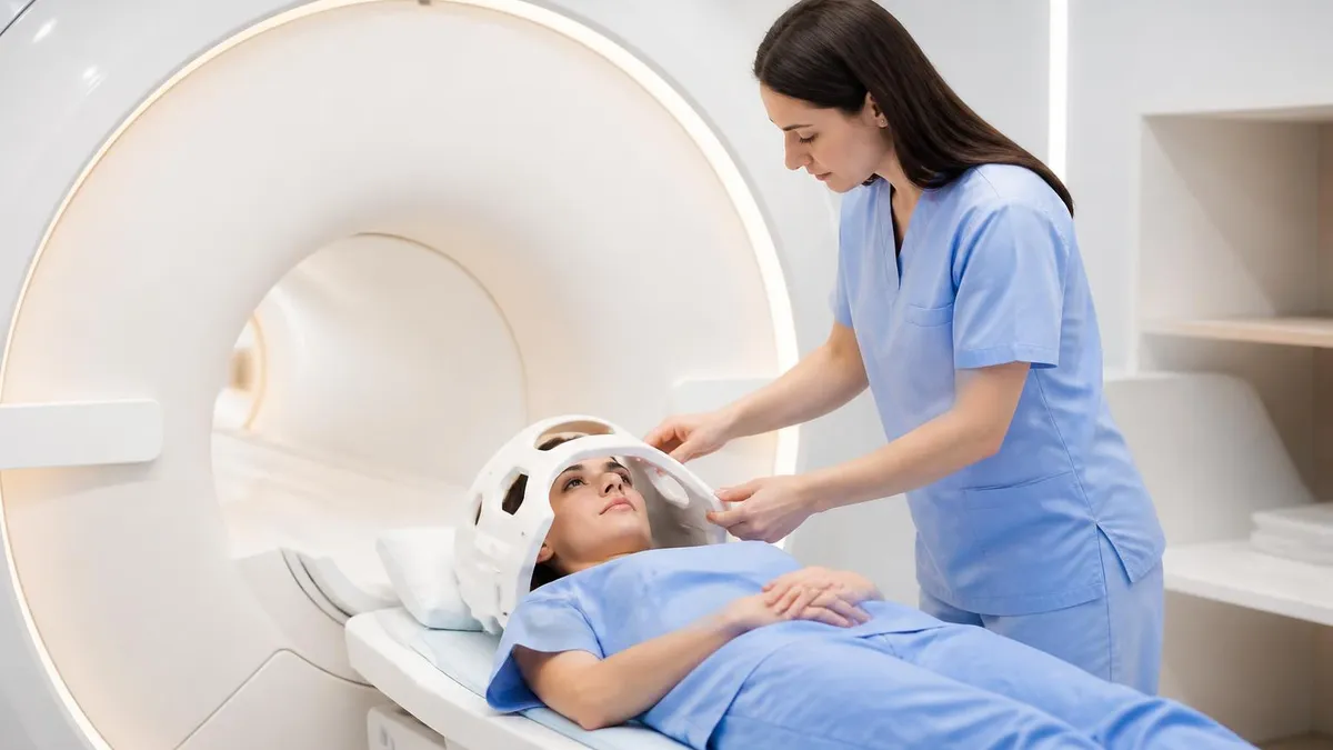
Major MRI Accident Categories
Most dangerous category. Steel objects pulled into Zone IV become projectiles. Oxygen tanks, IV poles, scissors, hairpins, jewelry, clothing buttons, contractor tools. Screening at Zone IV access is the primary defense.
Skin burns from conductive loops formed by patient positioning, coiled leads, transdermal patches, or jewelry. Usually second-degree. Preventable with proper coil placement and pre-scan inspection.
Aneurysm clips made before 1995, certain pacemakers, neurostimulators, cochlear implants. Some implants are conditionally safe at certain field strengths only. Patient history is critical.
Gadolinium-based contrast agents can cause allergic reactions and, rarely, nephrogenic systemic fibrosis (NSF) in patients with poor kidney function. Screening for eGFR is standard.
MRI scanners produce 100-130 dB during sequences. Hearing protection is mandatory. Inadequate protection can cause temporary or permanent hearing loss.
Not technically an accident, but a real adverse event. Can cause patients to move during scan, requiring sedation or repeat imaging. Open-bore and 3T short-bore scanners help.
The ACR MRI safety zone system is what most major U.S. hospitals follow, and it's worth understanding even as a patient. Zone I is unrestricted public access — the hospital corridor leading to the imaging department. Zone II is a transition area, typically where patients change clothes and complete screening forms. Zone III is the access-controlled area immediately outside the scan room — only trained staff and screened patients pass through. Zone IV is the scan room itself, where the magnetic field is always active and access must be physically controlled.
Where is MRI restriction zone 4? It's the scan room — the room containing the MRI scanner. The boundary of Zone IV is the physical doorway into that room, and the door should be locked or staffed continuously. Modern facilities use card-access systems, interlocks tied to the scanner's RF cage door, and in some cases handheld or doorway ferromagnetic detectors that alert staff to metal entering the zone.
The strict definition matters because the magnetic fringe field extends beyond the door — typically 5-10 feet for a 1.5T scanner — and ferromagnetic objects can be pulled in from outside the technical boundary.
This is why the access protocol uses the door as the control point even though the physics doesn't have a sharp boundary there.
Where should I run a code in the MRI environment? Outside Zone IV. If a patient in the scanner suffers cardiac arrest or other emergency, the standard protocol is to remove the patient from the bore using the table's emergency release, exit Zone IV with the patient, close the Zone IV door behind you, and then begin resuscitation in Zone III or in a code response area equipped with normal medical equipment.
Code carts contain ferromagnetic items (oxygen tanks, monitor leads, intubation equipment) that cannot enter Zone IV under any circumstances. Running a code inside Zone IV with a normal code cart is exactly the type of emergency-mode protocol breakdown that has caused fatal accidents. ACR safety guidelines are explicit on this point and most centers run periodic drills.
Implant screening is the area where patient cooperation matters most. Patients with pacemakers, defibrillators (ICDs), neurostimulators, deep-brain stimulators, cochlear implants, insulin pumps, or programmable shunts need to disclose every device, ideally with the manufacturer model number.
Many of these devices are now "MR-conditional" — they're safe at specific field strengths and under specific scanning parameters, but only if those parameters are followed. Older devices, especially pre-2010 pacemakers, are often MR-unsafe and require careful evaluation before any scan. The risk is real: a non-MR-safe pacemaker exposed to the static field can have its programming altered or be physically displaced, which can be fatal.
Tattoos with iron-oxide pigments are a smaller but real risk. The FDA receives a few dozen burn reports annually involving tattoos. Modern professional tattoo inks largely avoid iron-oxide pigments, but older tattoos, amateur tattoos, and tattoos from certain regional sources may contain enough ferromagnetic material to heat up. Permanent makeup (eyeliner, eyebrow tattoos) is similar. The risk is generally limited to first-degree burns rather than serious injury, but the technologist should know about all tattoos before the scan, especially large ones in the imaging area.
For decades, pacemakers were an absolute contraindication for MRI. That changed starting around 2008 with the introduction of "MR-conditional" pacemakers from Medtronic, Biotronik, and Boston Scientific. These devices can be scanned at 1.5T (some at 3T) when specific conditions are met: the device must be programmed to a specific MR mode before the scan, the scan parameters must stay within defined limits, and a cardiologist must be involved in the pre-scan evaluation. Even with MR-conditional pacemakers, the screening process is involved — not a casual decision. mri and pacemakers compatibility depends on the specific model, and patients should bring their device implant card to the MRI department before scheduling.
Stents are a common patient concern but generally lower risk than people assume. Coronary stents implanted since roughly 2000 are made of MRI-compatible materials (316L stainless steel, nitinol, cobalt-chromium alloys) and are considered safe at 1.5T and 3T after the initial healing period. ACR guidelines generally allow MRI immediately after stent placement at 1.5T, and after 6 weeks at 3T for some older stent designs. mri and stents compatibility is well-documented; the manufacturer's implant card or surgical record will identify the specific stent model if there's any doubt.
Aneurysm clips are a different story. Older ferromagnetic clips (used widely until the mid-1990s) can be displaced or rotated by the magnetic field, potentially causing serious neurological injury. Modern titanium and titanium-alloy clips are MR-safe at 1.5T and most 3T fields. But the safety determination depends on the specific clip — patients with intracranial aneurysm clips need to have the clip's manufacturer and model verified before any MRI is scheduled. "My doctor said it's fine" is not adequate documentation when the clip is sitting inside someone's skull near a critical artery.
Dermal piercings and surface piercings are an underappreciated issue. Standard ear, nose, and navel piercings can usually be removed for a scan, but dermal piercings (anchored under the skin) and certain surface piercings cannot be easily removed and may need to be left in place. If the jewelry is non-ferromagnetic (titanium, niobium, certain stainless steels), it's generally safe.
If it's ferromagnetic, it presents a heating risk and potentially a displacement risk depending on the anchor's geometry. mri and dermal piercings guidance varies by facility — some will scan with the piercing in place after testing it with a handheld magnet, others will postpone the scan until the piercing can be replaced with non-ferromagnetic jewelry.
Braces and orthodontic appliances are another common question. Modern braces are typically non-ferromagnetic stainless steel and are MR-conditional — meaning they don't pose a major safety risk, but they can cause significant image artifacts. mri and braces on teeth usually means a successful scan with some image degradation in the area of the braces; the brain and most body imaging is unaffected.
If the scan is of the head or neck (and especially if you're imaging the jaw, sinuses, or skull base) the artifacts can make the scan less useful. In those cases the radiologist or ordering doctor may want the braces removed temporarily, but for most other imaging, braces stay in place.
Patient screening forms should ask about: surgical history (any implants, clips, plates, screws), tattoos and permanent makeup, body piercings, medication patches, pregnancy status, eye injuries involving metal, hearing aids, dentures and bridgework, and any prior MRI experiences. The form is not a formality — every question is on it because that exact answer has caused a problem at some point. Patients who fill it out quickly without thinking are exactly the patients who later turn out to have a forgotten metal fragment in an eye or a pacemaker they didn't mention. Slow down on screening forms.
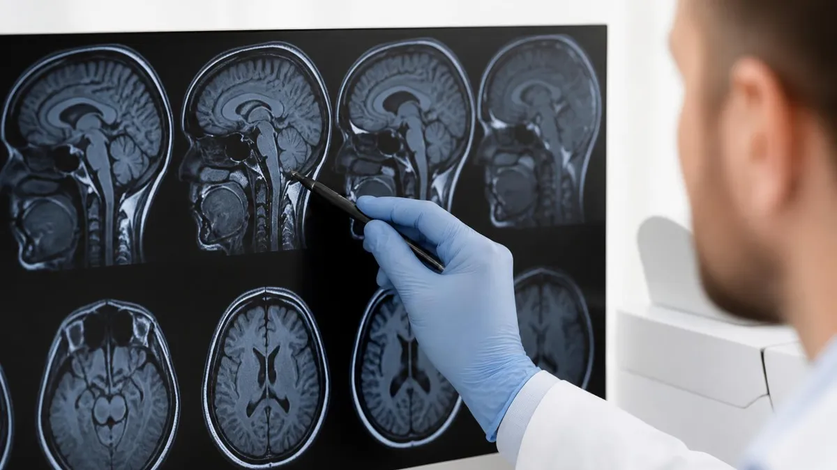
MRI Risks by Implant or Object Type
- Pre-2008 devices: Generally considered MR-unsafe. Most cardiologists and radiologists will not scan these patients without extensive review.
- MR-conditional pacemakers (2008+): Medtronic Revo MRI, Biotronik ProMRI, Boston Scientific Ingenio MRI, and successors. Safe at 1.5T (some 3T-rated) with proper programming.
- Required steps: Cardiology pre-evaluation, device reprogramming to MR mode, scan parameter limits, post-scan reprogramming.
- ICDs (defibrillators): Similar conditional safety profile. More devices are MR-conditional than patients realize.
- Patient action: Bring the implant identification card to scheduling. The model number determines the safety profile.
What about mri accident video footage online? Search results turn up dramatic videos of test demonstrations (hospital staff intentionally bringing items into Zone IV to show what happens) and a handful of actual accident reconstructions. The educational videos are genuinely useful for understanding the force involved — watching a steel pipe pulled across a room at 30+ mph drives home why screening matters. The reconstructions, often from FDA or hospital safety committee reports, walk through specific accidents and the screening failures that allowed them.
The famous "oxygen tank at Westchester" reconstruction from the 2001 fatal accident is one of the most-shown teaching videos in MRI safety training. It typically shows a small ferromagnetic oxygen cylinder being released into Zone IV and accelerating into the scanner bore — illustrating both the force and the trajectory that injured Michael Colombini. Most major centers show this video during MRI staff onboarding.
Patient-facing educational videos at scan facilities cover the same material at a less technical level, focusing on what the patient needs to do (remove all metal, fill out screening forms accurately, hold still during the scan, communicate via the squeeze ball if there's a problem).
The MRI safety information that actually changes patient outcomes is generally not the dramatic projectile content but the screening checklist. Did you complete the form honestly? Did you mention every implant, patch, and jewelry item? Are you wearing only the provided gown — no street clothes with hidden metal fasteners? Did you take out all jewelry, including dermal piercings if you have them? Did you confirm your pregnancy status (gadolinium contrast in early pregnancy has potential fetal effects)? Did you discuss any claustrophobia concerns before the scan so the technologist can plan for it?
The technologist's job during the scan includes monitoring you continuously via camera and microphone. If you feel anything unusual — burning, tingling, pain, sudden anxiety — squeeze the call button. The scan can be stopped immediately. Patients sometimes hesitate to interrupt a scan because they don't want to delay the imaging or seem difficult. This is exactly wrong; the technologists want to know immediately if anything feels off. Interrupting a scan because your hand started tingling is the right call. Continuing because you're trying to be polite is how minor issues become injuries.
The post-scan questions matter too. Did you feel any unusual heat during specific sequences? Any sensation under jewelry, tattoos, or patches that you weren't sure about? Any unusual sounds (the loud knocking is normal, but anything sharp or different from sequence to sequence is worth mentioning)? Most facilities ask these questions as part of discharge, but the questions are easy to brush off. Take a minute.
Patient Steps for a Safe MRI
Complete the screening form honestly
Bring implant documentation
Remove all metal and patches
Wear the provided gown
Communicate during the scan
Report anything unusual after the scan
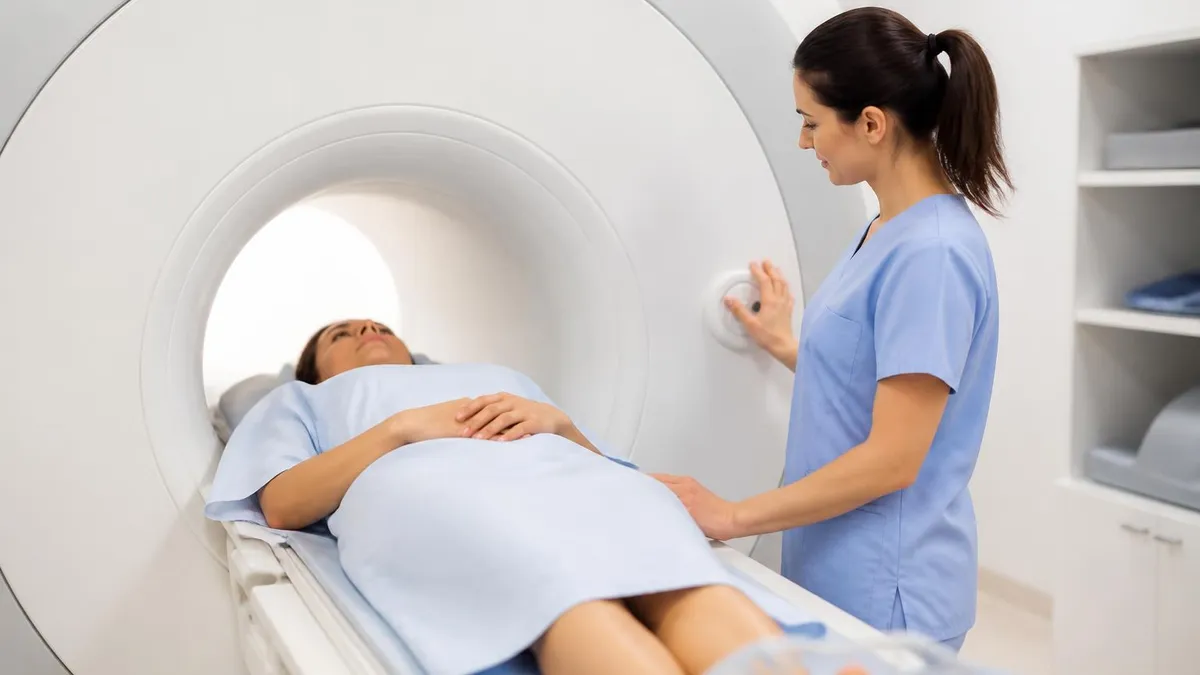
Gadolinium-based contrast agents (GBCAs) used in MRI are generally well-tolerated, but two issues matter: (1) Nephrogenic Systemic Fibrosis (NSF) is a serious complication that can occur in patients with severe kidney dysfunction (eGFR <30). Screening for kidney function is standard before contrast. (2) Gadolinium deposition in brain tissue has been observed after repeated contrast exams, especially with linear (vs. macrocyclic) agents. FDA guidance now favors macrocyclic agents and recommends limiting unnecessary repeated contrast. Patients with prior gadolinium reactions, severe kidney disease, or pregnancy should discuss contrast specifically with the ordering physician.
The FDA MAUDE database is a public record of medical device adverse events, including MRI-related incidents. Searching it for MRI accidents returns thousands of reports across the past decade — most are minor (small burns, anxiety reactions, equipment malfunctions) but the serious ones are documented in detail. The reports include the device manufacturer's investigation, the facility's response, and any FDA follow-up. They're an underused resource for understanding real-world failure modes.
Reading through MAUDE reports reveals patterns that don't show up in textbook safety training. The most common screening failure isn't ignorance — it's time pressure. Late-day scans where the technologist is rushing to finish before the shift ends. Add-on emergency scans that bypass normal screening. Trauma patients who can't reliably answer questions. Pediatric patients whose parents may not know full implant history. Patients with cognitive impairment or language barriers. These are the categories where screening fails most often, and they're predictable — facilities that recognize them and build extra checks for these situations have better safety records.
The other pattern that shows up repeatedly is staff turnover. Many of the serious incidents involve newer staff members — within the first few months at a facility — who hadn't fully internalized the Zone IV access protocols. Standard MRI safety training works, but it takes repetition and reinforcement.
The Westchester case noted that the anesthesiologist who brought the oxygen tank into the scan room was newly assigned to MRI duties. The Mumbai case involved a relative who'd never been trained on MRI safety. Both followed instructions that seemed reasonable given their training level — and both were standing at Zone IV with ferromagnetic objects.
For patients looking at mri accidents pictures or news coverage of specific incidents, the takeaway shouldn't be that MRI is dangerous. Over 30 million scans happen each year in the U.S., and serious adverse events run roughly 1 per 100,000 — comparable to or lower than the adverse event rate for many other common medical procedures.
The risk is real but the absolute rate is low, and almost all serious incidents trace back to identifiable screening failures rather than inherent equipment risk. A center with rigorous screening, trained safety officers, ferromagnetic detection at Zone IV, and a culture that treats safety protocol violations seriously is essentially safe for routine scans.
The pre-scan conversation with your technologist is your last opportunity to surface concerns. Don't rush through it. Don't downplay an old surgery, a forgotten piercing, or a medication patch you put on yesterday. Don't say "it's probably fine" when you're not sure — say "I'm not sure, can you check?" That single phrase is what separates safe scans from the rare incidents in the FDA database. The technologist has tools (handheld detector, manufacturer databases, radiologist consultation) to resolve uncertainty quickly. You just need to flag it.
What to Verify Before Your MRI
Pacemaker, ICD, neurostimulator, cochlear implant, aneurysm clip, joint replacement, stent — bring the manufacturer's implant card. Model number matters.
Any prior surgery involving plates, screws, or wires? Bring the surgical record if you can. Pre-2000 hardware especially needs verification.
Mention all tattoos, especially in the area being scanned. Permanent makeup (eyeliner, eyebrows) counts. Iron-oxide pigments can heat.
Fentanyl, lidocaine, nicotine, hormones, glucose monitors. All transdermal patches with metallic backing must be removed before scan.
Confirm pregnancy status before scheduling. Most non-contrast MRI is considered safe in pregnancy, but gadolinium contrast has potential fetal effects.
Welding, grinding, or metalworking occupational history? You may need an orbital X-ray before MRI to check for embedded fragments.
MRI Pros and Cons
- +MRI has a publicly available content blueprint — you know exactly what to prepare for
- +Multiple preparation pathways accommodate different schedules and budgets
- +Clear score reporting shows specific strengths and weaknesses
- +Study communities share current insights from recent test-takers
- +Retake policies allow recovery from a difficult first attempt
- −Tested content scope requires substantial preparation time
- −No single resource covers everything optimally
- −Exam-day performance can differ from practice test performance
- −Registration, prep, and retake costs accumulate significantly
- −Content changes between versions can make older materials less reliable
MRI Questions and Answers
Final word on MRI safety: the failure modes are well-documented and most accidents are preventable. The risk profile is lower than most patients realize — over 30 million scans per year in the U.S. with serious adverse events running about 1 per 100,000.
The risk that does exist concentrates in identifiable screening failures: ferromagnetic objects entering Zone IV without clearance, conductive loops on the patient's body that aren't repositioned before the scan, implants that the patient didn't disclose or that the facility didn't verify, and contrast administered without checking kidney function. Each of these failure modes has a screening defense that works when staff and patients both engage with it.
For patients, the practical message is simple: take the screening form seriously, bring your implant documentation, mention every piercing and tattoo and patch, and speak up during the scan if anything feels off. For healthcare staff, the message is also straightforward: maintain Zone IV access discipline even during emergencies, use ferromagnetic detectors when available, double-check implant identification rather than assuming, and treat new staff orientation as a recurring requirement rather than a one-time event. The accidents that show up in the FDA database have repeating patterns — and the patterns are addressable with the protocols that already exist.
About the Author

Medical Laboratory Scientist & Clinical Certification Expert
Johns Hopkins UniversityDr. Sandra Kim holds a PhD in Clinical Laboratory Science from Johns Hopkins University and is certified as a Medical Technologist (MT) and Medical Laboratory Scientist (MLS) through ASCP. With 16 years of clinical laboratory experience spanning hematology, microbiology, and molecular diagnostics, she prepares candidates for ASCP board exams, MLT, MLS, and specialist certification tests.
Join the Discussion
Connect with other students preparing for this exam. Share tips, ask questions, and get advice from people who have been there.
View discussion (8 replies)