
Weight Limit for MRI Machine: Table Ratings, Bore Sizes, and Open MRI Options
🎯 Weight limit for MRI machine explained: typical 350–550 lb table limits, bore sizes, open MRI options, plus pelvic MRI CPT code basics for US patients.
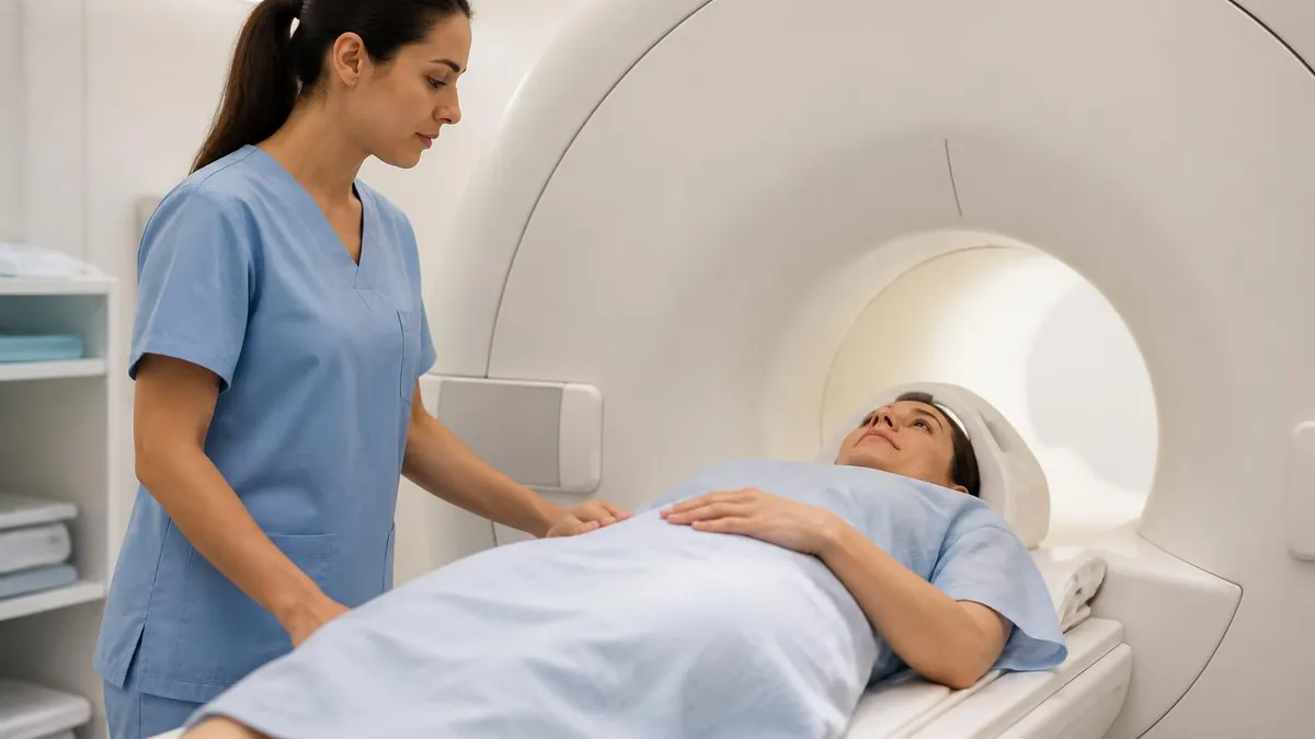
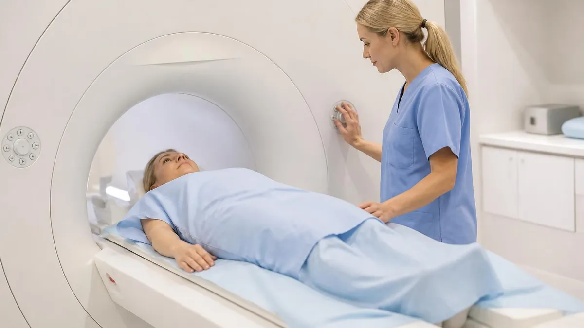
The weight limit for MRI machine tables is one of the first questions larger patients ask, and the honest answer is that it depends on the scanner. Most clinical systems in the United States list a patient table capacity between 350 and 550 pounds, while a smaller number of wide-bore and bariatric-ready units go higher. Knowing your number before you book prevents cancelled appointments, wasted trips, and the stress of being turned away at the front desk.
Weight is only half of the equation. The scanner's bore, the tunnel you slide into, limits how much body girth fits, and a person under the table rating can still be too wide for a 60 cm opening. Technologists therefore weigh both numbers together with your height and the body part being scanned. This guide covers table ratings, bore diameters, open MRI alternatives, and ways to find a compatible facility without a dozen phone calls.
Manufacturers publish these limits in site planning documents, and imaging centers repeat them in their scheduling notes. A scanner that advertises a 70 cm bore and a 550 pound table is not automatically available at your local hospital, because many sites still run older 1.5 tesla systems bought a decade ago. Equipment age, not just brand, explains why two facilities in the same city can give you completely different answers about the same scan.
Understanding the machine also helps you read your paperwork. When a physician orders a scan, the order carries a billing code. A pelvic mri cpt code such as 72195, 72196, or 72197 tells the facility which region and contrast protocol to schedule, which in turn affects how long you lie on the table. Longer exams change comfort, positioning, and whether a wide-bore room is the smarter choice for your body.
If you want a broader view of how scanners differ from other imaging tools, our comparison of mri machine specifications and CT hardware explains why MRI relies on a powerful magnet instead of radiation, and why that design creates different physical limits. A CT gantry and an MRI bore are not interchangeable, so a weight rating that works for one scanner may not apply to the other at all.
Throughout this article you will find practical numbers rather than vague reassurance. We cover typical table ratings by scanner class, what happens when a patient exceeds them, which open and wide-bore options exist, and how to prepare for the call to the scheduling desk. We also show how pelvic exams are coded, including the gynecologic variant people search as mri pelvis w wo gyn cpt, so your insurance paperwork matches the scan you receive.
Everything here is general education for United States patients and students, not medical advice. Always confirm the exact limit with the specific scanner your facility will use, because one center may operate three machines with three different ratings. A short call that names your weight, height, and body region gets a reliable answer within minutes and keeps your appointment on track, whether you are booking a spine, knee, brain, or pelvic study.
MRI Weight Limits by the Numbers
How to Confirm Your Scanner's Weight Limit
Know Your Real Numbers
Ask the Scheduler for the Model
Verify Against the Manufacturer
Request Written Confirmation
Have a Backup Plan
A table weight rating is an engineering limit, not a suggestion. The patient table must hold a static load, glide in and out of the magnet on motors, and stop within a millimeter of the programmed position. Typical ratings fall into bands: older 1.5 tesla closed systems often list 350 to 450 pounds, mainstream wide-bore scanners list 440 to 550 pounds, and some bariatric configurations advertise 600 pounds or more.
Bore diameter is the second specification that decides who fits. A conventional bore measures about 60 centimeters, roughly 24 inches, while wide-bore systems open to 70 centimeters, about 28 inches. Those extra ten centimeters transform the experience for larger patients, people with shoulder limitations, and anyone anxious about confined spaces. Some newer designs push to 80 centimeters, though they remain uncommon outside major academic centers and a handful of dedicated bariatric imaging programs.
Field strength is a different axis entirely. Most hospital scanners run at 1.5 tesla or 3 tesla, and one tesla equals 10,000 gauss, tens of thousands of times stronger than Earth's magnetic field. Higher field produces stronger signal and finer detail, but it does not change the table rating on its own. A 3 tesla wide-bore scanner can carry the same load as a 1.5 tesla sibling from the same manufacturer.
Magnet length matters too. Many modern short-bore designs shorten the tunnel to roughly 125 to 160 centimeters, which means your head can sometimes stay outside the magnet during pelvic, hip, or knee studies. Combined with a wider opening, this improves comfort significantly. Shorter magnets are not always better for imaging, since field uniformity is harder to maintain, but modern engineering has closed most of that gap for routine musculoskeletal and abdominal protocols.
Open MRI scanners take a different approach. Instead of a tunnel, they use two flat magnet plates with open space on the sides, in C-shaped or vertical-field designs. Field strength usually ranges from 0.2 to 1.2 tesla, and table ratings commonly land between 350 and 550 pounds. Patients who search for the closest open mri to my location are often motivated by claustrophobia, but body size is an equally common reason for choosing one.
Open systems come with tradeoffs. Lower field strength can mean longer scan times, sometimes 45 to 60 minutes for studies that take 25 minutes on a 1.5 tesla magnet, and image detail for small structures such as the pituitary, brachial plexus, or tiny pelvic lesions may be reduced. Radiologists weigh these limits against the benefit of getting any diagnostic image at all, and many facilities reserve open scanners for patients who truly cannot use a tunnel.
Weight also enters the software. At the console, the technologist types your weight, and the scanner calculates specific absorption rate, the radiofrequency energy deposited in tissue. An incorrect entry can distort safety limits or force sequence changes that degrade quality. That is one more reason to give your true weight when scheduling. Honest numbers protect you, protect image quality, and let the facility assign the right room from the very first call.
Pelvic MRI CPT Code Guide: 72195, 72196, and 72197
The pelvic mri cpt code 72195 describes magnetic resonance imaging of the pelvis without contrast material. It is the simplest protocol and is often chosen for patients with kidney concerns, pregnancy-related questions, or a contrast allergy. The technologist acquires T1 and T2 weighted images in several planes, usually axial, sagittal, and coronal, covering the bladder, reproductive organs, rectum, and surrounding lymph nodes.
Because no injection is needed, the exam is typically the shortest of the three, often 30 to 40 minutes on the table. That matters for larger patients, who may find extended time on a firm table uncomfortable. Radiologists can answer many questions about fibroids, ovarian cysts, and anatomy without contrast, so your physician may begin here and escalate only if the images are inconclusive.
Wide-Bore and Open MRI: Are They Worth Choosing?
- +Higher table ratings, often 500 to 550 pounds, accommodate more patients
- +A 70 cm bore reduces claustrophobia and positioning stress
- +Short-bore designs let your head stay outside during pelvic or leg exams
- +Open designs allow a family member or technologist to stay beside you
- +Easier entry and exit for patients with limited shoulder or hip mobility
- +Often cuts the need for sedation, saving cost and recovery time
- −Fewer facilities own wide-bore or open scanners, so travel may be needed
- −Low-field open magnets can mean longer scans and softer image detail
- −Appointment waits can be longer because capacity is limited
- −Some insurers require documentation before approving a non-standard scanner
- −Certain advanced protocols are available only on 1.5T or 3T tunnel systems
- −Prices at hospital-based wide-bore rooms can be higher than at outpatient sites
Checklist: Find the Closest Open MRI to My Location
- ✓Search maps for open MRI or wide-bore MRI within 25 miles.
- ✓Ask your referring physician which nearby centers they trust.
- ✓Call and request the scanner model, not just the center name.
- ✓Confirm the table weight limit in pounds for that exact model.
- ✓Ask for the bore diameter and whether an open design is offered.
- ✓Check that the facility holds ACR accreditation for MRI.
- ✓Ask whether they scan pelvic studies, including the 72197 protocol.
- ✓Confirm they accept your insurance and ask about prior authorization.
- ✓Compare freestanding center prices against hospital outpatient prices.
- ✓Book the earliest slot and save your confirmation in writing.
Weight and Width Are Two Different Limits
A patient can be under a 500 pound table rating and still not fit a 60 cm bore. Always ask for both the table limit and the bore diameter, and mention your widest body measurement when you call the scheduler.
What happens if your weight or girth exceeds the rating? Facilities will not override the limit, because overloading a motorized table risks mechanical failure and patient injury, and manufacturers void warranties for exceeding specifications. The scheduler will usually redirect you to a center with a higher-capacity or wider scanner. That redirect is routine and not a judgment about you; it is simply how responsible imaging departments protect both patients and very expensive equipment.
Start by asking whether a wide-bore 70 cm scanner with a 500 to 550 pound table is available. Many outpatient centers installed these systems over the past ten years, and a quick call often finds one within an hour's drive. Ask for the exact model, since two scanners at the same address may have different ratings, and request the earliest appointment on the larger machine so the delay does not slow your care.
Another option is an open or standing MRI. Vertical-field open systems and upright scanners let patients sit, stand, or recline, and some list capacity around 500 pounds or more. They are especially useful for spine and joint imaging in weight-bearing positions. However, availability is limited, protocols are narrower, and some insurers expect the referring physician to document why a conventional tunnel will not work for you.
Hospital-based bariatric programs deserve a look. Large medical centers sometimes maintain imaging suites with reinforced tables, ceiling lifts, wider stretchers, and staff trained in safe transfers. Ask the radiology department whether they have a bariatric coordinator. These teams can also arrange extended appointment slots, which relieves pressure when positioning takes extra time, and they can coordinate sedation or anesthesia if anxiety is the barrier rather than body size.
Sometimes a different test answers the clinical question. CT scanners often carry table limits of 450 to 650 pounds, depending on the model, and their gantry openings range from about 70 to 85 centimeters, so they accommodate more patients. Ultrasound has no practical weight limit, though image quality can drop with deeper tissue. Your physician decides whether an alternative is acceptable, and you should raise the question directly instead of assuming MRI is the only route.
Image quality is another consideration. Larger body habitus means more tissue between the coil and the target, which lowers signal and may require longer acquisitions or different coil choices. Pelvic phased-array coils wrap around the body, and if the coil cannot close, technologists substitute smaller surface coils or the built-in body coil. Radiologists adapt protocols, but being upfront about your size lets them select the sequence set that gives the best result.
Finally, prepare for the day itself. Wear comfortable clothing without metal, remove jewelry, and bring your insurance card and physician order. Practice slow breathing, ask about earplugs or headphones, and request a mirror or prism glasses so you can see out of the bore. Arrive 20 minutes early, because screening forms, weighing, and coil fitting all take time. A calm, well-informed patient moves through the process faster and with less stress.
Do not assume a scanner can hold you based on a website photo or a center's general description. Table limits vary by model, and arriving over the limit means a wasted trip, a lost appointment slot, and possible delays to your diagnosis. Confirm by phone first.
Billing codes are the language that connects your order, the scanner, and your insurer. CPT codes are maintained by the American Medical Association, and three of them cover standard pelvic magnetic resonance imaging: 72195 without contrast, 72196 with contrast, and 72197 without contrast followed by contrast. The right code depends on what your physician wants to see, not on the machine, but the protocol changes how long you spend inside it.
Gynecologic referrals most often use 72197. Common indications include suspected endometriosis, adenomyosis, uterine fibroids that need mapping before surgery, complex adnexal masses found on ultrasound, congenital uterine anomalies, and staging of cervical or endometrial cancers. When a patient or billing specialist searches for mri pelvis w wo gyn cpt, this is the code family they are looking for. The two-phase design lets the radiologist see tissue before and after enhancement.
Prior authorization is where many patients hit delays. Most commercial plans and Medicare Advantage plans require approval before an advanced imaging study, and the ordering office submits the CPT code along with an ICD-10 diagnosis code, such as those for endometriosis, uterine leiomyoma, or an ovarian cyst. If the code on the authorization does not match the code the facility performs, claims can be denied, so ask both offices to confirm the same number.
Costs vary widely by setting. Independent imaging centers frequently quote cash prices from roughly $400 to $1,500 for pelvic studies, while hospital outpatient departments can bill several times more for the identical exam. Contrast material is often billed on a separate line. If you are paying out of pocket or facing a high deductible, request a written estimate that lists the CPT code, contrast charge, and radiologist reading fee before you commit.
Preparation for a pelvic exam differs from other scans. Many facilities ask you to fast for four to six hours to reduce bowel motion, and some give an antispasmodic such as glucagon to calm the intestines and sharpen images. A moderately full bladder is often requested, so you may be told to avoid emptying it for an hour beforehand. Always follow your center's specific instructions, because protocols vary by radiologist.
Tell the technologist about any intrauterine device, surgical clips, or implants. Many IUDs are labeled MR conditional, which means they can be scanned safely under defined conditions, but the facility must verify the exact model. Also mention pregnancy or the chance of it. For larger patients, ask whether the pelvic phased-array coil fits comfortably, because a well-seated coil raises signal and shortens the time you need to hold still.
Finally, treat the paperwork as part of the procedure. Ask for a copy of your order, note the CPT code, and keep the authorization number in your phone. After the scan, request that the radiologist's report and images be sent to your physician and to a patient portal or disc. If a claim is denied, a documented code match and a letter of medical necessity are your strongest tools for a successful appeal.
Practical preparation starts with a short script for your first phone call. Say your weight, height, and the body part to be scanned, then ask which scanner you would be assigned and its table limit in pounds. Add that you want the bore diameter as well. Writing down the name of the person you spoke with and the date gives you something concrete to reference if the story changes later.
To locate the closest open mri to my location, combine several sources rather than relying on one. Use map searches for open MRI and wide-bore MRI, check your insurer's provider directory, and ask your referring physician which centers send them clear reports. Imaging centers sometimes list their equipment on their own websites, and some post model names in the about section, which saves you a phone call.
Prepare five questions before you ring any facility: What is the scanner model? What is the table limit? What is the bore diameter? Is an open or short-bore option available? How soon can I be scheduled? These questions take under two minutes and expose nearly every mismatch in advance. If the scheduler cannot answer, ask to speak with the lead technologist, who will know the equipment well.
Look for accreditation. The American College of Radiology accredits MRI facilities that meet standards for personnel, equipment performance, and image quality, and many payers prefer or require it. Accreditation does not guarantee a high weight limit, but it signals that the site audits its scanners and protocols. Pair accreditation with the specific model answers, and you will have a trustworthy picture of whether the center is right for you.
Safety screening deserves equal attention. Tell the staff about pacemakers, cochlear implants, aneurysm clips, neurostimulators, metal fragments, or recent surgery, and bring implant cards if you have them. Many modern devices are MR conditional, but conditions such as field strength, such as 1.5 tesla only, apply. Tattoos, medication patches, and some makeup contain metal and can heat. Disclose everything, because the magnet is always on, even when no scan is running.
Students and aspiring technologists can use these same specifications as study material. Registry-style exams ask about field strength, gradient performance, bore design, specific absorption rate, and patient positioning, and the memory hooks are simple: 1 tesla equals 10,000 gauss, standard bore is 60 cm, wide bore is 70 cm. Pair reading with timed practice quizzes, and review every missed question so errors become strengths before test day.
After your scan, follow through. Confirm when results will reach your physician, usually within 24 to 72 hours for routine studies, and schedule the follow-up visit rather than waiting for a call. Keep a copy of the images and report for your records, particularly if you may switch providers. Good documentation turns one scan into a lasting reference for your care, and helps any future facility choose the right scanner.
MRI Questions and Answers


How this page was made: written with AI tools and published by Nathan Beja, founder of Practice Test Geeks, who is responsible for its content. Exam facts are checked against the official sources listed below. Not reviewed by a subject-matter expert. Editorial policy
Sources & references
Exam facts on this page (fees, format, dates, passing scores) are checked against these official sources; the date is our last check. Editorial policy
- arrt.org — Passing score, score release, fees, exam information · verified Oct 2026
- assets-us-01.kc-usercontent.com — Renewal, score validity, test delivery, eligibility, exam format, test dates, time limit · verified Oct 2026