
MRI Dye Contrast: Complete Guide to Gadolinium, Safety, and When It's Used
MRI dye contrast explained: how gadolinium works, when it's needed, safety risks, and prep tips. ✅ Complete 2026 September guide for patients and MRI techs.
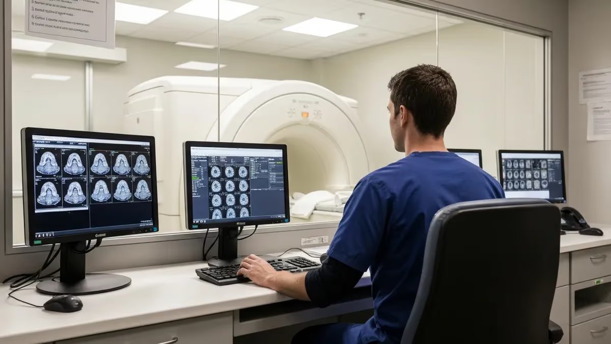
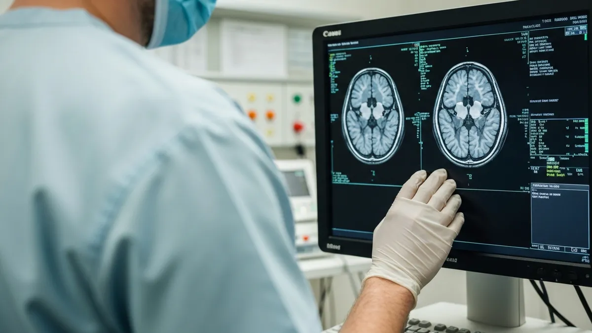
MRI dye contrast — most often a gadolinium-based contrast agent (GBCA) — is injected intravenously to make specific tissues, blood vessels, and abnormalities stand out far more clearly on the final images. Unlike the iodinated contrast used in CT scans, gadolinium works by altering the magnetic relaxation properties of nearby hydrogen protons, producing regions of increased signal brightness on T1-weighted sequences. Radiologists rely on this enhancement to detect tumors, inflammation, infection, and vascular disease that would be nearly invisible on non-contrast scans alone.
The decision to use mri dye contrast is never automatic. Physicians weigh the diagnostic benefit against patient-specific factors such as kidney function, allergy history, pregnancy status, and whether the clinical question can realistically be answered without enhancement. In straightforward musculoskeletal studies or routine lumbar spine evaluations, contrast is often unnecessary. However, for brain metastases, multiple sclerosis lesion activity, or post-surgical tumor follow-up, gadolinium is essentially indispensable for confident interpretation.
Gadolinium itself is a rare-earth metal that is toxic in its free ionic form. Pharmaceutical manufacturers bind it tightly to organic chelate molecules — either linear or macrocyclic in structure — to create a stable complex that the kidneys excrete efficiently. The chelation strategy dramatically reduces the risk of gadolinium dissociating inside the body, though this risk is not zero, a fact that has driven significant regulatory scrutiny and changes to clinical practice over the past decade.
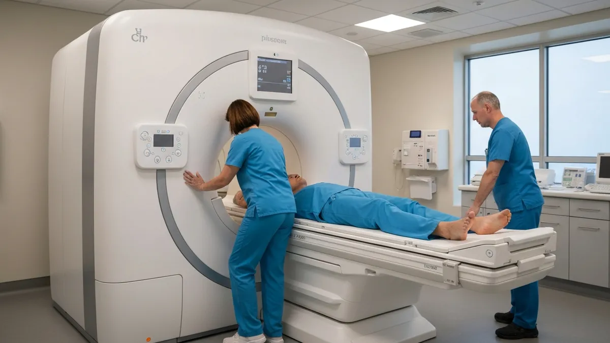
From the MRI technologist's perspective, contrast administration involves far more than simply pushing a syringe. You must verify the physician's order, confirm renal function labs (typically eGFR within 30 to 60 days), screen the patient for prior contrast reactions and known allergies, establish IV access with an appropriate gauge catheter, and document the lot number, dose, and injection time in the patient record. Some facilities use power injectors for dynamic vascular studies, while others rely on hand injection for standard brain or spine protocols.
The timing of contrast injection relative to image acquisition is critical and varies by study type. A brain MRI for tumor follow-up typically acquires post-contrast T1 sequences immediately after injection. A dynamic liver study using multiphase imaging captures arterial, portal venous, and delayed phases within tight time windows measured in seconds. Breast MRI relies on subtraction images that highlight the kinetic curve of contrast uptake and washout, with that curve pattern helping distinguish benign from malignant lesions.
Patients frequently arrive anxious about receiving contrast, often mixing up MRI gadolinium with CT iodine contrast and assuming they face the same risk of severe allergic reactions. While anaphylactoid reactions to GBCAs do occur, they are considerably rarer than with iodinated agents — estimated at roughly 0.03 to 0.1 percent of administrations. That said, any facility administering contrast must maintain emergency medications, resuscitation equipment, and staff trained to respond to adverse events, regardless of how low the baseline probability appears.
This guide covers every clinically relevant aspect of MRI contrast: how gadolinium agents are classified, when they are indicated or contraindicated, how nephrogenic systemic fibrosis and gadolinium retention have reshaped prescribing, what patients should expect on the day of the scan, and how MRI technologists and radiologists apply contrast knowledge in real-world practice. Whether you are a patient preparing for your first contrast MRI or a technologist studying for registry certification, the information here will give you a clear and accurate foundation.
MRI Contrast by the Numbers
Types of Gadolinium-Based Contrast Agents
Gadoterate (Dotarem), gadobutrol (Gadavist), and gadoteridol (ProHance) use a rigid ring structure that binds gadolinium more tightly, resulting in lower rates of gadolinium release and retention in tissues. These are now preferred in most clinical guidelines.
Gadopentetate dimeglumine (Magnevist) and gadoxetate (Eovist) have an open-chain structure that is less stable. Linear agents are associated with higher gadolinium deposition in brain, bone, and skin and carry greater NSF risk in renally impaired patients.
Gadodiamide (Omniscan) and gadoversetamide (OptiMARK) are the least stable GBCAs available. Many institutions have removed them from formularies after evidence linking them to the highest rates of nephrogenic systemic fibrosis in high-risk patients.
Gadoxetate (Eovist/Primovist) is taken up by hepatocytes and excreted into bile, providing a hepatobiliary phase roughly 20 minutes post-injection. This unique property makes it the agent of choice for characterizing small liver lesions and detecting bile duct communications.
Gadofosveset (Ablavar) binds reversibly to albumin, prolonging intravascular retention for high-resolution MR angiography. Though withdrawn from the US market, its pharmacokinetic principle remains important for understanding vascular contrast-enhanced MRA.
When a gadolinium-based contrast agent enters the bloodstream, it distributes rapidly into the extracellular fluid compartment, which includes both the intravascular space and the interstitial space between cells. Gadolinium does not cross intact cell membranes under normal circumstances, which is precisely what makes contrast enhancement such a powerful diagnostic tool. Pathological processes — tumors, active inflammation, infection, disruption of the blood-brain barrier — alter vascular permeability, allowing contrast to leak into regions where it would not normally accumulate, producing focal areas of increased T1 signal.
The mechanism of T1 shortening is central to understanding why gadolinium makes tissues appear brighter. Gadolinium is a paramagnetic substance with seven unpaired electrons, giving it an unusually strong magnetic moment. When gadolinium molecules tumble through tissue near water protons, they dramatically accelerate the longitudinal (T1) relaxation of those protons. A shorter T1 means the protons recover their magnetization faster between radiofrequency pulses, resulting in a higher signal on T1-weighted images when the repetition time (TR) is set appropriately short.
The blood-brain barrier (BBB) plays a particularly important clinical role in contrast enhancement. Under normal conditions, tight junctions between cerebral endothelial cells prevent gadolinium from entering the brain parenchyma, so healthy brain tissue does not enhance. When a tumor, abscess, or area of demyelination disrupts those tight junctions, gadolinium leaks across the BBB and accumulates in the abnormal tissue. This is why a ring-enhancing lesion on a post-contrast T1 brain MRI immediately raises suspicion for high-grade glioma, metastasis, or cerebral abscess — each of which disrupts the BBB in a characteristic pattern.
Dose calculations for gadolinium follow weight-based protocols, with the standard dose for most indications set at 0.1 mmol per kilogram of body weight, equivalent to 0.2 mL/kg of a 0.5 mol/L concentration agent. Some applications — particularly MR angiography and perfusion imaging — use double or triple doses to achieve adequate vascular enhancement or to prolong the enhancement window. However, regulatory agencies and professional societies have increasingly cautioned against high-dose protocols given concerns about gadolinium retention, pushing institutions toward single-dose approaches whenever diagnostically acceptable.
Post-contrast sequences are typically acquired within two to fifteen minutes of injection for most parenchymal studies, because gadolinium begins washing out of tissues relatively quickly as the kidneys excrete the agent. The timing window matters enormously: scan too early and the contrast has not yet distributed into interstitial spaces; scan too late and the concentration has fallen below the threshold needed to produce visible enhancement. Radiologists communicate specific protocol timing requirements to technologists through detailed scan protocols that specify acquisition order, delay times, and phase requirements.
Beyond standard T1 post-contrast imaging, contrast agents are fundamental to several advanced MRI techniques. Dynamic contrast-enhanced MRI (DCE-MRI) uses rapid sequential acquisition to map the kinetics of contrast uptake and washout, generating quantitative parameters like Ktrans (volume transfer coefficient) that reflect tissue perfusion and vascular permeability. These pharmacokinetic maps are used in oncology to assess tumor vascularity, monitor treatment response, and characterize breast lesions. MR perfusion imaging in the brain uses gadolinium's susceptibility effects during first-pass transit to measure cerebral blood volume and flow in stroke and tumor evaluation.
Understanding contrast physiology is not just academic for MRI registry candidates — it is the conceptual foundation for answering protocol questions, troubleshooting poor enhancement, explaining scan findings to patients, and advocating for appropriate contrast use in your clinical environment. Technologists who understand why contrast behaves the way it does make better clinical decisions than those who simply follow a checklist without grasping the underlying science.
Contrast Indications, Contraindications, and Special Populations
Contrast is indicated when the clinical question cannot be reliably answered by unenhanced sequences alone. Classic indications include evaluation of primary brain tumors and metastases, detection of active multiple sclerosis plaques, characterization of spinal cord lesions, assessment of post-surgical tumor recurrence versus treatment changes, evaluation of infection and abscess, MR angiography of the aorta and peripheral vessels, and breast MRI for cancer screening in high-risk women. In abdominal imaging, contrast is essential for liver lesion characterization, pancreatic mass evaluation, and renal mass assessment when CT is contraindicated.
The referring physician determines the clinical indication, but the MRI technologist and radiologist share responsibility for confirming that the indication is appropriate and documented. Many institutions have implemented contrast utilization review programs that flag orders without clear indications, both to control costs and to minimize unnecessary gadolinium exposure. Technologists should feel empowered to contact the ordering provider or radiologist when the indication is unclear, because administering contrast without a legitimate clinical justification exposes the patient to risk without benefit.
MRI With Contrast vs. MRI Without Contrast: Key Trade-offs
- +Reveals tumors, metastases, and active lesions invisible on non-contrast sequences
- +Defines blood-brain barrier disruption with high sensitivity for neurological diseases
- +Enables dynamic vascular imaging (MRA) without ionizing radiation
- +Differentiates viable tumor from post-treatment necrosis or fibrosis
- +Improves detection of small enhancing lesions in MS follow-up
- +Supports quantitative perfusion and permeability mapping in research and oncology
- −Requires IV access, increasing patient discomfort and scan preparation time
- −Gadolinium retention in brain and other tissues occurs even with normal renal function
- −NSF risk in severe renal impairment limits use in a significant patient subset
- −Additional cost compared to non-contrast examinations
- −Rare but serious anaphylactoid reactions require emergency preparedness
- −Extends total scan time due to pre- and post-contrast sequence requirements
MRI Contrast Patient Preparation Checklist
- ✓Verify the physician's order specifies contrast and documents the clinical indication.
- ✓Obtain or review eGFR lab results from within the past 30 days (60 days for stable chronic kidney disease patients).
- ✓Screen the patient verbally for prior contrast reactions, allergies, asthma, and atopy risk factors.
- ✓Confirm the patient has fasted for at least two hours before the exam (required at many institutions for abdominal contrast studies).
- ✓Establish patent IV access with an 18- to 22-gauge catheter in the antecubital fossa or forearm.
- ✓Select the appropriate GBCA type based on indication, renal function, and institutional formulary guidelines.
- ✓Calculate the correct weight-based dose (standard 0.1 mmol/kg) and prepare the syringe or power injector.
- ✓Explain the injection process to the patient, including expected sensations such as warmth and metallic taste.
- ✓Confirm the crash cart, emergency medications (epinephrine, diphenhydramine, atropine), and oxygen are accessible.
- ✓Document lot number, expiration date, dose administered, injection time, and patient tolerance in the scan record.
Macrocyclic Agents Are Now the Standard of Care
Following FDA safety communications in 2017 and updated ACR guidance through 2026, macrocyclic gadolinium agents (gadobutrol, gadoterate, gadoteridol) are preferred over linear agents for virtually all indications. Their rigid ring structure retains gadolinium far more effectively, minimizing tissue deposition. Institutions that have not yet updated their formularies to prioritize macrocyclic agents should do so as part of routine quality and safety review.
Nephrogenic systemic fibrosis (NSF) is a rare but devastating condition first described in 1997 in dialysis patients and definitively linked to gadolinium-based contrast agents by 2006. The disease causes progressive fibrosis of the skin, subcutaneous tissue, fascia, muscles, and in severe cases, internal organs including the heart, lungs, and kidneys. Affected patients experience debilitating skin thickening and tightening, joint contractures, and in the most severe cases, fatal cardiopulmonary involvement. The connection to gadolinium transformed how the medical community understood and managed contrast risk in vulnerable populations.
The pathophysiology of NSF centers on gadolinium dissociation from its chelate molecule. In patients with severely reduced renal clearance, the contrast agent remains in circulation far longer than the 1.5 to 2 hours typical in healthy individuals. Extended exposure time allows the thermodynamic equilibrium between bound and free gadolinium to shift, releasing free gadolinium ions that can deposit in tissues and trigger a fibrotic inflammatory cascade. Linear agents, with their less stable chelation chemistry, release gadolinium more readily than macrocyclic agents, explaining the strong epidemiological association between linear agent use and NSF cases.
The epidemiology of NSF shifted dramatically after 2007, when regulatory agencies required black-box warnings on high-risk GBCAs and clinical guidelines began restricting their use in patients with eGFR below 30 mL/min/1.73m². Reported NSF cases worldwide dropped from hundreds per year to nearly zero in subsequent years, demonstrating that risk stratification and agent selection are profoundly effective preventive measures. Today, NSF is largely a historical tragedy that shaped modern contrast safety standards rather than an ongoing clinical threat, provided guidelines are followed rigorously.
Separate from NSF, gadolinium retention in the brain gained attention starting in 2013 when researchers reported T1 signal hyperintensity in the dentate nucleus and globus pallidus of patients who had received multiple doses of linear GBCAs. Subsequent studies using mass spectrometry confirmed measurable gadolinium deposits in brain tissue of patients with no known renal impairment. Importantly, no neurological symptoms or clinical harm have been definitively attributed to brain gadolinium retention in the human population studied to date, but the finding prompted the FDA and EMA to take regulatory action and continues to drive ongoing research.
The clinical significance of gadolinium retention remains debated. Animal studies have shown behavioral changes at very high gadolinium doses, but translating those findings to human clinical practice is challenging given the dose disparities involved. Large epidemiological studies have not found increased rates of neurological disease, cognitive impairment, or mortality in patients who received multiple GBCA doses compared to matched controls. Nevertheless, the principle of minimizing gadolinium exposure when possible — using contrast only when it will meaningfully change clinical management — is now embedded in standard radiology practice guidelines.
For MRI technologists, understanding gadolinium retention means being able to explain the issue accurately to patients who ask, without either dismissing their concerns or amplifying anxiety beyond what the evidence warrants. Patients who have received multiple contrast MRIs for a chronic condition such as MS or a followed tumor often ask whether they should be worried about the gadolinium that has accumulated over years of scanning.
The honest answer is that the scientific community is still studying this question, that no clinical harm has been proven, and that the decision to continue contrast imaging should be made with the supervising radiologist and referring physician based on the patient's specific diagnostic needs.
Regulatory bodies continue to monitor gadolinium safety. The FDA's ongoing Gadolinium Retention and Exposure Working Group reviews new evidence periodically, and the ACR Manual on Contrast Media is updated regularly to reflect the current state of the science. Technologists and radiologists are expected to stay current with these guidelines as part of their professional responsibilities, and many registry examination questions test knowledge of the current regulatory and safety landscape surrounding gadolinium agents.
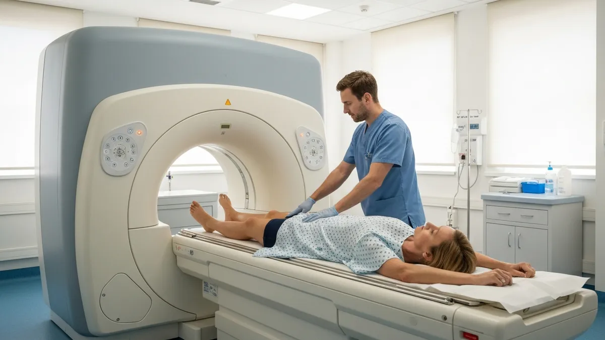
Never administer gadolinium-based contrast to a patient with unknown or severely impaired renal function without first consulting the supervising radiologist. An eGFR below 30 mL/min/1.73m² is a contraindication to high-risk linear agents and requires individualized risk-benefit assessment for any GBCA. Policies vary by institution, but the default must be to check labs and verify — not to proceed and document the risk later.
In day-to-day clinical practice, the decision pathway for contrast administration is more nuanced than any protocol document can fully capture. Consider a patient presenting for an MRI of the lumbar spine for low back pain and radiculopathy. Standard practice at most institutions is to perform this examination without contrast, since disc herniations, nerve root compression, and degenerative changes are clearly visible on T2-weighted sequences without gadolinium. Contrast would be added if the clinical question involved infection, tumor, or post-surgical evaluation for epidural fibrosis versus recurrent disc herniation — a distinction that unenhanced sequences cannot reliably make.
Brain MRI protocols illustrate the complexity even more vividly. A first-time seizure workup in a young adult might be performed without contrast if the clinical suspicion for a structural lesion is low, relying on FLAIR and T2 sequences to detect cortical dysplasia or hippocampal sclerosis.
The same patient presenting with a new focal neurological deficit would receive contrast from the outset to exclude tumor, abscess, or demyelinating disease. A patient with known primary brain tumor returning for a six-week post-radiation follow-up receives contrast as a matter of protocol, even when clinically stable, because the imaging itself drives management decisions about further treatment.
Vascular applications of MRI contrast have expanded considerably in recent years. Contrast-enhanced MR angiography (CE-MRA) of the renal arteries, carotid arteries, and peripheral vasculature provides high-resolution images with excellent contrast-to-noise ratio that often equals or exceeds CT angiography, without ionizing radiation.
This makes CE-MRA particularly valuable in younger patients, patients with iodine contrast allergy, and those requiring serial follow-up of treated aneurysms or vascular malformations. The technologist's role in CE-MRA includes precise bolus timing — either using a test bolus technique or automated bolus-triggering software — to ensure the contrast arrives in the target vessel at the exact moment of acquisition.
Breast MRI with contrast has become a critical tool in cancer detection and staging. The ACR recommends annual contrast-enhanced breast MRI screening for women with greater than 20 percent lifetime risk of breast cancer, including BRCA1 and BRCA2 mutation carriers. The scan uses a dedicated bilateral breast coil, rapid dynamic acquisition over approximately seven minutes post-injection, and subtraction imaging to remove background tissue signal and highlight enhancing lesions. Technologists performing breast MRI must be familiar with breast anatomy, compression positioning, and the kinetic analysis software used to generate enhancement curve data that radiologists use in BI-RADS assessments.
Cardiac MRI with contrast has transformed the evaluation of ischemic and non-ischemic cardiomyopathy. Late gadolinium enhancement (LGE) imaging, acquired ten to fifteen minutes after contrast injection, exploits the differential washout kinetics between viable myocardium and fibrotic or necrotic tissue.
In infarcted myocardium, the contrast agent washes out slowly because the damaged cells cannot maintain normal membrane integrity, resulting in persistent enhancement that delineates the infarct territory with sub-millimeter resolution. LGE pattern analysis — subendocardial versus transmural versus mid-wall — allows cardiologists to differentiate ischemic from non-ischemic causes of reduced ejection fraction and guides decisions about revascularization, implantable defibrillator placement, and prognosis.
The growing application of artificial intelligence to contrast-enhanced MRI interpretation is an emerging frontier that technologists and radiologists alike should understand. AI algorithms trained on large datasets of contrast-enhanced brain MRIs can now detect and measure enhancing lesions with speed and reproducibility that rivals human readers in controlled studies.
AI-based perfusion analysis tools are FDA-cleared for stroke triage, automatically generating color-coded maps of brain tissue at risk from raw DSC perfusion data within minutes of acquisition. These tools augment — rather than replace — clinical judgment, and technologists who understand the underlying contrast physiology are better positioned to recognize when algorithm outputs should be questioned.
For registry exam candidates, the contrast material section of the ARRT MRI examination covers pharmacology, safety screening, adverse reaction management, and regulatory considerations. Questions frequently probe knowledge of the mechanism of gadolinium's T1 shortening effect, the difference between ionic and non-ionic agents, the eGFR thresholds that trigger different levels of caution, and the correct management algorithm for moderate and severe contrast reactions. Practicing with targeted question sets that mirror registry-style clinical vignettes is the most efficient way to solidify this knowledge and build the pattern recognition that the exam rewards.
If you want more targeted practice, the mri abdomen and pelvis cpt code page covers the same material with additional questions and explanations.
If you want more targeted practice, the open mri near me page covers the same material with additional questions and explanations.
Practical preparation for a contrast-enhanced MRI begins well before the patient arrives at the scanner. Technologists reviewing the schedule the day before should flag any patients with documented renal disease, prior contrast reactions, diabetes (a surrogate risk factor for renal impairment), or age over 60 for proactive lab review. Many institutions use an automated lab interface that populates the eGFR directly into the radiology information system, allowing technologists to identify at-risk patients before the day of the scan and alert the radiologist when a case requires individualized review.
On the day of the examination, a structured pre-scan interview is your primary safety tool. Speak with the patient privately, use plain language, and give them time to answer. Ask specifically whether they have ever had a reaction to contrast dye during a previous MRI or CT scan — not just allergies in general, since patients may not think of contrast reactions as allergies.
Ask about their kidney function history. Ask about sickle cell disease, which can interact adversely with contrast agents and magnetic fields. Ask whether they are pregnant or breastfeeding. Document every answer. This conversation is not a formality; it is the last reliable safety checkpoint before injection.
IV access for contrast administration requires thoughtful site selection. Antecubital veins are preferred for power injection because they can accommodate higher flow rates without rupture. If you must use a hand or wrist vein, use a smaller gauge needle and hand-inject at a slow rate.
Always flush the IV line with normal saline before and after contrast injection to confirm patency, to prevent contrast mixing with other medications, and to clear the tubing efficiently. If you are using a power injector, program the saline flush volume (typically 20 to 30 mL) to follow the contrast bolus, which helps push the contrast column forward and reduce the dose needed to achieve adequate enhancement.
Post-injection monitoring is mandatory at all facilities. Keep the patient in the scanner room or an adjacent recovery area for at least 15 minutes after contrast administration. The majority of acute adverse reactions occur within this window. Have clear escalation protocols so that any technologist who observes a reaction can immediately summon a radiologist and, if needed, activate emergency medical services. Never leave a post-contrast patient unobserved in the MRI suite or discharge them from the department before the minimum monitoring period is complete.
For patients who report prior mild reactions to gadolinium, many radiologists will pre-medicate with corticosteroids and antihistamines using a standardized protocol — for example, methylprednisolone 32 mg orally at 12 hours and 2 hours before injection, plus diphenhydramine 50 mg IV or oral one hour before. Pre-medication reduces but does not eliminate the risk of recurrent reactions. Patients with prior severe anaphylaxis to GBCAs should be discussed with the ordering physician about whether contrast is truly necessary, whether a different agent class can be used, and whether the examination should be performed in a setting with full anesthesia support available.
Documentation cannot be overemphasized as a practical and professional responsibility. Your contrast administration record should include the agent name and concentration, lot number and expiration date, dose administered in milliliters and millimoles, route of administration, injection rate if power-injected, time of injection, patient tolerance, any adverse events and their management, and the name of the radiologist who authorized the administration. This record protects the patient by creating a complete safety trail, protects you professionally, and provides essential data for quality improvement programs tracking adverse event rates.
Finally, build your contrast knowledge continuously. The field evolves: new agents are introduced, safety data accumulates, guidelines are updated, and clinical applications expand. Read ACR guidance documents and ARRT practice standards annually. Participate in department continuing education on contrast safety. Review your facility's contrast quality assurance data. Technologists who stay current not only provide better patient care — they pass their registry exams with confidence and advance into leadership roles where they shape protocol development and safety culture for their entire department.
MRI Questions and Answers
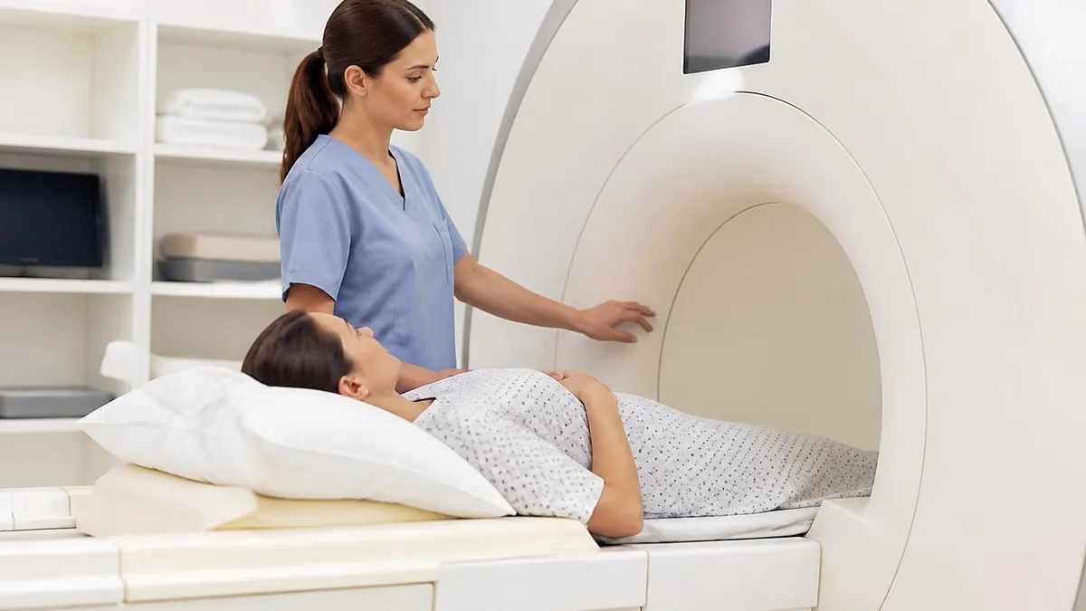
DWI MRI: Diffusion-Weighted Imaging Explained (2026 Guide)
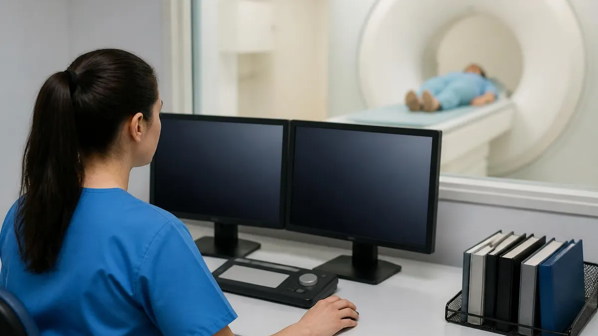
MRI Lumbar Spine Without Contrast: Complete Guide to the Scan, What It Shows, and How to Prepare
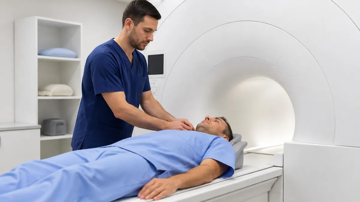
MRI History: From Nuclear Resonance to Modern Scanners

MRI of Cervical Spine: Complete Guide to Imaging, Anatomy, Pathology, and Patient Preparation
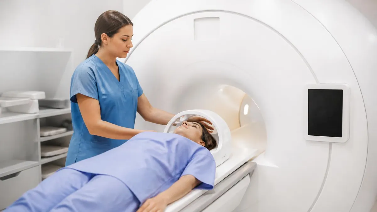
Where to Get an MRI: Facilities, Costs, and How to Choose
About the Author

Medical Laboratory Scientist & Clinical Certification Expert
Johns Hopkins UniversityDr. Sandra Kim holds a PhD in Clinical Laboratory Science from Johns Hopkins University and is certified as a Medical Technologist (MT) and Medical Laboratory Scientist (MLS) through ASCP. With 16 years of clinical laboratory experience spanning hematology, microbiology, and molecular diagnostics, she prepares candidates for ASCP board exams, MLT, MLS, and specialist certification tests.
Join the Discussion
Connect with other students preparing for this exam. Share tips, ask questions, and get advice from people who have been there.
View discussion (8 replies)