How Does MRI Work? The Science Behind the Scan Explained 2026 July
MRI uses magnetic fields and radio waves to create detailed images of organs and tissues. 📚 Learn how the technology works, what it detects, and why
How Does MRI Work?
You've probably had an MRI, know someone who's had one, or you're about to get one and want to understand what's actually happening inside that big noisy machine. MRI is one of medicine's most important diagnostic tools, but most people — including many healthcare professionals — have only a vague understanding of the physics behind it.
The explanations tend to be either oversimplified ('it takes pictures using magnets') or impenetrably technical. This guide sits in the middle: accurate enough to be genuinely informative, clear enough that you don't need a physics background or medical training to follow along and genuinely understand what's happening during your own actual scan experience in the machine.
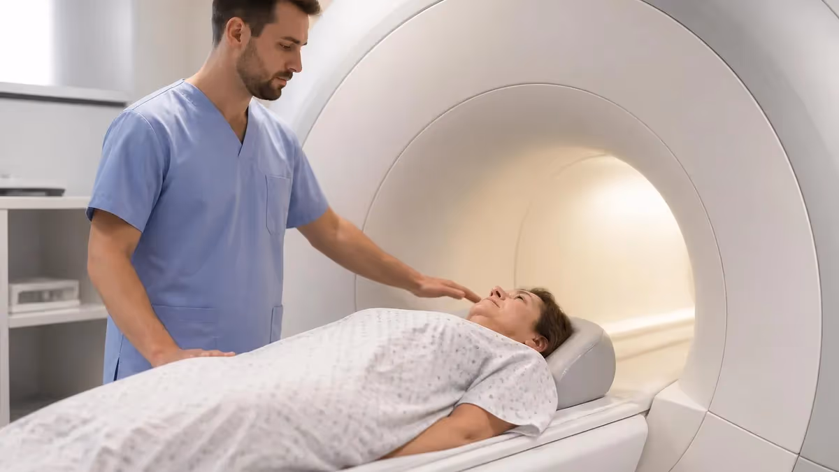
MRI — Magnetic Resonance Imaging — creates detailed pictures of the inside of your body using powerful magnetic fields and radio waves. No X-rays, no radiation, no surgery. You lie inside a large cylindrical magnet, the machine sends radio wave pulses into your body, and your body's hydrogen atoms respond by emitting signals that a computer processes into incredibly detailed cross-sectional images. The entire process is based on physics rather than chemistry — it exploits a natural property of hydrogen atoms called nuclear magnetic resonance to distinguish between different types of tissue.
If you've ever wondered why MRI produces such remarkably clear images of soft tissues — brains, spinal cords, muscles, tendons, internal organs — while X-rays and CT scans show bones much better, the answer lies in what each technology measures. X-rays measure how much radiation passes through tissue (dense tissue like bone blocks more radiation). MRI measures how hydrogen atoms in different tissues respond to magnetic fields — and since different tissues contain different amounts of water (which contains hydrogen), MRI can distinguish between tissues that look identical on X-ray.
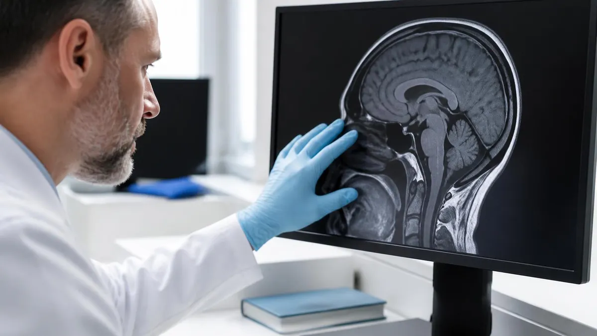
The result is the most detailed soft-tissue imaging available in medicine. A brain MRI can show individual structures within the brain with sub-millimetre resolution. A knee MRI can visualise the internal structure of ligaments and cartilage. A cardiac MRI can show the heart muscle contracting in real time. This imaging capability has transformed how doctors diagnose conditions ranging from brain tumours and spinal cord injuries to meniscus tears and liver disease — often eliminating the need for exploratory surgery that would have been required decades ago.
Understanding how MRI works doesn't require a physics degree. The core concepts — magnetic alignment, radio wave excitation, signal detection, and image reconstruction — can be explained in terms anyone can follow. This guide walks through the science step by step, explains why different scan types exist, and addresses the practical questions patients and students commonly ask about MRI technology.
- What it uses: A powerful magnetic field (1.5 or 3 Tesla) + radio frequency (RF) pulses — NO ionising radiation
- What it measures: How hydrogen atoms in your body's water and fat respond to magnetic fields and RF energy
- Why it's detailed: Different tissues contain different amounts of water — MRI detects these differences with high resolution
- Image types: T1-weighted (anatomy), T2-weighted (fluid/pathology), FLAIR (brain lesions), DWI (stroke detection), and many others
- Magnet strength: 1.5 Tesla (standard) or 3 Tesla (high-resolution) — roughly 30,000–60,000 times Earth's magnetic field
- Scan time: 15–90 minutes depending on body part and sequences ordered
- Safety: No radiation exposure — safe for repeated imaging. Contraindicated with certain metallic implants
How MRI Creates an Image: Step by Step
Step 1: Magnetic Alignment
Step 2: Radio Frequency Excitation
Step 3: Signal Emission (Relaxation)
Step 4: Signal Detection and Spatial Encoding
Step 5: Image Reconstruction
T1 vs T2 Weighted Images: Why MRI Takes Multiple Sequences
When you have an MRI, the machine doesn't take just one picture — it runs multiple sequences, each producing a different type of image that highlights different tissue characteristics. The two most fundamental image types are T1-weighted and T2-weighted, and understanding the difference explains why MRI is so much more informative than a single X-ray.
T1-weighted images emphasise anatomical detail. On a T1 image, fat appears bright (white) and water appears dark. This makes T1 ideal for showing normal anatomy — the structure of the brain, the layers of an organ, the boundaries between different tissues. T1 images are also the standard for post-contrast imaging: when gadolinium contrast dye is injected, areas that take up the contrast (like tumours or inflamed tissue) 'light up' bright on T1, making pathology easier to identify against the background of normal anatomy.
T2-weighted images emphasise fluid and pathology. On a T2 image, water appears bright (white) and fat appears darker. This makes T2 particularly useful for detecting abnormalities: most pathological processes — inflammation, infection, oedema, tumours, cysts — involve increased water content in the affected tissue. On T2, these abnormal areas appear bright against the darker normal tissue, essentially highlighting the problem areas. Radiologists often say 'pathology is bright on T2' as a general principle.
FLAIR (Fluid-Attenuated Inversion Recovery) is a modified T2 sequence that suppresses the signal from free-flowing fluid (like cerebrospinal fluid in the brain). This makes brain lesions that would be hidden by the bright CSF on standard T2 images visible as bright spots against the now-dark CSF. FLAIR is essential for detecting multiple sclerosis plaques, small strokes, and other brain pathology that sits near the fluid-filled ventricles.
Diffusion-Weighted Imaging (DWI) detects the movement of water molecules at the cellular level. In acute stroke, brain cells swell and restrict water movement — DWI detects this restriction within minutes of the stroke occurring, making it the fastest MRI sequence for diagnosing acute brain ischemia. DWI has transformed stroke diagnosis because it identifies affected brain tissue much earlier than CT or standard MRI sequences.
Each MRI exam includes a combination of sequences chosen by the radiologist based on the clinical question. A brain MRI might include T1, T2, FLAIR, DWI, and post-contrast T1. A knee MRI might include T1, T2, and proton density sequences. The sequences are chosen to answer specific diagnostic questions — there's no single 'standard' MRI that works for every body part and every condition.
Key Components of an MRI Machine
The large cylindrical structure that the patient lies inside. Most clinical MRI magnets are superconducting electromagnets cooled with liquid helium to near absolute zero (-269°C). The magnetic field is always on — 24 hours a day, 365 days a year — even when no scans are being performed. The field strength is measured in Tesla: 1.5T is standard, 3T provides higher resolution, and research magnets reach 7T or higher. For reference, 1.5 Tesla is approximately 30,000 times stronger than Earth's magnetic field.
Three sets of gradient coils create small, controlled variations in the magnetic field in the x, y, and z directions. These gradients encode spatial information into the MRI signals — without them, the machine would know that hydrogen atoms are responding but wouldn't know where they are. The rapid switching of gradient coils is what produces the loud knocking and buzzing sounds during an MRI scan. The speed and strength of gradient coils affect image quality and scan speed.
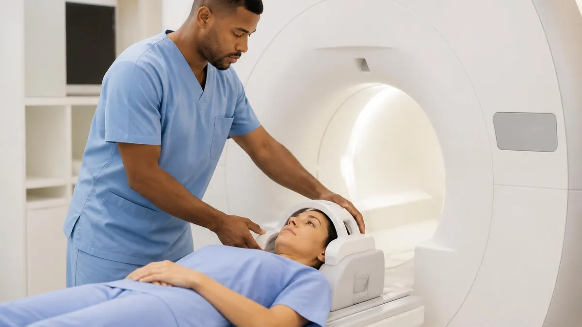
Radio frequency coils serve two functions: transmitting RF pulses that excite hydrogen atoms and receiving the signals that hydrogen atoms emit as they relax. Some coils do both; in many modern systems, the body coil transmits and dedicated surface coils receive. Surface coils (the devices placed over or around the body part being scanned — head coil, knee coil, spine coil) are designed to optimise signal reception for specific anatomical areas, producing clearer images than a generic whole-body coil.
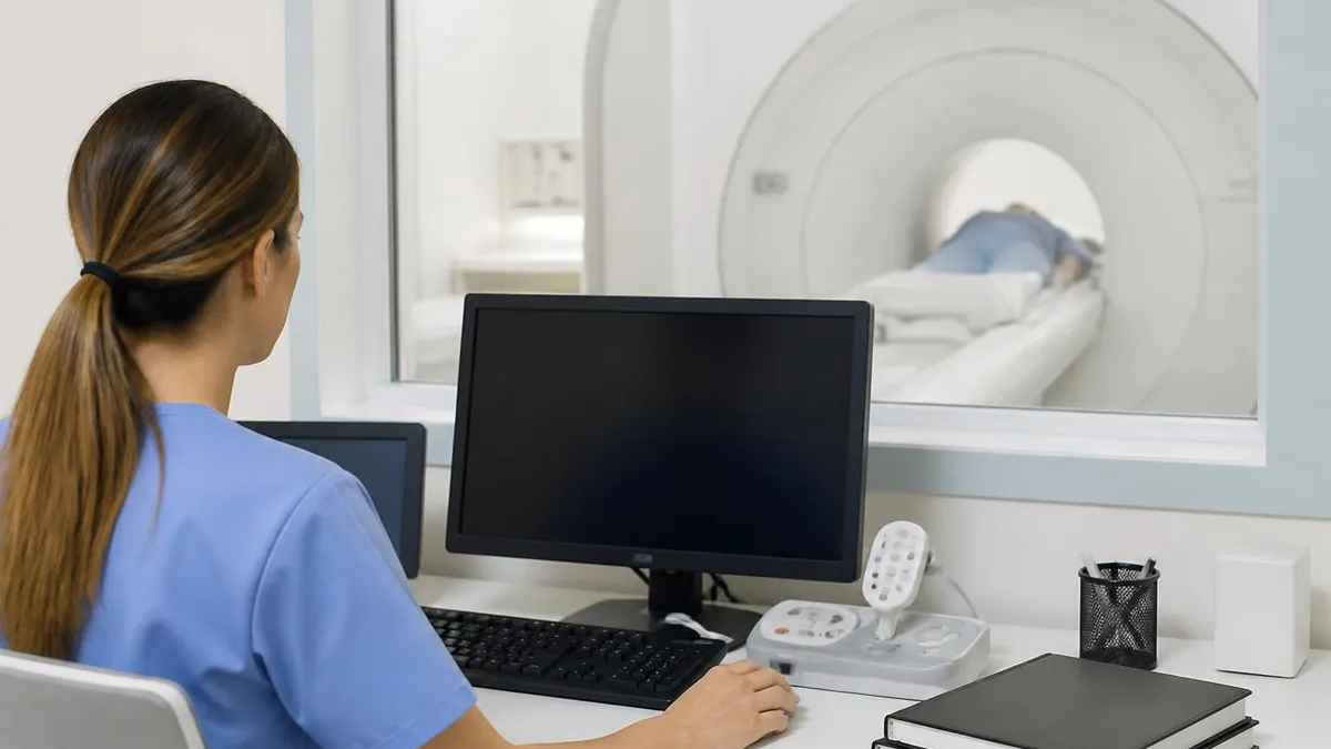
The MRI's computer controls the timing of RF pulses and gradient activations (the 'pulse sequence'), processes the raw signal data using Fourier transform mathematics, and reconstructs the final images. Modern MRI computers process millions of data points to produce each image slice. The computer also manages quality control, stores patient data, and sends images to the PACS (Picture Archiving and Communication System) where radiologists review them.
Why MRI Is Safe (and Its Limitations)
MRI is fundamentally safe because it uses magnetic fields and radio waves — not ionising radiation like X-rays or CT scans.
- No radiation dose: Unlike CT and X-ray, MRI doesn't expose you to ionising radiation. There's no cumulative radiation risk from repeated MRI scans
- Safe for children: MRI is the preferred imaging modality for paediatric patients when soft tissue detail is needed, because there's no radiation exposure
- Safe during pregnancy (with caution): MRI is generally considered safe after the first trimester, though gadolinium contrast is avoided during pregnancy unless absolutely necessary
- The magnetic field itself: Decades of research and clinical use have found no harmful biological effects from the static magnetic fields used in clinical MRI scanners (1.5T and 3T)
- RF energy: The radio frequency pulses deposit a small amount of energy in tissue as heat (measured as SAR — Specific Absorption Rate). MRI systems monitor and limit SAR to safe levels
Why MRI Makes So Much Noise
The loud banging, clicking, and buzzing sounds during an MRI scan are caused by the rapid switching of gradient coils inside the machine. Gradient coils are electromagnets that create small variations in the magnetic field to encode spatial information. When electrical current flows through these coils, the interaction between the current-carrying wire and the main magnetic field creates a force (the Lorentz force) that physically vibrates the coil structure — similar to how a speaker cone vibrates to produce sound.
Each MRI sequence activates the gradient coils in a different pattern, which is why the sounds change throughout the scan — the knocking rhythm, pitch, and intensity shift between sequences. Some sequences are relatively quiet; others produce sounds exceeding 100 decibels (comparable to a jackhammer or a rock concert). This is why earplugs or noise-cancelling headphones are mandatory during MRI scans — prolonged exposure to these sound levels without protection can damage hearing.
MRI manufacturers have developed quieter scanning techniques that reduce gradient switching speed, but these 'quiet MRI' sequences often take longer to acquire images and may produce slightly lower image quality. The trade-off between noise reduction and image quality or scan speed is an active area of engineering development. Modern 3T scanners tend to be louder than 1.5T scanners because the stronger magnetic field amplifies the Lorentz force on the gradient coils, making the vibrations — and the noise — more intense.
The sound patterns during an MRI are specific to each pulse sequence being run — experienced MRI technologists can often identify which sequence is running just by listening to the characteristic rhythm and pitch. Some patients find it helpful to think of the sounds as the machine 'working through its checklist' of different image types rather than as random noise. Knowing that each sound pattern represents a different sequence collecting different information can make the experience feel more purposeful and less chaotic.
What MRI Can and Can't Do
- ✓MRI excels at soft tissue imaging — brain, spinal cord, muscles, tendons, ligaments, internal organs, and joint structures are shown in exceptional detail
- ✓MRI detects tumours, inflammation, infection, and fluid collections by identifying tissues with abnormal water content or contrast enhancement
- ✓MRI shows blood vessels without injection (MR angiography) — useful for evaluating stroke risk, aneurysms, and vascular malformations
- ✓MRI is less effective for bones than CT — while MRI can show bone marrow and stress fractures, CT provides better detail of cortical bone structure
- ✓MRI cannot image patients with certain metallic implants — ferromagnetic devices, some older pacemakers, and metallic foreign bodies are contraindications
- ✓MRI is sensitive to motion — breathing, swallowing, and voluntary movement degrade image quality. Sedation may be needed for patients who can't stay still
- ✓MRI doesn't show real-time dynamic function as well as ultrasound or fluoroscopy — although cardiac MRI and functional MRI (fMRI) can capture some dynamic information
MRI vs CT vs X-ray: When MRI Is the Right Choice
- +No ionising radiation — MRI can be repeated without cumulative radiation risk, making it ideal for monitoring conditions over time and for imaging children
- +Unmatched soft tissue contrast — MRI distinguishes between different types of soft tissue (muscle, fat, cartilage, fluid, tumour) better than any other imaging modality
- +Multiplanar imaging — MRI produces images in any plane (axial, sagittal, coronal, oblique) without repositioning the patient, providing comprehensive anatomical views
- +Functional imaging capabilities — fMRI can map brain activity, cardiac MRI can assess heart function, and diffusion MRI can detect acute stroke within minutes
- −Longer scan times — MRI takes 15–90 minutes versus 5 minutes for CT, increasing patient discomfort and the chance of motion artefacts
- −More expensive — MRI costs $400–$3,500 versus $200–$1,500 for CT and $50–$300 for X-ray, making it less accessible for routine screening
- −Metal contraindications — patients with certain implants, pacemakers, or metallic foreign bodies cannot safely undergo MRI, requiring alternative imaging
- −Claustrophobia and noise — the enclosed tube and loud sounds make MRI uncomfortable or impossible for some patients without sedation or open-bore alternatives
Gadolinium Contrast: How and Why It's Used in MRI
Some MRI exams include an injection of gadolinium-based contrast agent, which enhances the visibility of certain structures and pathology. Gadolinium is a paramagnetic substance that shortens the T1 relaxation time of nearby hydrogen atoms, making tissues that absorb the contrast appear bright on T1-weighted images. This enhancement helps radiologists distinguish between tissues that would look similar without contrast — for example, identifying a tumour's boundaries within surrounding brain tissue, or detecting areas of active inflammation versus old scarring.
Gadolinium is injected intravenously during the scan — typically partway through, so the radiologist can compare pre-contrast and post-contrast images of the same area. The contrast circulates through the bloodstream and concentrates in areas with increased blood supply or disrupted barriers (like the blood-brain barrier in brain tumours). After the scan, gadolinium is cleared by the kidneys over the following 24–48 hours.
Gadolinium contrast is generally safe, with a much lower risk of allergic reaction than the iodinated contrast used in CT scans. However, patients with severe kidney disease are at risk for a rare but serious condition called nephrogenic systemic fibrosis (NSF), which is why kidney function is checked (via blood test) before contrast administration in patients with known or suspected kidney impairment. Mild side effects (brief headache, nausea, a metallic taste) occur in a small percentage of patients and resolve quickly.
Not all MRI exams require contrast — many diagnostic questions can be answered with non-contrast sequences alone. Your ordering physician decides whether contrast is needed based on the clinical question and what information the MRI needs to provide. If contrast isn't necessary, you won't receive it.
Some patients worry about gadolinium retention — traces of gadolinium have been detected in the brain tissue of patients who received multiple contrast-enhanced MRIs over time. Research on this is ongoing, and regulatory agencies (FDA, EMA) have concluded that the clinical benefits of gadolinium contrast outweigh the known risks for patients who need it. Current guidance recommends using gadolinium only when clinically indicated rather than as a routine part of every MRI — and most radiologists follow this principle, ordering contrast only when it will meaningfully change the diagnostic information the scan provides.
MRI Technology: Key Numbers
Advanced MRI Techniques
Beyond standard anatomical imaging, MRI technology has evolved to provide functional, metabolic, and microstructural information that goes far beyond what a simple picture can show.
Functional MRI (fMRI) detects brain activity by measuring changes in blood oxygenation. When a brain region is active, it consumes more oxygen, causing a detectable change in the MRI signal (the BOLD — Blood Oxygen Level Dependent — effect). fMRI is used in neuroscience research to map which brain areas are involved in specific tasks, and clinically to map critical brain functions (language, motor control) before neurosurgery, helping surgeons avoid damaging essential brain areas.
Diffusion Tensor Imaging (DTI) maps the white matter tracts in the brain — the bundles of nerve fibres that connect different brain regions. By measuring how water molecules diffuse along nerve fibres (water moves more easily along a fibre than across it), DTI creates a three-dimensional map of the brain's wiring. This is used in neurological research, pre-surgical planning, and evaluating conditions like traumatic brain injury and multiple sclerosis that damage white matter connections.
MR Spectroscopy measures the chemical composition of tissue rather than its physical structure. Different chemicals produce distinct spectral peaks in the MRI signal, allowing the radiologist to identify abnormal chemical concentrations — elevated choline (a marker of cell proliferation) in a brain mass suggests malignancy; elevated lactate in brain tissue suggests ischemia. MR spectroscopy adds metabolic information to the anatomical picture that standard MRI provides.
Cardiac MRI uses ECG-gated sequences synchronised to the heart's electrical cycle to create images of the beating heart. This allows assessment of heart chamber size and function, heart muscle thickness, areas of scarring or inflammation (using late gadolinium enhancement), and blood flow through heart valves. Cardiac MRI is considered the gold standard for quantifying heart function and characterising heart muscle disease.
Unlike most medical equipment that's turned on for procedures and off between them, an MRI's main magnetic field is always active — 24 hours a day, 7 days a week. The superconducting magnet maintains its field continuously, even when no patients are being scanned. This means metal safety precautions apply at ALL times in the MRI suite, not just during scans. Ferromagnetic objects brought near the scanner — wheelchairs, oxygen tanks, tools, scissors — can be violently pulled into the bore by the magnetic force, potentially injuring anyone in the path. The strict metal screening protocols before every MRI aren't just precautions — they prevent genuinely dangerous situations that have caused serious injuries in hospitals where screening was inadequate.
The Future of MRI Technology
MRI technology continues advancing in ways that will make scans faster, quieter, more detailed, and more accessible in the coming years.
Artificial intelligence is being integrated into MRI reconstruction algorithms, allowing scanners to produce diagnostic-quality images from less data — which means shorter scan times. AI-accelerated MRI can reduce a 30-minute scan to 10–15 minutes without sacrificing image quality. This benefits patients (less time in the machine), hospitals (more patients per scanner per day), and healthcare systems (lower per-scan costs). Several AI-accelerated MRI systems have received FDA clearance and are entering clinical use.
Low-field MRI systems (0.064T — much weaker than standard 1.5T scanners) are being developed as portable, affordable alternatives to traditional MRI. These bedside MRI systems can be wheeled to a patient's room in the ICU, eliminating the need to transport critically ill patients to a fixed MRI suite. The image quality is lower than standard MRI but sufficient for many clinical questions, and the dramatically lower cost (under $100,000 versus $1–3 million for a standard scanner) could make MRI accessible in settings and countries where traditional scanners are unaffordable.
Ultra-high-field MRI (7 Tesla and above) pushes resolution beyond what's possible at standard field strengths. 7T MRI can visualise brain structures at sub-millimetre resolution, detect tiny lesions invisible at 3T, and provide metabolic and functional information with unprecedented detail. Currently limited to research centres, 7T is gradually entering clinical practice for specific applications like neuroimaging and musculoskeletal imaging where the additional resolution changes diagnostic outcomes.
Hybrid imaging systems that combine MRI with other modalities are also advancing. PET-MRI scanners combine the metabolic information from positron emission tomography with the anatomical detail of MRI in a single examination session, providing complementary diagnostic data without requiring two separate scans. This hybrid approach is particularly valuable in oncology, where PET shows metabolic activity of tumours while MRI shows their exact anatomical location and relationship to surrounding structures. As these combined systems become more widely available, they'll likely become the standard for comprehensive cancer staging, treatment planning, and ongoing treatment response monitoring in oncology centres worldwide.
How Does MRI Work Questions and Answers
About the Author
Medical Laboratory Scientist & Clinical Certification Expert
Johns Hopkins UniversityDr. Sandra Kim holds a PhD in Clinical Laboratory Science from Johns Hopkins University and is certified as a Medical Technologist (MT) and Medical Laboratory Scientist (MLS) through ASCP. With 16 years of clinical laboratory experience spanning hematology, microbiology, and molecular diagnostics, she prepares candidates for ASCP board exams, MLT, MLS, and specialist certification tests.
Join the Discussion
Connect with other students preparing for this exam. Share tips, ask questions, and get advice from people who have been there.
View discussion (6 replies)