ICD 10 Abnormal MRI Brain: Complete Guide to Coding, Findings, and Clinical Interpretation in 2026 July
ICD 10 abnormal MRI brain coding guide: 🧠 R90.89, common findings, white matter lesions, lesion interpretation, and clinical workflow for radiologists.

The icd 10 abnormal mri brain code most often used in clinical practice is R90.89, which captures other abnormal findings on diagnostic imaging of the central nervous system. This code becomes essential when a brain MRI reveals incidental lesions, nonspecific white matter changes, or unexplained signal abnormalities that require further workup but lack a confirmed diagnosis. Radiologists, neurologists, and medical coders rely on this designation thousands of times each day across emergency rooms, outpatient imaging centers, and neurology clinics throughout the United States.
Understanding how abnormal MRI brain findings translate into accurate ICD-10 coding matters for three reasons: reimbursement, longitudinal patient tracking, and quality reporting. When a 52-year-old patient presents with headaches and the MRI reveals scattered T2 hyperintensities, the coder must decide whether to assign R90.89, a more specific demyelinating code, or the underlying symptom code. Each choice has downstream consequences for insurance authorization, follow-up imaging, and population health metrics that drive value-based care contracts.
This guide walks through every dimension of abnormal brain mri icd 10 MRI interpretation and coding. We cover the most common abnormal findings — white matter lesions, microhemorrhages, mass lesions, atrophy patterns, and diffusion restriction — alongside the specific ICD-10 codes that map to each scenario. We also explain how to differentiate incidental findings from clinically significant abnormalities, when to recommend follow-up imaging, and which red flags demand same-day neurological consultation.
Brain MRI accounts for roughly 12 million scans annually in the US, and studies estimate that 15 to 30 percent of these reveal at least one unexpected finding. Many are benign — small arachnoid cysts, pineal cysts, or developmental venous anomalies. Others, like aneurysms, low-grade gliomas, or early multiple sclerosis plaques, alter the patient's trajectory dramatically. Knowing how to triage these findings is one of the most consequential skills a radiologist or referring clinician develops over a career.
For technologists preparing for the ARRT MRI registry exam, this material is core content. Pathology recognition appears throughout the registry blueprint, and understanding the link between imaging findings and ICD-10 documentation deepens your value to the radiology team. If you're studying, we recommend pairing this article with structured practice tests like the What Is an MRI Test? How Magnetic Resonance Imaging Scans Diagnose Disease in 2026 overview to reinforce the connection between physics, anatomy, and clinical findings.
Throughout this guide we use real case examples drawn from typical emergency and outpatient practice. Each section includes the imaging description, the differential diagnosis, the appropriate ICD-10 code (or code family), and the recommended next step. By the end you should be able to read a brain MRI report, identify the level of abnormality, select an accurate ICD-10 code, and communicate the urgency to ordering providers with confidence.
Finally, coding practices change every October when CMS releases the annual ICD-10-CM update. The 2026 update introduced refinements to several R90 subcategories and clarified guidance around incidental cerebral microbleeds in patients on anticoagulation. We flag the 2026-specific changes throughout the article so that practicing coders and clinical teams can update their templates and macros accordingly.
Abnormal MRI Brain by the Numbers
Primary ICD-10 Code Categories for Brain MRI Abnormalities
R90.0 covers intracranial space-occupying lesions found on imaging without a confirmed diagnosis. R90.89 captures other abnormal CNS imaging findings, the workhorse code for nonspecific white matter changes and incidentalomas awaiting workup.
G35 (multiple sclerosis), G93.9 (brain disorder unspecified), and G91 (hydrocephalus) apply when imaging confirms a specific neurological diagnosis. Use these only when the radiologist provides a definitive interpretation, not for suspected disease.
I63 (cerebral infarction), I61 (intracerebral hemorrhage), I67.5 (Moyamoya), and I67.89 (small vessel ischemic disease) cover vascular pathology. Document acuity, location, and laterality to support the most specific I-code possible.
C71 covers malignant brain neoplasms by location; D33 covers benign primary brain tumors; D43 covers neoplasms of uncertain behavior. Many lesions on initial MRI fall into D43 until biopsy or follow-up confirms the histology.
Q04 (congenital malformations of brain), Q07 (other congenital nervous system malformations), and related codes cover Chiari malformations, agenesis of the corpus callosum, and developmental venous anomalies found incidentally on adult MRI.
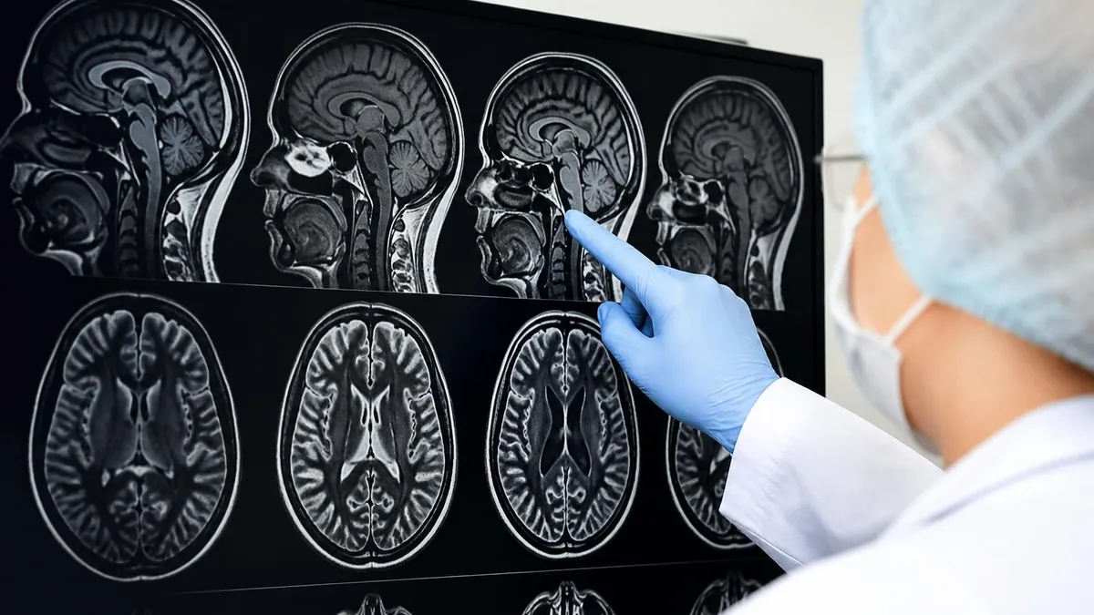
Common abnormal MRI brain findings span a wide spectrum from clinically insignificant incidentalomas to life-threatening pathology requiring immediate intervention. The most frequent abnormality reported on adult brain MRI is nonspecific T2/FLAIR hyperintensities in the subcortical and periventricular white matter. These lesions appear in roughly 50 percent of patients over age 60 and reflect chronic small vessel ischemic disease, migraine-associated changes, or normal aging. When isolated and asymptomatic, they typically code to R90.89 with secondary documentation of vascular risk factors.
Cerebral microhemorrhages, visible only on susceptibility-weighted imaging (SWI) or gradient echo (GRE) sequences, appear in 5 to 25 percent of older adults. Lobar microbleeds suggest cerebral amyloid angiopathy, while deep microbleeds at the basal ganglia and thalamus point to hypertensive vasculopathy. The 2026 ICD-10 update clarified that incidental microbleeds in anticoagulated patients should be coded with R90.89 plus the appropriate Z code for long-term anticoagulant use, alerting the prescriber to reassess bleeding risk.
Mass lesions present a major diagnostic challenge. A well-circumscribed extra-axial mass with dural tail enhancement typically represents a meningioma (D32.0), while a ring-enhancing intra-axial lesion in an immunocompromised patient could be toxoplasmosis, lymphoma, or metastasis. Initial reports often use D49.6 (neoplasm of unspecified behavior of brain) until tissue diagnosis is available. Always document size in three dimensions, location, mass effect, and surrounding edema — these elements drive the choice between observation, biopsy, and surgical resection.
Atrophy patterns provide critical information for dementia workup. Generalized cortical atrophy with hippocampal volume loss suggests Alzheimer disease (G30.9), while focal frontotemporal atrophy points to FTD (G31.09). Disproportionate ventricular enlargement with relatively preserved sulci raises concern for normal pressure hydrocephalus (G91.2). When the pattern is nonspecific, use R90.89 and let the neurologist correlate with clinical and neuropsychological testing.
Diffusion restriction on DWI with corresponding low ADC values indicates acute cytotoxic edema, most commonly from acute ischemic stroke. The pattern of restriction — wedge-shaped in a vascular territory, gyriform along cortical ribbons, or scattered punctate foci — narrows the differential. Acute stroke codes to I63 with the specific vascular territory documented. The companion article on MRI Medical Abbreviation: What MRI Stands For and Why It Matters reviews the sequence terminology that appears throughout these reports.
Demyelinating lesions in multiple sclerosis follow a characteristic distribution: periventricular, juxtacortical, infratentorial, and spinal cord. Dawson fingers — ovoid lesions oriented perpendicular to the lateral ventricles — are nearly pathognomonic. Active lesions enhance with gadolinium. When the 2017 McDonald criteria are met (dissemination in space and time), the diagnosis codes to G35; otherwise, use G37.9 (demyelinating disease of CNS, unspecified) or R90.89 if the radiologist describes the findings without a definitive interpretation.
Posterior fossa abnormalities deserve special attention because subtle findings have major consequences. A Chiari I malformation with greater than 5 mm tonsillar ectopia codes to Q07.02. Cerebellar atrophy in an alcoholic patient codes to G31.2. A new posterior fossa mass in an adult should raise concern for metastasis (C79.31) given that the cerebellum is a common site of secondary tumors, especially from lung and breast primaries. Always describe brainstem involvement carefully — it affects surgical planning.
MRI Practice Test Questions
Prepare for the MRI - Magnetic Resonance Imaging exam with our free practice test modules. Each quiz covers key topics to help you pass on your first try.
MRI Knowledge
MRI Exam Questions covering Knowledge. Master MRI Test concepts for certification prep.
MRI Physics
Free MRI Practice Test featuring Physics. Improve your MRI Exam score with mock test prep.
MRI Anatomy and Pathology
MRI Test Prep for MRI Anatomy and Pathology. Practice MRI Quiz questions and boost your score.
MRI Anatomy and Positioning
MRI Questions and Answers on MRI Anatomy and Positioning. Free MRI practice for exam readiness.
MRI Contrast Agents
Free MRI Quiz on MRI Contrast Agents. MRI Exam prep questions with detailed explanations.
MRI Patient Care and Positioning
MRI Practice Questions for MRI Patient Care and Positioning. Build confidence for your MRI certification exam.
Abnormal MRI Brain Findings by Category
White matter lesions are the most common abnormal MRI brain finding, appearing as T2 and FLAIR hyperintensities scattered through the periventricular, deep, and subcortical white matter. The Fazekas score grades severity from 0 (none) to 3 (confluent), guiding clinical interpretation. Mild punctate lesions in patients under 40 typically reflect migraine, while confluent periventricular caps in patients over 60 indicate chronic small vessel ischemic disease, coded to I67.89 when the radiologist confirms the etiology.
The differential for white matter lesions includes demyelinating disease, vasculitis, Lyme neuroborreliosis, neurosarcoidosis, and CADASIL. Lesion location matters enormously: callosal involvement, juxtacortical lesions, and spinal cord lesions raise suspicion for MS, while temporal pole and external capsule involvement suggests CADASIL. When the pattern is indeterminate, R90.89 with a recommendation for clinical correlation is the appropriate code, prompting neurology consultation and possible lumbar puncture for oligoclonal bands.
Is R90.89 the Right Code for Your Abnormal Brain MRI?
- +Captures nonspecific abnormalities awaiting further workup
- +Supports medical necessity for follow-up imaging and consultation
- +Avoids premature commitment to a specific diagnosis
- +Allows tracking of incidentalomas across longitudinal care
- +Accepted by most payers for short-interval follow-up MRI
- +Useful when radiologist provides differential rather than diagnosis
- −Vague code may trigger payer denials for advanced imaging
- −Less specific than diagnosis codes, lowers quality scoring
- −Doesn't drive appropriate care pathway alerts in the EHR
- −Cannot be used as principal diagnosis on inpatient claims
- −Risk of being a default catch-all that masks documentation gaps
- −Inadequate when imaging clearly shows a treatable pathology
ICD 10 Abnormal MRI Brain Documentation Checklist
- ✓Confirm the radiologist's final impression names a specific finding or assigns 'abnormal, nonspecific'
- ✓Document lesion location (lobe, vascular territory, deep gray, white matter, posterior fossa)
- ✓Record lesion size in three dimensions (mm) for any mass or significant abnormality
- ✓Note enhancement pattern after gadolinium when contrast was administered
- ✓Identify diffusion restriction status with corresponding ADC value findings
- ✓Capture susceptibility-weighted findings including microbleeds, calcifications, or hemorrhage
- ✓List secondary findings such as mass effect, midline shift, hydrocephalus, or edema
- ✓Cross-reference patient symptoms with imaging findings to select symptom + R90.89 when needed
- ✓Add Z codes for relevant history: anticoagulation, prior stroke, malignancy, immunocompromise
- ✓Recommend specific follow-up imaging interval or specialist consultation in the report impression
R90.89 is a placeholder, not a permanent diagnosis
When R90.89 appears on three or more consecutive imaging studies for the same patient without progression to a specific diagnosis code, internal audit systems should flag the record. Either the workup is incomplete, the radiologist needs to provide a more definitive interpretation, or the finding is truly stable and warrants a different chronicity descriptor. Treat R90.89 as a starting point that demands closure within 6 to 12 months.
Red flag findings on abnormal MRI brain demand immediate communication with the ordering provider, regardless of the time of day. Acute stroke with diffusion restriction in a patient who may be within the thrombectomy window (24 hours from last known well) requires the radiologist to call the ED or neurology stroke team directly, document the call in the report addendum, and update the ICD-10 code to I63 with vascular territory specificity. Time-to-treatment metrics depend on this communication occurring within minutes, not hours.
Unruptured aneurysms larger than 7 mm, posterior circulation aneurysms of any size, and aneurysms with daughter sacs or irregular morphology warrant prompt neurosurgical or neurointerventional consultation. The ICD-10 code I67.1 captures cerebral aneurysm without rupture, while I60 series codes apply to subarachnoid hemorrhage from a ruptured aneurysm. Document the aneurysm size, location, and neck morphology to facilitate treatment planning between clipping, coiling, and flow diversion options.
Mass lesions with significant mass effect — midline shift greater than 5 mm, effacement of the basal cisterns, or transtentorial herniation — require same-day neurosurgical evaluation. The 2026 ICD-10 updates emphasize coding the mass effect itself when it drives clinical urgency, using G93.5 (compression of brain) alongside the mass code. This dual coding supports the medical necessity of emergent intervention and improves capture of severity for risk-adjustment models in value-based contracts.
Intracranial hemorrhages of all types require urgent triage. Subdural hematomas in elderly patients on anticoagulation, epidural hematomas with the classic lens-shaped configuration, intraparenchymal hematomas with intraventricular extension, and subarachnoid hemorrhage in the basal cisterns all map to specific I60 through I62 codes. The choice between operative drainage, blood pressure management, and reversal of anticoagulation depends on hematoma volume, location, and underlying etiology — information the imaging report must clearly convey.
Infectious processes including brain abscess, meningitis with hydrocephalus, and herpes encephalitis with characteristic temporal lobe involvement demand immediate antimicrobial therapy. Code G06.0 for brain abscess, G00 series for bacterial meningitis, and B00.4 with G05.1 for herpes encephalitis. The temporal lobe asymmetric edema with restricted diffusion that defines HSV encephalitis is one of the most important pattern recognition skills for any radiologist reading neuro MRI, because acyclovir within 6 hours of presentation dramatically alters outcomes.
Spinal cord and brainstem findings deserve emphasis because they sit at the periphery of routine brain MRI fields of view but carry enormous clinical weight. Acute brainstem strokes in the basilar artery distribution, central pontine myelinolysis from rapid sodium correction, and cervicomedullary junction lesions all change management urgently. The radiologist should explicitly comment on the visualized cord segments and recommend dedicated spine imaging when abnormalities extend below the standard brain coverage.
For technologists, recognizing red flag findings during image acquisition allows you to add appropriate sequences before the patient leaves the magnet. SWI for suspected microbleeds, MRA for vascular questions, post-contrast 3D T1 for mass characterization, and DWI repeats with different b-values for ambiguous restriction patterns are all add-ons that save callbacks and accelerate diagnosis. Familiarity with neuroanatomy through resources like Knee MRI Images: A Complete Guide to Reading, Understanding, and Interpreting Knee Scans builds the cross-anatomical literacy that strengthens overall imaging judgment.
The ACR Practice Parameter for Communication of Diagnostic Imaging Findings requires direct, documented communication for critical results within a defined timeframe. For brain MRI, any acute stroke, hemorrhage, mass with significant edema, or impending herniation must be communicated to the ordering provider within 30 minutes. Failure to document this communication exposes the radiologist and institution to malpractice liability, regardless of the accuracy of the ICD-10 code assigned.
Reporting templates and best practices for abnormal MRI brain studies have evolved substantially over the past decade, driven by structured reporting initiatives, voice recognition macros, and increasingly sophisticated EHR integration. Modern radiology workflows benefit from templated reports that capture the essential anatomic regions in sequence: ventricles, cisterns, parenchyma by lobe, basal ganglia, thalami, brainstem, cerebellum, vasculature, and extracranial soft tissues. Each template should automatically pull in priors for comparison and prompt the reader to address each region before signing.
Standardized lexicons reduce coding ambiguity. The ACR Common Data Elements (CDE) for brain MRI define specific terms for white matter disease severity (Fazekas grade), atrophy patterns, and lesion characterization. When radiologists use CDE language consistently, downstream coders can map findings to ICD-10 codes more reliably, and natural language processing tools can extract structured data for registries, quality metrics, and research databases. This consistency also supports the longitudinal tracking that R90.89 originally intended to enable.
Macros for common abnormal findings save dictation time and standardize follow-up recommendations. A typical macro for incidental nonspecific white matter lesions might include the Fazekas score, a statement that findings are nonspecific but most commonly reflect chronic small vessel ischemic disease in an older adult, a recommendation to optimize vascular risk factors, and a suggestion for clinical correlation. Coders can then apply R90.89 with confidence, knowing the radiologist's intent is clearly documented for audit purposes.
Follow-up recommendations should follow evidence-based guidelines whenever available. The ACR Incidental Findings Committee publishes white papers for common incidentalomas including aneurysms, meningiomas, pituitary lesions, and white matter changes. Following these guidelines protects against both undertreatment of significant findings and overtreatment of benign incidentalomas. The recommendations should appear explicitly in the impression section with intervals stated as 'follow-up MRI in 6 months' rather than vague 'as clinically indicated' language.
Communication with referring providers benefits from peer-to-peer relationships, regular case conferences, and feedback loops that close the diagnostic process. When the radiologist learns that an indeterminate mass was ultimately diagnosed as a meningioma, that knowledge sharpens future interpretation. Many institutions now use closed-loop communication platforms that document the radiologist's call to the provider, the provider's acknowledgment, and the eventual disposition of the finding — improving both patient safety and medicolegal protection.
Quality improvement programs should monitor radiologist coding patterns. Outliers who use R90.89 disproportionately may need additional training on specific diagnosis codes, while those who never use R90.89 may be over-committing to diagnoses without sufficient evidence. The right balance varies by subspecialty and patient population, but transparent peer benchmarking creates a culture of accurate, accountable coding that benefits the entire department's reimbursement and quality metrics.
Finally, patient communication is increasingly part of the radiologist's role. With the 21st Century Cures Act requiring immediate patient access to imaging reports, the language we use matters more than ever. Plain-language summaries appended to technical reports, careful wording around incidental findings, and clear next-step recommendations all reduce patient anxiety and improve adherence to follow-up. Background on imaging history, like our piece on The History of MRI: From Discovery to Modern Medicine, helps patients understand the technology behind their results.
Practical tips for final preparation in coding and interpreting abnormal MRI brain studies start with building a reliable reference library. Keep an updated ICD-10-CM codebook accessible during dictation, bookmark the ACR Incidental Findings white papers, and maintain a personal cheat sheet of the 25 most common codes you assign. The 2026 codebook reorganized several R90 subcategories, so make sure your macros reflect the current code descriptors rather than legacy text from prior editions that may trigger payer rejections.
Develop a habit of systematic review before signing every report. Use a checklist that includes ventricular size, midline structures, gray-white differentiation, vascular flow voids, posterior fossa visualization, paranasal sinuses, mastoids, orbits, and the visualized cervical spine. Missing extracranial findings is one of the most common sources of malpractice claims in neuroradiology. The few extra seconds spent reviewing these regions protect against both clinical and legal consequences while ensuring complete documentation for accurate ICD-10 code assignment.
For technologists, attention to acquisition technique directly affects diagnostic accuracy. Motion-degraded studies, incomplete coverage, missing sequences, and inadequate fat suppression all compromise the radiologist's ability to render definitive diagnoses. When you suspect pathology during the scan, communicate with the radiologist before the patient leaves the scanner so additional sequences can be added. This collaborative workflow turns ambiguous R90.89 reports into specific diagnostic codes that better serve patients.
Continuing education in neuroradiology pays dividends throughout a career. Attend at least one major neuroradiology course annually — ASNR, ASHNR, or regional offerings — and maintain a personal library of teaching cases. Online resources like Radiopaedia, STATdx, and the ACR Case in Point series build pattern recognition over time. The investment in education translates directly into more confident, more specific diagnoses that improve both patient care and coding accuracy across thousands of future studies.
For coders working from radiology reports, develop relationships with the reading radiologists. Regular feedback sessions where coders flag ambiguous reports and radiologists clarify their intended meaning improve documentation quality over time. Many academic centers now embed coders within radiology departments to enable real-time query resolution, dramatically reducing rework and accelerating revenue cycle metrics. These embedded models also catch documentation gaps that pure retrospective coding workflows miss.
Stay current with payer policies, which evolve faster than the codebook. Medicare local coverage determinations (LCDs) define which ICD-10 codes support medical necessity for follow-up brain MRI in various clinical scenarios. Commercial payers often follow Medicare with a lag but introduce their own variations. A monthly review of LCD updates and your top denial reasons prevents repetitive errors and identifies opportunities to refine your coding templates for better first-pass approval rates.
Finally, remember that ICD-10 coding for abnormal MRI brain is fundamentally about telling the patient's story accurately. The code is not the end product — it is shorthand for a clinical situation that affects treatment decisions, insurance authorization, public health surveillance, and research data. When you select a code, ask yourself whether it would help the next clinician understand what the imaging showed and what needs to happen next. If the answer is yes, you have coded well.
MRI Questions and Answers
What Is an MRI Test? How Magnetic Resonance Imaging Scans Diagnose Disease in 2026

MRI Medical Abbreviation: What MRI Stands For and Why It Matters

The History of MRI: From Discovery to Modern Medicine

Knee MRI Images: A Complete Guide to Reading, Understanding, and Interpreting Knee Scans

Noise of MRI Machine: Why MRI Scanners Are So Loud and What to Expect
About the Author

Medical Laboratory Scientist & Clinical Certification Expert
Johns Hopkins UniversityDr. Sandra Kim holds a PhD in Clinical Laboratory Science from Johns Hopkins University and is certified as a Medical Technologist (MT) and Medical Laboratory Scientist (MLS) through ASCP. With 16 years of clinical laboratory experience spanning hematology, microbiology, and molecular diagnostics, she prepares candidates for ASCP board exams, MLT, MLS, and specialist certification tests.
Join the Discussion
Connect with other students preparing for this exam. Share tips, ask questions, and get advice from people who have been there.
View discussion (6 replies)