
Brain MRI Findings: Understanding Ischemic Changes and Common Abnormalities on MRI
💡 Ischemic changes in brain MRI explained: learn how DWI, FLAIR, and T2 reveal stroke, white matter disease, and common brain findings on MRI scans.
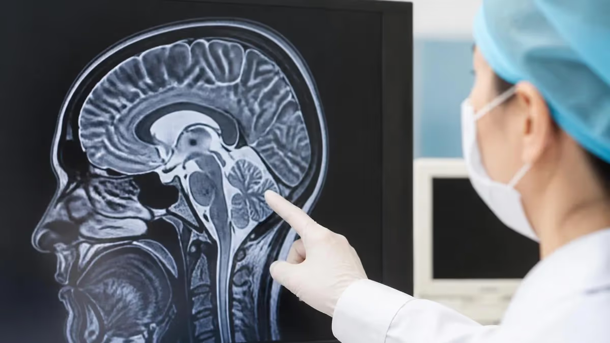
Ischemic changes in brain MRI are among the most frequently reported findings in modern neuroimaging, appearing on scans of patients ranging from acute stroke emergencies to older adults undergoing routine evaluation for headaches or memory loss. Magnetic resonance imaging detects these changes earlier and more precisely than computed tomography, making it the gold standard for assessing reduced blood flow to brain tissue. Understanding how ischemia appears across different MRI sequences helps technologists, students, and clinicians recognize patterns that guide diagnosis, treatment, and prognosis with remarkable accuracy.
When brain tissue loses adequate oxygen and glucose supply, a cascade of cellular events begins within minutes. Diffusion-weighted imaging, or DWI, captures the earliest signal of this injury by detecting restricted movement of water molecules inside swollen, energy-starved cells. A bright DWI signal paired with a dark apparent diffusion coefficient map confirms acute ischemia, often before any other sequence shows abnormality. This combination is the cornerstone of stroke imaging and explains why MRI has become indispensable in emergency neurology departments across the United States.
Not all ischemic changes represent acute events. Chronic small vessel ischemic disease produces scattered or confluent white matter hyperintensities on FLAIR and T2-weighted images, reflecting years of cumulative microvascular damage. Radiologists frequently describe these as nonspecific, age-related, or consistent with chronic microangiopathy. While often clinically silent, extensive white matter disease correlates with cognitive decline, gait disturbance, and increased future stroke risk, so accurate characterization carries genuine importance for long-term patient management and counseling.
The strength of MRI lies in its multiparametric approach. No single sequence tells the whole story; instead, technologists acquire DWI, FLAIR, T1, T2, gradient echo or susceptibility-weighted imaging, and sometimes perfusion and angiographic sequences. Each contributes a different piece of the puzzle, allowing the interpreting physician to distinguish acute from chronic, ischemic from hemorrhagic, and reversible from irreversible injury. This layered information is what separates a confident diagnosis from an ambiguous one in time-critical clinical situations.
For imaging professionals, recognizing ischemic patterns is only part of the picture. A complete brain MRI report addresses many other findings, including masses, demyelination, inflammation, and developmental variants. Knowing how ischemia coexists with or mimics these entities sharpens interpretation and reduces error. Familiarity with normal anatomy across sequences is equally essential, since subtle asymmetries or signal changes are only meaningful when measured against a reliable baseline of what healthy brain tissue should look like.
This guide walks through the full landscape of brain MRI findings, with special emphasis on ischemic changes and how they evolve over time. We cover the sequences that matter, the timeline of stroke evolution, the distinction between acute and chronic disease, and the common abnormalities that appear alongside or in place of ischemia. Whether you are preparing for a registry exam or simply deepening your clinical knowledge, the sections ahead offer practical, structured insight you can apply immediately.
Brain Ischemia on MRI by the Numbers
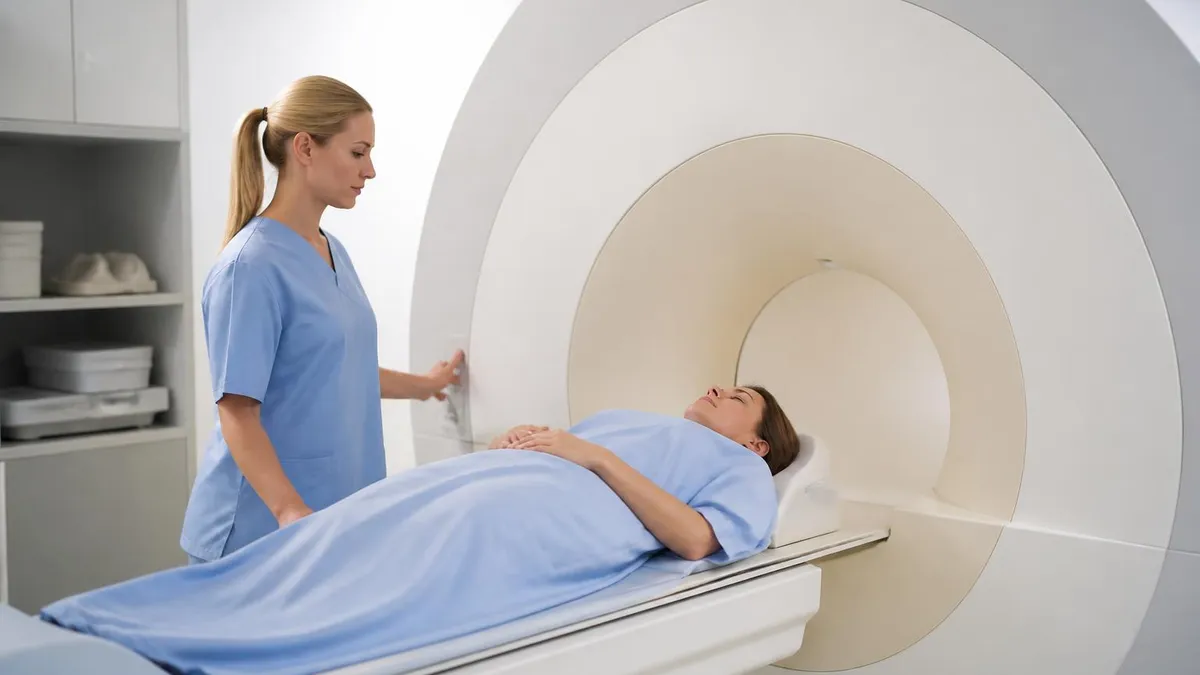
MRI Sequences That Reveal Ischemic Changes
The most sensitive sequence for acute ischemia. Restricted water movement in injured cells produces bright signal within minutes, making DWI the first-line tool for confirming early stroke before other sequences change.
A quantitative map paired with DWI. True acute ischemia shows low (dark) ADC, distinguishing genuine restricted diffusion from T2 shine-through artifact that can falsely brighten DWI images.
Fluid-attenuated inversion recovery suppresses cerebrospinal fluid and highlights white matter hyperintensities. It reveals chronic small vessel disease and subacute infarcts, becoming positive several hours after symptom onset.
Shows edema and gliosis as high signal. Useful for chronic infarcts, established lacunes, and characterizing the age of an ischemic lesion alongside FLAIR and DWI findings.
Susceptibility-weighted and gradient echo sequences detect blood products and microbleeds. They help exclude hemorrhagic transformation and identify chronic microhemorrhages associated with vascular disease.
The evolution of an ischemic stroke on MRI follows a predictable timeline that interpreting physicians use to estimate when an event occurred. In the hyperacute phase, within the first six hours, DWI lights up brightly while the ADC map turns dark, signaling cytotoxic edema. At this stage conventional T2 and FLAIR sequences may appear completely normal, which is precisely why diffusion imaging revolutionized acute stroke care and enabled treatment decisions that older imaging methods could never support.
As hours pass into the acute phase, vasogenic edema develops and FLAIR signal begins to rise. By roughly six to twenty-four hours, the infarct becomes visible on FLAIR and T2 as a region of increased signal, often conforming to a specific vascular territory such as the middle cerebral artery distribution. The combination of bright DWI and emerging FLAIR signal strongly suggests an infarct that is hours old rather than days, refining the clinical picture considerably.
During the subacute phase, spanning several days to two weeks, the lesion reaches peak edema and may show mass effect. Importantly, DWI signal begins to fade through a phenomenon called pseudonormalization, typically around ten to fourteen days, as the ADC value normalizes and then rises. A radiologist who sees normal DWI but bright FLAIR understands the event is likely subacute, not acute, a distinction that meaningfully changes patient counseling and treatment urgency.
In the chronic phase, weeks to months later, the infarcted tissue undergoes encephalomalacia and gliosis. T1-weighted images show low signal volume loss, T2 and FLAIR show high signal gliotic change, and the affected region may collapse into a fluid-filled cavity. These chronic findings are permanent and reflect tissue that can no longer recover, contrasting sharply with the reversible appearance seen in the earliest minutes after vessel occlusion.
Lacunar infarcts deserve special attention because they are so common in patients with hypertension and diabetes. These small deep infarcts, usually under fifteen millimeters, occur in the basal ganglia, thalamus, internal capsule, and pons. Acutely they restrict diffusion like any other infarct, but chronically they leave tiny CSF-filled cavities surrounded by a rim of gliosis. Recognizing lacunes helps build the broader picture of small vessel ischemic burden in an individual patient.
Perfusion imaging adds another dimension by mapping blood flow and identifying the ischemic penumbra, the tissue at risk but not yet infarcted. When perfusion deficit exceeds the diffusion abnormality, a mismatch exists, indicating salvageable brain that may benefit from intervention. This diffusion-perfusion mismatch concept has reshaped how stroke teams select patients for thrombectomy, extending treatment windows for carefully chosen candidates well beyond traditional limits in many comprehensive stroke centers.
Understanding these temporal patterns transforms MRI from a static snapshot into a dynamic story. By correlating DWI, ADC, FLAIR, and perfusion together, the interpreter can confidently place an ischemic event in time, distinguish it from chronic disease, and communicate findings that directly influence whether a patient receives thrombolysis, thrombectomy, or conservative management in the critical hours after symptom onset.
MRI Practice Test Questions
Prepare for the MRI - Magnetic Resonance Imaging exam with our free practice test modules. Each quiz covers key topics to help you pass on your first try.
MRI Knowledge
MRI Exam Questions covering Knowledge. Master MRI Test concepts for certification prep.
MRI Physics
Free MRI Practice Test featuring Physics. Improve your MRI Exam score with mock test prep.
MRI Anatomy and Pathology
MRI Test Prep for MRI Anatomy and Pathology. Practice MRI Quiz questions and boost your score.
MRI Anatomy and Positioning
MRI Questions and Answers on MRI Anatomy and Positioning. Free MRI practice for exam readiness.
MRI Contrast Agents
Free MRI Quiz on MRI Contrast Agents. MRI Exam prep questions with detailed explanations.
MRI Patient Care and Positioning
MRI Practice Questions for MRI Patient Care and Positioning. Build confidence for your MRI certification exam.
Acute vs Chronic Ischemic Changes in Brain MRI
Acute ischemic changes appear within minutes to hours of vessel occlusion. The defining feature is restricted diffusion: bright signal on DWI with corresponding dark ADC. Conventional sequences often lag behind, with FLAIR and T2 remaining normal in the first hours. This DWI-FLAIR mismatch is clinically valuable because it suggests a recent event still within the treatment window for thrombolysis or mechanical thrombectomy.
Acute infarcts typically follow a vascular territory and may show early mass effect as edema accumulates. Identifying the affected artery, whether anterior, middle, or posterior cerebral, helps confirm an embolic or thrombotic mechanism. Prompt recognition of these acute signals enables stroke teams to act within the narrow timeframe that determines whether brain tissue can be salvaged or lost permanently.
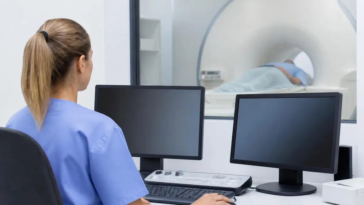
MRI vs CT for Detecting Brain Ischemia
- +Detects acute ischemia within minutes using DWI
- +Far superior sensitivity for small and lacunar infarcts
- +No ionizing radiation exposure to the patient
- +Excellent posterior fossa and brainstem visualization
- +Characterizes lesion age through sequence correlation
- +Identifies salvageable penumbra with perfusion imaging
- −Longer scan times than CT, harder in unstable patients
- −Limited availability for emergent off-hours imaging
- −Contraindicated with many implants and devices
- −Motion artifact degrades images in agitated patients
- −Higher cost than non-contrast head CT
- −Claustrophobia limits some patients without sedation
Pros and Cons at a Glance
| Pros | Cons |
|---|---|
| Detects acute ischemia within minutes using DWI | Longer scan times than CT, harder in unstable patients |
| Far superior sensitivity for small and lacunar infarcts | Limited availability for emergent off-hours imaging |
| No ionizing radiation exposure to the patient | Contraindicated with many implants and devices |
| Excellent posterior fossa and brainstem visualization | Motion artifact degrades images in agitated patients |
| Characterizes lesion age through sequence correlation | Higher cost than non-contrast head CT |
| Identifies salvageable penumbra with perfusion imaging | Claustrophobia limits some patients without sedation |
Checklist for Reporting Ischemic Changes in Brain MRI
- ✓Review DWI first for any region of restricted diffusion.
- ✓Confirm true restriction with a corresponding dark ADC map.
- ✓Assess FLAIR and T2 for edema, gliosis, and lesion age.
- ✓Determine whether findings fit a specific vascular territory.
- ✓Check SWI or gradient echo for hemorrhagic transformation.
- ✓Distinguish acute infarct from chronic small vessel disease.
- ✓Grade white matter hyperintensity burden using the Fazekas scale.
- ✓Identify lacunar infarcts in deep gray and brainstem structures.
- ✓Note any mass effect, midline shift, or herniation risk.
- ✓Correlate imaging findings with clinical timing and symptoms.
Bright DWI plus dark ADC equals acute ischemia
The single most important rule in stroke imaging is to always pair DWI with the ADC map. Bright DWI alone can result from T2 shine-through, but genuine acute ischemia shows bright DWI with truly low ADC signal. This pairing prevents misclassification and confirms cytotoxic edema within minutes of onset.
Ischemic changes rarely exist in isolation on a brain MRI, and a thorough interpretation must account for the many other abnormalities that can appear on the same study. Mass lesions, including primary brain tumors and metastases, often present with surrounding vasogenic edema, enhancement after contrast, and mass effect. Distinguishing a tumor from a subacute infarct can be challenging, but tumors usually do not respect vascular territories and frequently demonstrate irregular enhancement patterns that infarcts in their typical evolution do not produce.
Demyelinating disease, especially multiple sclerosis, generates white matter lesions that can mimic chronic small vessel ischemia on FLAIR. The key distinguishing features include the characteristic ovoid periventricular plaques oriented perpendicular to the ventricles, known as Dawson's fingers, along with involvement of the corpus callosum, juxtacortical regions, and spinal cord. Active demyelinating lesions may enhance and occasionally restrict diffusion at their edges, requiring careful correlation with patient age and clinical history for accurate classification.
Hemorrhage is a critical finding that MRI characterizes with remarkable precision through its evolving signal on T1, T2, and susceptibility-weighted sequences. The age of a bleed can be estimated from the predictable transformation of hemoglobin breakdown products, from oxyhemoglobin to deoxyhemoglobin, methemoglobin, and finally hemosiderin. Recognizing hemorrhage is essential before any consideration of thrombolytic therapy, since converting an ischemic stroke into a hemorrhagic one represents one of the most feared complications in acute stroke management.
Infectious and inflammatory processes also enter the differential for brain MRI findings. Encephalitis, particularly herpes simplex, characteristically involves the temporal lobes and limbic system with edema and restricted diffusion that can superficially resemble infarction. Abscesses produce a classic appearance of central restricted diffusion within a ring-enhancing cavity, a pattern that helps separate them from necrotic tumors. Awareness of these entities prevents the tunnel vision that can occur when an interpreter focuses solely on vascular causes.
Normal variants and benign findings round out the spectrum and must not be overcalled as pathology. Enlarged perivascular spaces, also called Virchow-Robin spaces, follow CSF signal on all sequences and can be mistaken for lacunar infarcts by the inexperienced eye. Developmental venous anomalies, choroid plexus calcifications, and incidental cysts appear regularly. Distinguishing these harmless findings from true disease protects patients from unnecessary anxiety, additional testing, and potential overtreatment based on misinterpreted images.
Atrophy and volume loss represent another common observation, particularly in older patients and those with neurodegenerative conditions. Generalized atrophy widens sulci and enlarges ventricles, while focal atrophy may point to specific dementias such as Alzheimer's disease, which classically affects the hippocampus and medial temporal lobes. Although atrophy itself is not ischemic, it frequently coexists with white matter hyperintensities, and the combined burden offers meaningful insight into a patient's overall brain health and cognitive trajectory.
Bringing these findings together requires a systematic search pattern that examines every brain region, every sequence, and every potential pathology rather than stopping at the first abnormality. A disciplined approach ensures that an obvious acute infarct does not distract the interpreter from a coexisting tumor, microbleed, or developmental lesion. This comprehensive mindset is what elevates competent image review into genuinely expert neuroradiologic interpretation that serves patients well.
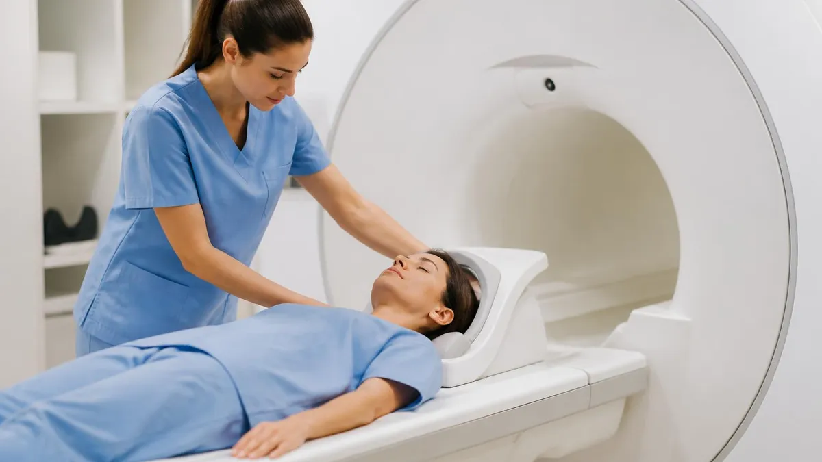
Before any thrombolytic therapy, hemorrhage must be definitively excluded. Susceptibility-weighted and gradient echo sequences are highly sensitive to blood products. Administering clot-busting drugs to a patient with an unrecognized bleed can be catastrophic, making this one of the most time-sensitive decisions in acute stroke imaging.
Even experienced readers encounter pitfalls when interpreting ischemic changes on brain MRI, and recognizing these traps is a hallmark of expertise. The most notorious is T2 shine-through, where a chronic lesion with high T2 signal appears bright on DWI not because of restricted diffusion but because the underlying T2 contrast carries through into the diffusion image. Always confirming the ADC map prevents misreading old gliotic tissue as a new acute stroke and avoids unnecessary, potentially harmful interventions.
Pseudonormalization is another well-known trap. As a subacute infarct ages past roughly ten to fourteen days, the initially dark ADC value rises and crosses back through normal before becoming elevated. During this window the DWI signal can appear deceptively unremarkable, leading an unwary reader to underestimate the lesion. Correlating DWI with FLAIR, which remains bright in subacute infarcts, resolves this ambiguity and keeps the lesion's true age firmly in view.
Distinguishing chronic small vessel disease from demyelination causes frequent uncertainty, particularly in middle-aged patients. Vascular white matter hyperintensities tend to be symmetric, peripheral, and spare the corpus callosum, while demyelinating plaques are often perpendicular to the ventricles and involve the callosum and juxtacortical white matter. Patient age, clinical history, and lesion distribution together usually clarify the diagnosis, but overlap exists and warrants honest acknowledgment of diagnostic limits in the report.
Artifacts can masquerade as pathology if not recognized. Motion degrades DWI and can create ghosting that mimics lesions, while susceptibility artifact near the skull base, sinuses, and metallic hardware can obscure or simulate abnormalities. Magnetic field inhomogeneity and chemical shift add further complexity. A reader who understands the physics behind each sequence can confidently separate true findings from technical artifacts, which underscores why a solid grounding in MRI physics remains essential for accurate interpretation.
Vascular territory knowledge prevents both over- and under-calling infarcts. An acute infarct should generally conform to the supply region of a specific artery, and a lesion that crosses multiple territories or fails to match any vascular distribution should prompt consideration of alternative diagnoses such as tumor, demyelination, or venous infarction. Cerebral venous thrombosis in particular produces atypical, often hemorrhagic infarcts that do not follow arterial boundaries and require dedicated venographic sequences to confirm.
Finally, clinical correlation cannot be overstated. Imaging findings gain meaning only in the context of symptom onset, neurological examination, and risk factors. A bright DWI lesion in a patient with sudden weakness carries different weight than an identical finding discovered incidentally. Communicating clearly with the referring clinician, documenting the estimated lesion age, and flagging any findings that could alter urgent management transforms a technically accurate report into one that genuinely improves patient outcomes.
Building these habits takes deliberate practice and repeated exposure to varied cases. Reviewing prior studies for comparison, maintaining a consistent search pattern, and revisiting difficult cases after the clinical outcome is known all accelerate skill development. Over time, the patterns of ischemia and its mimics become second nature, and the interpreter learns to navigate the subtle distinctions that separate confident, defensible diagnoses from the uncertain reads that frustrate clinicians and patients alike.
Putting brain MRI interpretation into daily practice starts with developing a reliable, repeatable workflow. Begin every study by confirming patient identity, the clinical indication, and the available sequences, then orient yourself with a quick survey of the whole brain before drilling into specifics. Establishing this routine prevents satisfaction of search, the cognitive error in which finding one abnormality causes a reader to stop looking, and it ensures that subtle ischemic changes are never overlooked because attention drifted to an obvious lesion.
When ischemia is suspected, anchor your review on the diffusion images and immediately cross-reference the ADC map. Train your eye to ask three questions for every bright DWI focus: is the ADC truly dark, does the lesion fit a vascular territory, and does the FLAIR signal support an acute or subacute timeframe. Answering these consistently builds the pattern recognition that allows accurate aging of an infarct, which in turn drives appropriate clinical decisions about treatment and prognosis.
For those preparing for registry or board examinations, practice questions are invaluable for reinforcing these concepts under realistic conditions. Working through scenario-based items that ask you to identify the most sensitive sequence, estimate lesion age, or distinguish ischemia from a mimic strengthens recall far more effectively than passive reading. Repeated testing exposes knowledge gaps early, allowing focused study of the sequences, timelines, and distinguishing features that examiners emphasize most heavily on the actual test.
Comparison with prior imaging is one of the most powerful tools available and is frequently underused. A white matter hyperintensity that is unchanged over several years is reassuringly chronic, while a new lesion in the same region demands explanation. Whenever possible, pull old studies and document interval change explicitly. This simple habit resolves much of the ambiguity that surrounds nonspecific findings and dramatically improves the clinical usefulness of your interpretation for the referring team.
Communication skills matter as much as image interpretation. A precise, well-organized report that states the location, age, and vascular territory of an infarct, grades the chronic small vessel burden, and clearly flags any urgent findings serves the patient far better than a vague description. When findings could change immediate management, such as a treatable acute infarct or an unsuspected hemorrhage, a direct phone call to the clinician closes the loop and reflects the professionalism expected of imaging experts.
Staying current is the final ingredient in sustained competence. Stroke imaging continues to evolve, with expanding thrombectomy windows, advanced perfusion software, and refined protocols reshaping how scans are acquired and interpreted. Following reputable continuing education, reviewing updated guidelines, and discussing challenging cases with colleagues keep your skills sharp. The fundamentals of DWI, ADC, and FLAIR remain constant, but the clinical applications grow richer, rewarding those who commit to ongoing learning throughout their careers.
Ultimately, mastering brain MRI findings is a journey of disciplined observation, physics knowledge, and clinical context applied together. Each scan offers a chance to refine your eye and deepen your understanding of how ischemia and its many mimics appear across sequences. With a structured approach, consistent practice, and clear communication, you can interpret these studies with the confidence and accuracy that patients depend on in moments when accurate imaging truly changes the course of their care.
Sample MRI - Magnetic Resonance Imaging Practice Questions
Try these questions from our free MRI - Magnetic Resonance Imaging practice tests. The correct answer and an explanation follow each question.
MR spectroscopy (MRS) of the brain measures metabolite concentrations. Which metabolite, reduced in neuronal loss and gliosis, is considered a marker of neuronal integrity?
- A. Choline (Cho)
- B. Creatine (Cr)
- C. N-acetylaspartate (NAA)
- D. Lactate (Lac)
Answer: C. N-acetylaspartate (NAA)
NAA (N-acetylaspartate) is located predominantly in neurons; its reduction indicates neuronal loss or dysfunction, making it a key spectroscopic marker of neuronal integrity.
Which anatomical landmark is used to prescribe the standard axial plane for brain MRI?
- A. Frankfurt plane
- B. Anterior commissure–posterior commissure (AC-PC) line
- C. Hard palate line
- D. Orbitomeatal baseline
Answer: B. Anterior commissure–posterior commissure (AC-PC) line
Axial brain MRI slices are typically aligned parallel to the AC-PC line for standardized anatomical coverage and comparison.
Gadolinium retention in the brain has been observed particularly in which structure on unenhanced T1-weighted images in patients receiving multiple GBCA doses?
- A. Hippocampus
- B. Dentate nucleus of the cerebellum
- C. Thalamus
- D. Caudate nucleus
Answer: B. Dentate nucleus of the cerebellum
Gadolinium deposition in the dentate nucleus and globus pallidus manifests as T1 hyperintensity on unenhanced brain MRI after multiple GBCA administrations.
For a brain MRI, which standard plane best demonstrates the corpus callosum in its entirety?
- A. Axial
- B. Coronal
- C. Sagittal
- D. Oblique
Answer: C. Sagittal
The midsagittal plane shows the entire corpus callosum from the genu to the splenium as a continuous C-shaped structure.
Take the full MRI - Magnetic Resonance Imaging practice test
MRI Questions and Answers

MRI of Cervical Spine: Complete Guide to Imaging, Anatomy, Pathology, and Patient Preparation
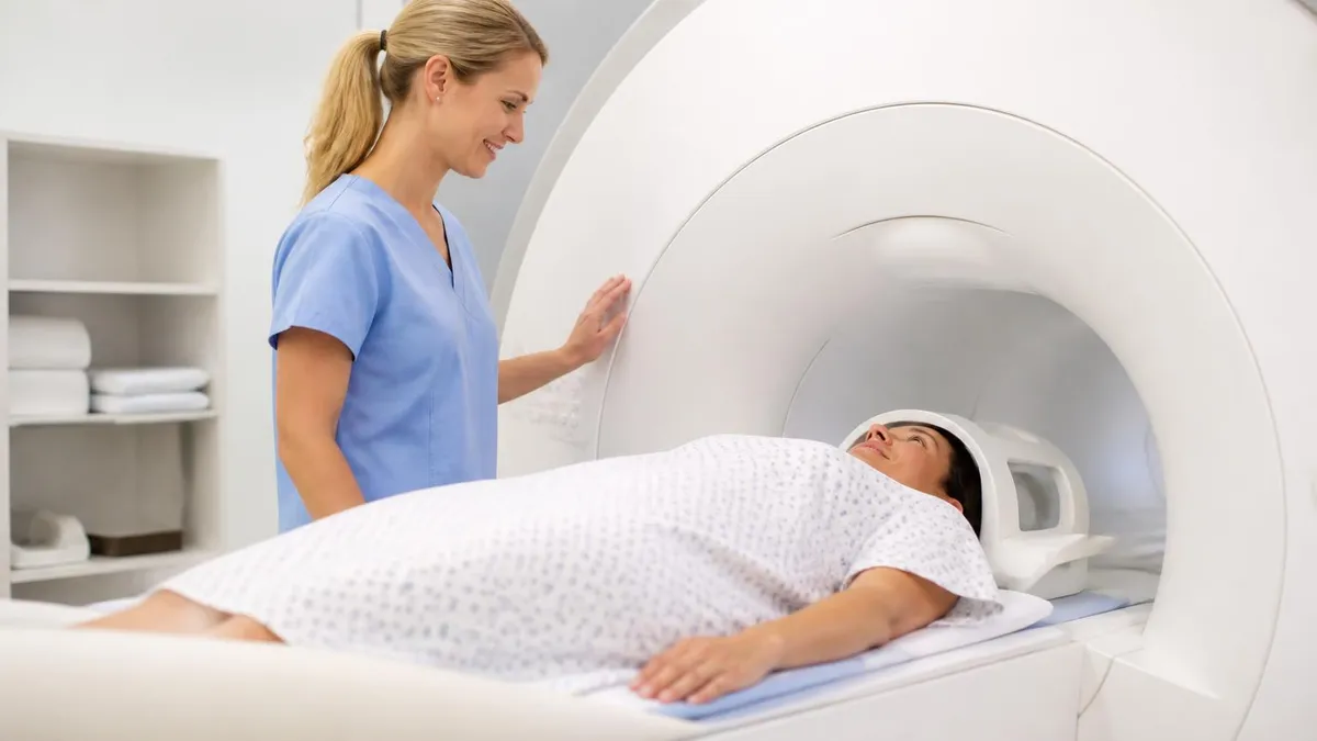
MRI Medical Abbreviation: What MRI Stands For and Why It Matters

Knee MRI Images: A Complete Guide to Reading, Understanding, and Interpreting Knee Scans
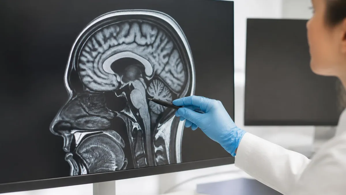
ICD 10 Codes for MRI: Complete Guide to Abnormal MRI Coding, Documentation, and Reimbursement
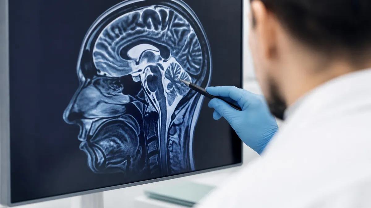
Is Nickel Titanium MRI Compatible? A Complete Guide to MRI Safety Materials
About the Author

Medical Laboratory Scientist & Clinical Certification Expert
Johns Hopkins UniversityDr. Sandra Kim holds a PhD in Clinical Laboratory Science from Johns Hopkins University and is certified as a Medical Technologist (MT) and Medical Laboratory Scientist (MLS) through ASCP. With 16 years of clinical laboratory experience spanning hematology, microbiology, and molecular diagnostics, she prepares candidates for ASCP board exams, MLT, MLS, and specialist certification tests.
Join the Discussion
Connect with other students preparing for this exam. Share tips, ask questions, and get advice from people who have been there.
View discussion (8 replies)