
ERP vs EEG: What Each Test Measures, How They Differ, and What to Expect
🧠 Learn what an EEG test measures vs ERP, how long it takes, costs, side effects, and what results mean. Complete 2026 September guide.

An EEG test — short for electroencephalogram — is one of the most widely ordered neurological evaluations in modern medicine, recording the brain's electrical activity through small sensors placed on the scalp. When clinicians and researchers discuss erp vs eeg, they are drawing a distinction between two related but importantly different applications of the same core electrode technology: the routine clinical EEG test used to diagnose seizure disorders, and the event-related potential (ERP) technique used primarily in neuroscience research to study cognitive processing.
Understanding what an EEG medical test actually measures helps clarify why these two approaches serve such different purposes. During a standard EEG test, electrodes capture spontaneous, ongoing electrical oscillations produced by millions of neurons firing in coordinated patterns. Clinicians look at the overall rhythm and amplitude of these waves — delta, theta, alpha, beta, and gamma bands — to identify abnormalities associated with epilepsy, encephalopathy, sleep disorders, and brain injuries. The signal is continuous and reflects the brain's baseline state rather than its response to a specific stimulus.
An event-related potential, by contrast, is derived from the same raw EEG signal but is extracted through a technique called signal averaging. A participant is presented with dozens or hundreds of repeated stimuli — a sound, a visual flash, a decision prompt — while the EEG records continuously. Because random neural noise cancels out across many trials while the stimulus-locked response stays consistent, averaging reveals tiny voltage deflections that occur at precise time points after each stimulus. These deflections, labeled by polarity and latency (P300, N200, N400), reflect specific stages of sensory and cognitive processing invisible in ordinary EEG recordings.
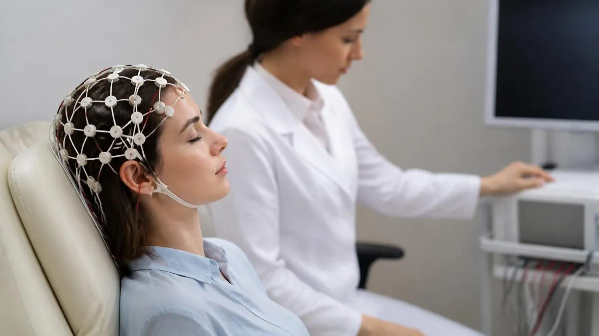
For patients wondering what is an EEG test and whether it involves any of this complexity, the reassuring answer is that a routine clinical EEG is straightforward and painless. Gel-filled electrodes are applied to the scalp following the standardized 10-20 system, you relax or sleep for 20 to 40 minutes, and the technologist monitors brain waves in real time. No needles, no radiation, and no recovery period are required. The ERP variant adds controlled stimulus delivery and longer recording sessions but is otherwise equally non-invasive.
The practical relevance of distinguishing ERP from EEG extends beyond academic curiosity. Neurologists ordering an EEG medical test for suspected epilepsy need the spontaneous rhythm data that a standard recording provides; a researcher studying working memory or language comprehension needs the averaged ERP waveforms that reveal millisecond-level cognitive events. Conflating the two can lead to confusion about what a particular test result means, what equipment is required, and why the cost and duration differ between clinical and research contexts.
This guide walks through both techniques in detail — covering what an EEG test involves, how long an EEG test takes, what EEG test costs look like in 2026, potential EEG test side effects, and exactly how ERP methodology builds on and differs from standard EEG acquisition. Whether you are a patient preparing for a routine recording, a student studying for a neurodiagnostic certification, or a researcher new to electrophysiology, you will find practical, accurate information organized to answer your most pressing questions about this foundational brain-imaging technology.
Because the EEG test remains the gold standard for real-time, millisecond-resolution brain activity monitoring — offering spatial coverage unavailable to single-electrode measures and temporal precision unavailable to fMRI — mastering the distinction between clinical EEG and ERP recording is an essential step in understanding modern neurological and cognitive neuroscience practice alike.
EEG Test by the Numbers

How EEG and ERP Recording Work
Both EEG and ERP recordings use the international 10-20 electrode placement system, positioning 19 to 256 electrodes at standardized scalp locations. Consistent placement allows recordings to be compared across sessions, labs, and patients worldwide, ensuring clinical reliability.
A standard clinical EEG captures continuous, spontaneous brain oscillations over 20 to 40 minutes. Technologists instruct patients to relax, open and close their eyes, breathe deeply, or sleep to elicit different brain states for diagnostic comparison.
ERP extraction requires presenting dozens to hundreds of repeated stimuli and mathematically averaging the resulting EEG epochs. Averaging cancels random noise and reveals consistent voltage deflections — P100, N200, P300 — locked to stimulus onset with millisecond precision.
Both techniques require careful artifact rejection to remove eye blinks, muscle activity, and electrode noise. ERP research demands more rigorous preprocessing pipelines — including independent component analysis — because cognitive components are far smaller than typical clinical EEG signals.
EEG amplifiers boost microvolt-scale brain signals to measurable levels while filtering out powerline interference. Clinical systems typically sample at 256 Hz; research-grade systems use 512 Hz to 2,000 Hz to capture the fine temporal structure required for ERP component analysis.
Understanding what a standard EEG test involves from the patient's perspective begins well before the appointment. Most neurologists advise washing your hair the night before and avoiding conditioners, oils, or styling products that create a greasy barrier between the scalp and the electrode gel. You should also get a normal night's sleep unless your doctor specifically orders a sleep-deprived EEG, which deliberately captures the drowsy-to-sleep transition to provoke abnormal discharges that only appear during that state transition.
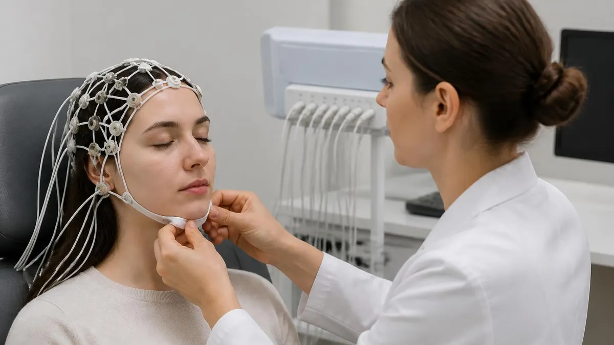
On the day of the test, a registered EEG technologist will measure your head and mark scalp positions with a soft pencil, then apply a small amount of conductive gel at each site before attaching the electrodes. The entire setup process typically takes 20 to 30 minutes.
Modern systems use flat metal disc electrodes held in place with adhesive paste or a cap embedded with pre-positioned sensors; neither option is painful, though some patients find the paste slightly cold or the cap mildly snug. Once the electrodes are connected to the amplifier, the technologist performs an impedance check to confirm that each electrode is making clean electrical contact with the scalp.
During the recording itself, you recline in a chair or lie on a table in a dimly lit room. The technologist will ask you to perform several activation procedures — opening and closing your eyes on command, breathing rapidly for three minutes (hyperventilation), and watching a strobe light flicker at varying frequencies (photic stimulation). These maneuvers are standard parts of what is an EEG test because they can provoke seizure activity in susceptible individuals that might not appear during resting recording.
Approximately 30 to 50 percent of people with epilepsy show abnormal discharges during hyperventilation, making it a diagnostically critical step.
For patients undergoing a sleep EEG or a prolonged recording, the session may last one to two hours. A 24-hour ambulatory EEG involves wearing a portable recorder at home, allowing neurologists to capture brain activity during the patient's natural sleep-wake cycle and to correlate any suspicious events with real-life triggers. Video-EEG monitoring, performed in an inpatient epilepsy monitoring unit, adds synchronized video recording so that clinical behaviors during recorded events can be directly compared with the simultaneous EEG pattern — the gold standard for characterizing seizure type and lateralizing the seizure focus before surgery.
Because the EEG test has been performed for nearly a century, its safety profile is exceptionally well established. EEG test side effects are rare and almost always minor: some patients experience a brief headache from the electrode cap pressure, others feel lightheaded after the hyperventilation phase (which is intentional and transient), and a small number develop a mild scalp irritation from the electrode gel.
Serious adverse events are exceedingly uncommon; the main clinical risk is that the activation procedures can provoke a seizure in patients with known or undiagnosed epilepsy, which is precisely the diagnostic goal rather than an accidental complication. Medical staff are trained to manage any seizure safely during the recording.
After the recording ends, the technologist removes the electrodes and helps you wash the gel from your hair — though most patients prefer to shower at home, as the gel is water-soluble and easy to remove. You can drive yourself home and return to all normal activities immediately because no sedation is used and there is no recovery period.
The raw EEG data is then reviewed by a board-certified neurologist or clinical neurophysiologist, who generates a formal report describing the dominant background rhythm, any focal or generalized abnormalities, the response to activation procedures, and an overall interpretation comparing your findings to age-matched norms.
One frequently asked question is whether an EEG can definitively diagnose epilepsy. The answer is nuanced: a normal EEG does not rule out epilepsy, because up to 50 percent of patients with clinically diagnosed epilepsy have a normal interictal (between-seizure) routine EEG. Conversely, about two percent of healthy adults show epileptiform discharges on EEG without ever experiencing clinical seizures. The EEG result must always be interpreted in the context of the patient's history, physical examination, and other diagnostic data — a principle that underscores why the EEG is a powerful but inherently probabilistic tool rather than a simple binary test.
EEG Test Cost, Duration, and What Affects Pricing
The EEG test cost in the United States typically ranges from $200 to $700 for a routine outpatient recording at a hospital or neurology clinic, though prices vary significantly by geography, facility type, and whether the study is read by an in-house neurologist or sent for remote interpretation. Uninsured patients paying out of pocket at community hospitals or independent diagnostic centers frequently find costs at the lower end of this range, while academic medical centers in high-cost cities may charge $500 to $700 before insurance adjustments. The EEG test price is separate from the neurologist's interpretation fee, which typically adds $100 to $250 to the total bill.
Extended studies cost considerably more. A 24-hour ambulatory EEG generally runs $500 to $1,500, and a video-EEG inpatient admission — which can last several days in an epilepsy monitoring unit — can exceed $10,000 before insurance. Medicare and most commercial insurance plans cover medically necessary EEGs when ordered for documented neurological indications such as seizure evaluation, altered consciousness, or cognitive decline workup. Patients should always verify their specific plan's deductible, copay, and whether the interpreting neurologist is in-network, as out-of-network interpretation is a common source of surprise billing in this specialty.

EEG vs ERP: Strengths and Limitations of Each Approach
- +Millisecond temporal resolution captures neural events faster than any other non-invasive brain imaging method
- +Completely non-invasive with no radiation, injections, or recovery time required
- +Portable EEG systems enable bedside monitoring in ICUs, ambulatory home studies, and intraoperative recordings
- +ERP averaging reveals cognitive components (P300, N400) invisible in spontaneous EEG, enabling precise cognitive research
- +Relatively low cost compared to MRI or PET neuroimaging for equivalent clinical indications
- +Continuous real-time monitoring is uniquely possible with EEG, enabling detection of subclinical seizures in comatose patients
- −Poor spatial resolution — standard 19-electrode systems cannot localize activity more precisely than a few centimeters
- −ERP requires many repeated trials, making it unsuitable for studying rare or non-repeatable cognitive events
- −Susceptible to artifacts from eye blinks, muscle tension, sweat, and electrode movement that can obscure or mimic brain signals
- −A normal EEG does not rule out epilepsy — up to 50% of epilepsy patients have normal interictal recordings
- −Volume conduction blurs the true source of scalp-recorded activity, complicating source localization without advanced modeling
- −ERP components lack diagnostic specificity outside research — no ERP biomarker is currently approved as a standalone clinical diagnostic tool
EEG Test Preparation Checklist: What to Do Before Your Appointment
- ✓Wash your hair the night before with regular shampoo and skip conditioner, oils, and styling products.
- ✓Follow your doctor's specific instructions about sleep — get a full night unless a sleep-deprived EEG was ordered.
- ✓Take all regular medications as prescribed unless your neurologist explicitly tells you to hold them.
- ✓Avoid caffeine for at least 8 hours before the test, as it alters brain wave patterns and can mask abnormalities.
- ✓Eat a light meal before arriving — low blood sugar can affect the EEG recording and may provoke symptoms.
- ✓Wear comfortable, loose-fitting clothing so you can relax easily during the 20-to-40-minute recording session.
- ✓Arrange a ride if you feel anxious about the test, though driving yourself is safe since no sedation is used.
- ✓Bring a list of all medications, supplements, and their doses for the technologist to document.
- ✓Inform the technologist of any scalp conditions, rashes, or piercings that might affect electrode placement.
- ✓Plan to arrive 15 minutes early to complete paperwork and allow the technologist adequate setup time.
The 50% Rule Every Patient Should Know
Up to 50% of people with clinically confirmed epilepsy have a completely normal routine EEG recorded between seizures. A single 30-minute recording captures only a small window of brain activity, and most patients do not happen to generate epileptiform discharges during that specific window. Serial EEGs, sleep-deprived recordings, and prolonged ambulatory or video-EEG monitoring substantially increase diagnostic yield and should be pursued when clinical suspicion remains high despite a normal initial result.
Reading and interpreting EEG results requires a thorough understanding of normal developmental variation as well as the full spectrum of pathological findings. The dominant background rhythm in a relaxed adult with eyes closed should be 8 to 13 Hz alpha activity, maximal over the posterior scalp regions.
This rhythm attenuates — becomes less prominent — when the eyes open or when mental activity increases, a phenomenon called alpha blocking or the alpha-desynchronization response. A posterior dominant rhythm below 8 Hz in an adult is considered abnormal and may indicate diffuse cerebral dysfunction, metabolic encephalopathy, or drowsiness that was not adequately accounted for during the recording.
Focal slowing — the presence of delta or theta waves localized to one scalp region — indicates a structural or functional abnormality in the underlying cortex. Common causes include cortical infarction, tumor, abscess, traumatic contusion, or focal epileptogenic tissue. Generalized slowing, by contrast, suggests a diffuse process affecting the whole brain: encephalopathy from metabolic derangements (liver failure, kidney failure, thyroid dysfunction), toxic exposures, severe sleep deprivation, or neurodegenerative disease. Distinguishing focal from generalized abnormalities is one of the most important contributions the EEG makes to the neurological evaluation because these two categories point toward fundamentally different diagnostic and management pathways.
Epileptiform discharges are the EEG findings most specifically associated with seizure disorders. These include interictal epileptiform discharges (IEDs) — spikes, sharp waves, and spike-wave complexes that appear between clinical seizures — and ictal patterns, which are the rhythmic or evolving EEG changes that occur during an actual seizure.
The morphology, distribution, and repetition rate of epileptiform discharges provide information about seizure type: three-per-second generalized spike-wave complexes are the hallmark of childhood absence epilepsy, focal spikes in the temporal region suggest mesial temporal lobe epilepsy (the most common adult focal epilepsy syndrome), and hypsarrhythmia — chaotic high-amplitude multifocal spikes — is the defining EEG pattern of West syndrome in infants.
Beyond epilepsy, the EEG contributes diagnostic value in several other clinical situations. In patients with altered consciousness or coma, the EEG reveals whether the brain is producing adequate cortical activity or whether subclinical seizures are perpetuating the comatose state.
In patients with dementia, characteristic EEG patterns can help differentiate Alzheimer's disease — which produces generalized slowing with posterior accentuation in advanced stages — from Creutzfeldt-Jakob disease, which produces periodic sharp wave complexes with a characteristic one-per-second periodicity that is highly specific for prion disease. The EEG also confirms brain death in some jurisdictions, where electrocerebral silence (a completely flat recording) combined with appropriate clinical criteria meets legal and ethical standards for brain death determination.
In the research context, ERP components serve as precise windows into cognitive processing stages. The P300 — a positive deflection occurring approximately 300 milliseconds after an infrequent or task-relevant stimulus — reflects processes of attention, working memory updating, and stimulus context evaluation.
Reduced P300 amplitude and delayed latency have been documented in schizophrenia, attention-deficit/hyperactivity disorder, mild cognitive impairment, and alcohol use disorder, making it one of the most widely studied candidate biomarkers in cognitive neuroscience. The N400, a negative deflection peaking around 400 milliseconds following a semantically incongruent word or image, reflects the ease of integrating a word into its surrounding context and has been used to study language comprehension, bilingualism, and semantic memory.
The mismatch negativity (MMN) component — an automatic, attention-independent response to a deviant stimulus within a series of standard stimuli — is particularly valuable in populations who cannot perform tasks, including comatose patients, infants, and individuals with severe cognitive or communication impairments.
Research using MMN has demonstrated residual auditory discrimination in some patients classified as being in a vegetative state, raising profound questions about consciousness and the degree to which ERP-based tools might eventually inform clinical decision-making in disorders of consciousness. These findings represent the frontier where the research ERP technique begins intersecting with clinical neurology in ways that may transform how neurologists assess awareness and prognosis in their most challenging patients.
Advances in source localization methods — including dipole modeling, independent component analysis, and high-density EEG combined with individual MRI-based head models — have substantially improved the spatial resolution of both clinical EEG and ERP recordings. While scalp EEG still cannot match the millimeter resolution of fMRI, modern source analysis techniques can localize epileptiform activity or ERP generators to specific cortical regions with accuracy approaching that of intracranial recordings in favorable cases, particularly when 64 or more electrodes are used and the source is superficial and well-circumscribed.

If your doctor ordered an EEG test, you will receive a clinical EEG — a spontaneous recording interpreted by a neurologist for diagnostic purposes. Event-related potential (ERP) studies are research tools conducted in university and hospital research labs, not ordered as routine clinical tests. Unless you are enrolled in a research study, you will not be asked to perform cognitive tasks or complete hundreds of stimulus trials during your diagnostic EEG appointment.
Event-related potential research has matured over six decades from a curiosity of sensory physiology into a mainstay methodology in cognitive neuroscience, clinical psychology, and increasingly in translational neurology. The technique's core strength is its ability to decompose human cognition into discrete, temporally ordered processing stages that map onto distinct neural events — a capability no other non-invasive imaging method currently matches.
When a participant reads a sentence ending in an unexpected word, the N400 response reveals not only that the brain detected the semantic mismatch but exactly how many milliseconds elapsed before that detection occurred and whether the detection was impaired by neurological disease, pharmacological manipulation, or developmental factors.
The relationship between ERP methodology and clinical EEG practice is best understood as a layered one: every ERP study begins with EEG acquisition, uses the same electrode systems and amplifiers, and applies the same basic principles of electrical neurophysiology. What differentiates them is the analytical strategy — averaging versus continuous spectrum analysis — and the experimental context — controlled stimulus presentation versus naturalistic recording.
A skilled neurodiagnostic technologist who understands the EEG signal at a deep level is well positioned to understand ERP methodology; conversely, a cognitive neuroscientist trained in ERP analysis brings sophisticated signal-processing expertise that complements clinical pattern recognition.
For patients exploring brain-computer interface (BCI) technologies, the P300 ERP component occupies a central role. The P300-based speller — developed in the 1980s and refined through decades of research — allows patients with amyotrophic lateral sclerosis (ALS), locked-in syndrome, or severe motor disabilities to communicate by focusing attention on rows and columns of letters that flash in sequence.
The system detects the P300 response when the target letter flashes and uses it to identify the intended letter with accuracy exceeding 90 percent in experienced users. This application illustrates how a research ERP component discovered in a laboratory setting can evolve into a practical clinical assistive technology, underscoring the translational value of fundamental ERP research.
Steady-state visual evoked potentials (SSVEPs) represent another bridge between clinical evoked potential testing and research ERP methodology. SSVEPs are elicited by flickering visual stimuli at specific frequencies (typically 6 to 30 Hz) and produce robust EEG responses at the stimulation frequency and its harmonics.
Clinically, SSVEPs are used to assess visual cortex integrity and are measured routinely as part of the visual evoked potential (VEP) battery in patients with multiple sclerosis, optic neuritis, and other conditions affecting the visual pathway. In research and BCI contexts, SSVEPs are harnessed as a control signal because they are large, frequency-specific, and do not require the participant to learn a new cognitive strategy — simply gazing at a flickering target is sufficient to generate a strong, classifiable signal.
The overlap between EEG and ERP methodology also extends to clinical applications of quantitative EEG (qEEG), in which digital signal processing techniques — spectral analysis, coherence, asymmetry measures, and connectivity metrics — extract numerical features from the continuous EEG signal to create topographic brain maps or to derive biomarkers for psychiatric conditions. While qEEG remains controversial in some clinical contexts, its use in monitoring anesthesia depth during surgery (using processed EEG indices such as the bispectral index, or BIS) is firmly established and represents one of the most widespread clinical applications of continuous quantitative EEG analysis.
Students and clinicians preparing for the ABRET (American Board of Registration of Electroencephalographic and Evoked Potential Technologists) examination will encounter questions about both routine EEG interpretation and evoked potential testing — including visual, auditory brainstem, and somatosensory evoked potentials, which are the clinical cousins of research ERPs.
Understanding how signal averaging, stimulus parameters, and electrode placement interact in evoked potential testing is directly tested on certification examinations, making the conceptual framework of ERP methodology practically important for anyone pursuing a career in neurodiagnostic technology. Practice questions focusing on activation procedures, electrode placement, and pattern recognition are an efficient way to solidify this knowledge base before examination day.
For anyone comparing the clinical value of the EEG test against other neuroimaging modalities, the EEG's unique combination of excellent temporal resolution, broad clinical applicability, low cost, and non-invasiveness ensures that it will remain indispensable even as higher-resolution technologies continue to improve. No other bedside tool can tell a neurologist in real time whether a comatose patient is experiencing subclinical seizures, approaching burst-suppression under anesthesia, or showing the periodic sharp wave complexes of prion disease — capabilities that make the EEG not merely a historical artifact of pre-MRI neurology but an actively essential tool in twenty-first-century neurological care.
Practical preparation strategies for neurodiagnostic technology certification begin with mastering the foundational knowledge domains that appear most frequently on ABRET examinations: electrode placement and the 10-20 system, normal EEG patterns across the lifespan, recognition of common artifact types, and identification of epileptiform and non-epileptiform abnormalities. These domains are not independent — understanding normal developmental EEG patterns from neonates through elderly adults provides the essential baseline against which every abnormality is judged, and artifact recognition is only reliable when you can confidently distinguish a genuine brain-generated signal from an electrode or patient-movement artifact.
One of the most efficient study strategies is repeated exposure to annotated EEG tracings from diverse patient populations and clinical contexts. Reviewing examples of absence epilepsy with classic three-per-second spike-wave complexes alongside examples of benign childhood epilepsy with centrotemporal spikes, Lennox-Gastaut syndrome with slow spike-wave, and the various encephalopathic patterns builds the pattern-recognition fluency that written descriptions alone cannot achieve. When supplementing tracing review with practice questions, focus on explaining why each answer is correct rather than simply memorizing the right choice — the examination tests understanding of underlying mechanisms, not rote recall of isolated facts.
For the ERP and evoked potential sections of neurodiagnostic certification examinations, concentrate on understanding the neural generators and clinical significance of the major components: the brainstem auditory evoked potential (BAEP) waves I through V and their localization to cochlear nerve and brainstem structures; the pattern-reversal visual evoked potential (PVEP) P100 wave and its prolongation in demyelinating disease; and the somatosensory evoked potential (SSEP) cortical N20 component as an index of thalamocortical pathway integrity. These are the clinically applied evoked potential measures that appear regularly in certification questions and in day-to-day neurodiagnostic practice.
Time management during EEG certification examination preparation matters as much as content coverage. Candidates who spread study over eight to twelve weeks consistently outperform those who compress preparation into intensive short sprints, because neurophysiology knowledge requires time to consolidate and interconnect across content domains. Building a weekly schedule that alternates between content review (reading, watching annotated EEG videos) and active recall (practice questions, teaching back concepts) leverages spaced repetition and retrieval practice — the two learning strategies with the strongest evidence base in educational neuroscience.
When preparing for the practical components of certification that involve real-time EEG acquisition, focus on troubleshooting common electrode problems during a live recording. Understanding how to identify and correct a high-impedance electrode (which appears as increased noise or artifact on that channel), how to recognize 60 Hz electrical interference and mitigate it through impedance reduction or notch filter application, and how to distinguish eye movement artifact from frontal slow waves are competencies that examiners specifically probe because they distinguish a technologist who can produce a high-quality recording from one who merely attaches electrodes and presses record.
Review the clinical contexts in which each EEG variant is indicated: routine outpatient EEG for initial seizure evaluation or treatment monitoring, sleep-deprived EEG when routine recording is non-diagnostic and clinical suspicion for epilepsy remains high, prolonged outpatient ambulatory EEG for characterizing event frequency and capturing typical episodes, and inpatient video-EEG monitoring for pre-surgical epilepsy evaluation or for definitively characterizing events that might be either epileptic or non-epileptic in origin. Each modality has specific preparation requirements, recording duration norms, and interpretive considerations that are tested in detail.
Finally, build familiarity with the legal, ethical, and safety dimensions of neurodiagnostic practice. ABRET certification examination content includes questions about infection control procedures between patients, electrical safety standards for EEG equipment in hospital environments, protocols for managing a seizure that occurs during a recording, and the technologist's scope of practice with respect to preliminary interpretation versus formal clinical reporting. These professional practice questions are easily overlooked by candidates who focus exclusively on the neurophysiology content, yet they represent a meaningful portion of the examination and reflect the real-world responsibilities of the registered EEG technologist in clinical practice.
EEG Questions and Answers
EEG vs ECG vs EKG: What Each Test Measures, How They Differ, and What to Expect
EEG Electroencephalography Practice Test PDF (Free Printable 2026)

Seizure EEG: What the EEG Test Shows, How It Works, and What to Expect
EEG Test Cost in 2026: Prices, Insurance Coverage, and How to Save

Invasive EEG: What the EEG Test Involves, How It Works, and What to Expect
About the Author

Educational Psychologist & Academic Test Preparation Expert
Columbia University Teachers CollegeDr. Lisa Patel holds a Doctorate in Education from Columbia University Teachers College and has spent 17 years researching standardized test design and academic assessment. She has developed preparation programs for SAT, ACT, GRE, LSAT, UCAT, and numerous professional licensing exams, helping students of all backgrounds achieve their target scores.