EEG and Sleep: How Brain Wave Testing Diagnoses Sleep Disorders 2026 July
Complete guide to EEG and sleep testing: 💡 what an EEG test measures, cost, duration, side effects, and how it diagnoses sleep disorders.
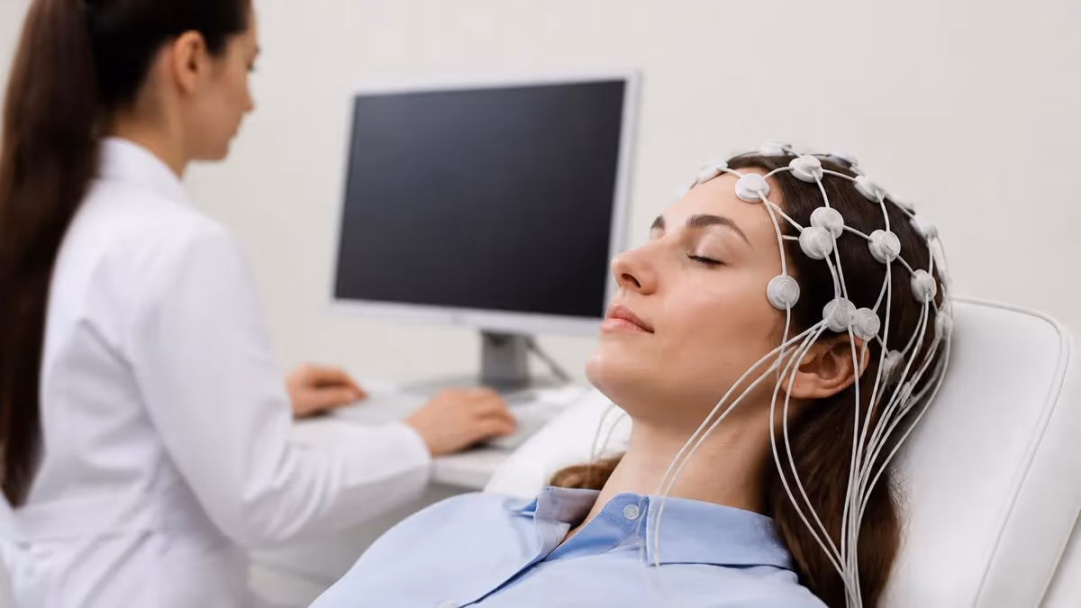
The relationship between EEG and sleep is one of the most important in clinical neurology, because brain wave recordings reveal what no other test can show: the precise architecture of consciousness as it shifts from wakefulness through light sleep, deep sleep, and REM. An EEG test, short for electroencephalogram, captures the electrical chatter of millions of cortical neurons through small sensors placed on the scalp, producing a continuous tracing that doctors interpret to diagnose seizures, sleep disorders, and other neurological conditions affecting how the brain rests and recovers.
Sleep medicine relies heavily on EEG because traditional symptoms like daytime fatigue, snoring, or insomnia cannot distinguish between obstructive sleep apnea, narcolepsy, parasomnias, or nocturnal seizures. Only by recording the brain directly can clinicians identify which sleep stage is disrupted, how long it lasts, and whether abnormal patterns are intruding where they should not be. This is why polysomnography, the formal sleep study, always includes a multi-channel EEG component alongside breathing, oxygen, and muscle monitoring.
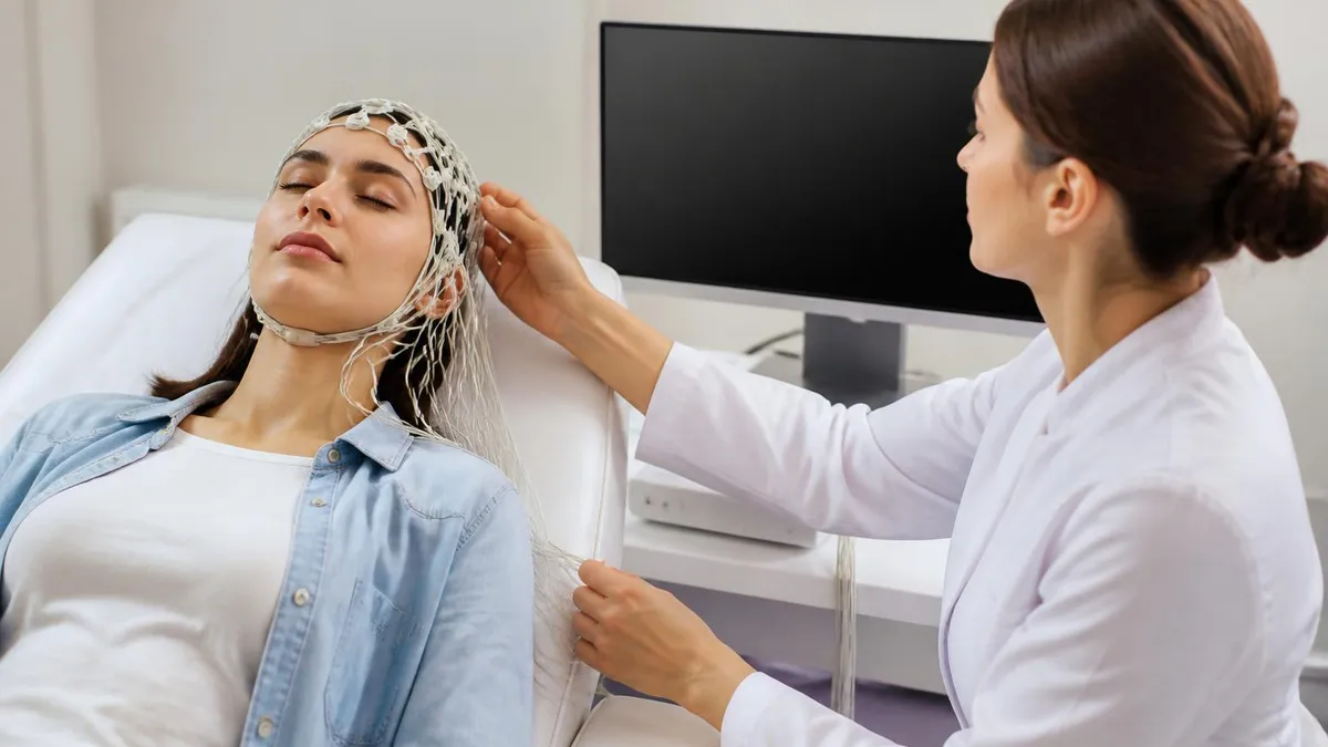
If you have been scheduled for a sleep-related EEG, you probably want to know exactly what is eeg test going to feel like, how long the recording takes, what the readings actually mean, and how much it will cost. The good news is that EEG is non-invasive, painless, and carries virtually no risks for healthy adults or children. The electrodes only listen; they never deliver electricity to the brain, and the gel used to attach them washes out with shampoo afterward.
This guide walks through everything you need to understand about EEG and sleep: the science behind the brain waves observed during each sleep stage, the differences between routine EEG, ambulatory EEG, and full overnight polysomnography, the typical price range across US clinics and hospitals, the rare side effects to watch for, and the practical preparation steps that improve recording quality. We will also cover how technologists score the study and how neurologists translate squiggly lines into a diagnosis.
Sleep-deprived EEG protocols are particularly common when doctors suspect epilepsy, because sleep deprivation provokes epileptiform discharges that might stay hidden during a normal recording. Patients are asked to stay awake most of the night before the test, then drift off naturally during the appointment. The combination of drowsiness and the natural sleep cycle increases diagnostic yield by 30 to 70 percent depending on the underlying condition, making this one of the most powerful tools in the neurologist's kit.
Whether your physician ordered the study to investigate suspected seizures, evaluate a sleep disorder, rule out narcolepsy with a multiple sleep latency test, or monitor cognitive recovery after a brain injury, the underlying technology is similar. By the end of this article you will know what to expect, how to prepare, what the results mean, and how to discuss findings with your care team in confident, informed language.
We will also touch on the career side, because EEG technologists who specialize in sleep studies represent a growing professional segment with strong job growth projected through the end of the decade. If you are exploring this field as a patient or a future tech, the same brain wave knowledge applies in both directions.
EEG and Sleep by the Numbers

Sleep Stages on EEG
Awake recordings show low-voltage fast beta activity with eyes open, transitioning to dominant 8-12 Hz alpha rhythm over posterior regions when eyes close. This baseline is critical for interpretation.
The first stage of sleep features alpha dropout, slow rolling eye movements, and the emergence of low-amplitude mixed-frequency activity. Vertex sharp waves may appear, lasting only minutes before deeper stages.
Characterized by 12-14 Hz sleep spindles and K-complexes, this stage accounts for nearly half of total sleep time. It plays a major role in memory consolidation and is required for restorative rest.
Slow-wave sleep dominated by high-amplitude delta waves below 4 Hz. This is the deepest, most restorative stage where growth hormone is released and the brain clears metabolic waste through glymphatic flow.
Rapid eye movement sleep produces low-voltage mixed frequencies resembling wakefulness, paired with muscle atonia and dreaming. REM disruptions are key markers in narcolepsy and REM behavior disorder.
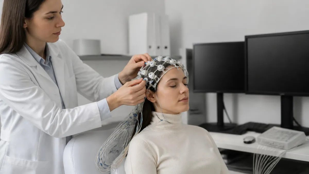
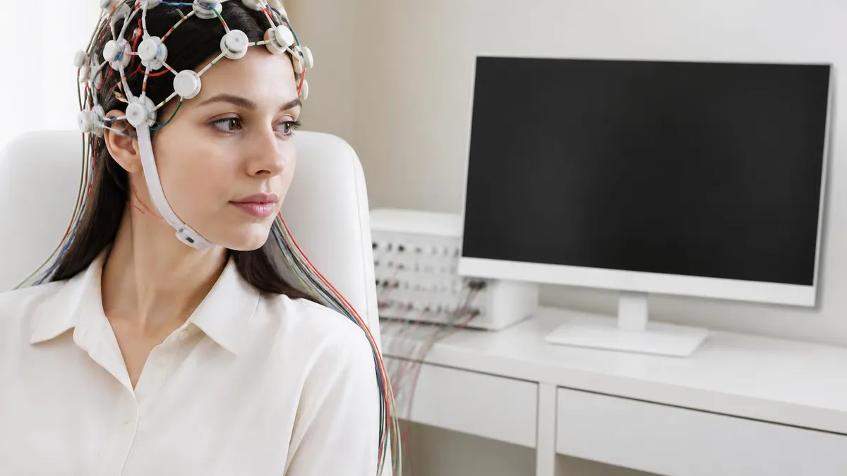
So eeg medical test recordings translate the brain's electrical activity into waveforms by amplifying the tiny voltages, measured in microvolts, that travel between cortical neurons. A standard clinical setup uses 21 electrodes placed according to the international 10-20 system, named because electrodes sit at intervals of 10 or 20 percent of the distance between standard skull landmarks. Each electrode reports a voltage relative to a reference electrode, and the differences are plotted in real time on screen or paper.
What is an EEG test measuring, precisely? It is not measuring thoughts or mental content, despite popular misconceptions. It captures summed postsynaptic potentials from pyramidal neurons in the cortex, particularly those oriented perpendicular to the scalp surface. These summed signals are coherent enough to penetrate skull and skin and reach the recording electrodes, where amplifiers boost them roughly 10,000 times so they can be visualized as oscillating waveforms with distinct frequencies and amplitudes.
Sleep introduces dramatic changes in this electrical signature. Wakefulness shows predominantly fast beta activity, with alpha rhythm emerging over the back of the head when eyes close. As drowsiness deepens, alpha breaks up and disappears, replaced by slower theta frequencies. Vertex sharp waves announce stage N1, followed by sleep spindles and K-complexes in N2. Slow delta waves of high amplitude define N3, the deepest sleep, and REM produces a paradoxically alert-looking pattern despite muscle paralysis.
EEG is uniquely valuable in sleep medicine because abnormal patterns surface most readily during sleep transitions. Many forms of epilepsy show epileptiform discharges only during drowsiness or light sleep, when cortical excitability is heightened. Nocturnal seizures, sleepwalking events, and REM behavior disorder can be distinguished from each other only by combining EEG with video recording and muscle tone monitoring. Without the brain wave channel, sleep disorders blur together clinically.
Modern digital EEG systems also support quantitative analysis, including spectral analysis, coherence measurements, and automated sleep staging algorithms that assist technologists in scoring overnight studies. While computers can flag patterns of interest, final interpretation always belongs to a board-certified neurologist or sleep medicine specialist who has trained to recognize artifacts, normal variants, and pathological patterns within the broader clinical context.
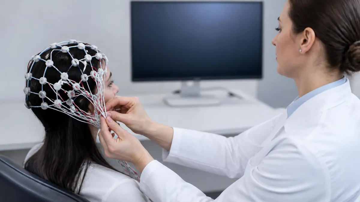
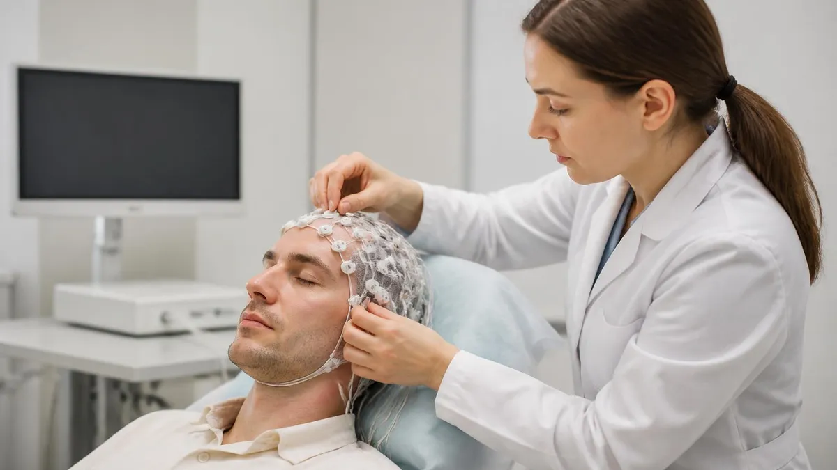
The recording itself is silent and painless. Electrodes are attached with conductive paste or collodion adhesive that holds them firmly against the scalp without restricting movement. Patients can blink, swallow, breathe, and shift position naturally, though excessive movement creates artifact that the technologist will ask you to minimize during critical recording windows. The overall experience is more like having stickers placed on your head than undergoing a medical procedure.
Results are typically available within a few days, sometimes longer for ambulatory studies that produce 24 to 72 hours of continuous data requiring careful review. Your neurologist will discuss findings in plain language, explaining whether the EEG was normal, showed minor non-specific changes, or revealed patterns consistent with a specific diagnosis. Most patients leave their first appointment with a clearer understanding of their condition.
EEG Practice Test Questions
Prepare for the EEG - Electroencephalography exam with our free practice test modules. Each quiz covers key topics to help you pass on your first try.
EEG Abnormal Epileptiform Patterns
EEG Exam Questions covering Abnormal Epileptiform Patterns. Master EEG Test concepts for certification prep.
EEG Activation Procedures
Free EEG Practice Test featuring Activation Procedures. Improve your EEG Exam score with mock test prep.
EEG Artifact Identification
EEG Test Prep for Artifact Identification. Practice EEG Quiz questions and boost your score.
EEG Electrode Placement and Montages
EEG Questions and Answers on Electrode Placement and Montages. Free EEG practice for exam readiness.
EEG - Electroencephalography Abnormal Epil...
EEG Mock Test covering - Electroencephalography Abnormal Epileptiform Patterns. Online EEG Test practice with instant feedback.
EEG - Electroencephalography Activation Pr...
Free EEG Quiz on - Electroencephalography Activation Procedures. EEG Exam prep questions with detailed explanations.
EEG - Electroencephalography Artifact Iden...
EEG Practice Questions for - Electroencephalography Artifact Identification. Build confidence for your EEG certification exam.
EEG - Electroencephalography Electrode Pla...
EEG Test Online for - Electroencephalography Electrode Placement and Montages. Free practice with instant results and feedback.
EEG - Electroencephalography Instrumentati...
EEG Study Material on - Electroencephalography Instrumentation and Calibration. Prepare effectively with real exam-style questions.
EEG - Electroencephalography Neuroanatomy ...
Free EEG Test covering - Electroencephalography Neuroanatomy and Physiology. Practice and track your EEG exam readiness.
EEG - Electroencephalography Non-Epileptif...
EEG Exam Questions covering - Electroencephalography Non-Epileptiform Abnormalities. Master EEG Test concepts for certification prep.
EEG - Electroencephalography Normal EEG Wa...
Free EEG Practice Test featuring - Electroencephalography Normal EEG Waveforms. Improve your EEG Exam score with mock test prep.
EEG - Electroencephalography Patient Safet...
EEG Mock Exam on - Electroencephalography Patient Safety and Ethics. EEG Study Guide questions to pass on your first try.
EEG - Electroencephalography Pediatric and...
EEG Test Prep for - Electroencephalography Pediatric and Neonatal EEG. Practice EEG Quiz questions and boost your score.
EEG ICU and Continuous EEG Monitoring
EEG Questions and Answers on ICU and Continuous EEG Monitoring. Free EEG practice for exam readiness.
EEG Instrumentation and Calibration
EEG Mock Test covering Instrumentation and Calibration. Online EEG Test practice with instant feedback.
EEG Neuroanatomy and Physiology
Free EEG Quiz on Neuroanatomy and Physiology. EEG Exam prep questions with detailed explanations.
EEG Non-Epileptiform Abnormalities
EEG Practice Questions for Non-Epileptiform Abnormalities. Build confidence for your EEG certification exam.
EEG Normal EEG Waveforms
EEG Test Online for Normal EEG Waveforms. Free practice with instant results and feedback.
EEG Patient Safety and Ethics
EEG Study Material on Patient Safety and Ethics. Prepare effectively with real exam-style questions.
Types of EEG Test for Sleep Evaluation
The routine EEG is a 20 to 40 minute recording performed in a quiet clinic room. It captures wakefulness, drowsy transitions, and often a brief period of light sleep, along with activation procedures like hyperventilation and photic stimulation to provoke abnormalities. It is the most common entry point for sleep-related EEG evaluation.
How long is an EEG test in this setup? Most appointments take 60 to 90 minutes total once you include electrode placement, the recording itself, and removal. The actual brain wave recording is shorter, but careful electrode application is what produces clean, interpretable data. Routine EEG is the most affordable option and works well for first-line screening.
Is an EEG Sleep Study Right for You?
- +Completely painless and non-invasive procedure
- +No radiation exposure unlike CT or X-ray imaging
- +Captures real-time brain activity during sleep stages
- +Can detect seizures, sleep disorders, and parasomnias
- +Relatively affordable compared to MRI or PET scans
- +Safe for children, pregnant patients, and older adults
- +Provides immediate raw data for technologist review
- −Requires sitting still for extended periods
- −Hair must be clean and free of styling products
- −Sleep-deprived protocols disrupt your normal schedule
- −Electrode paste leaves residue requiring shampoo to remove
- −Ambulatory units are bulky and somewhat noticeable
- −Results may show normal findings even when symptoms persist
- −Insurance preauthorization can delay scheduling by weeks
How to Prepare for Your EEG Test
- ✓Wash your hair the night before with regular shampoo only
- ✓Avoid conditioner, hair oils, gels, sprays, or styling products
- ✓Skip caffeine the morning of the test unless told otherwise
- ✓Eat a normal meal beforehand to prevent low blood sugar artifact
- ✓Continue prescription medications unless your neurologist says otherwise
- ✓Bring a list of current medications and recent symptom diary
- ✓Wear comfortable clothing that opens in the front for easy adjustment
- ✓Arrange a driver if sleep-deprived protocol is scheduled
- ✓Bring a pillow or blanket for comfort during the recording
- ✓Plan to wash hair again at home to remove conductive paste
Sleep deprivation boosts seizure detection by up to 70%
Studies consistently show that combining sleep deprivation with EEG recording dramatically increases the chance of capturing epileptiform discharges. If your neurologist orders a sleep-deprived protocol, the inconvenience of staying up most of the night is genuinely worth it for diagnostic accuracy.
The eeg test price varies enormously across the United States depending on geography, facility type, insurance coverage, and the specific protocol ordered. A routine 30-minute EEG at an outpatient neurology clinic typically runs between $200 and $700 without insurance, while hospital-based recordings often exceed $1,000 due to facility fees. Sleep-deprived studies cost roughly the same as routine recordings, but ambulatory EEG with 24 to 72 hours of monitoring can range from $1,500 to $3,000.
Full overnight polysomnography, which includes multi-channel EEG alongside respiratory and cardiac monitoring, is more expensive still, with cash prices commonly between $1,000 and $3,000 per night at accredited sleep centers. Home sleep tests are cheaper, often $200 to $500, but they include limited EEG channels and are designed primarily for screening obstructive sleep apnea rather than comprehensive sleep architecture analysis.
Insurance typically covers EEG when ordered by a physician for an appropriate clinical indication, such as suspected seizures, unexplained loss of consciousness, evaluation of sleep disorders, or follow-up of a known neurological condition. Most plans require prior authorization, particularly for ambulatory or extended studies. Your copay or coinsurance share depends on your deductible status and plan structure, but in-network EEG generally costs patients $50 to $300 out of pocket.
Medicare covers diagnostic EEG under Part B at 80 percent of the approved amount after the annual deductible. Medicaid coverage varies by state but generally includes EEG when medically necessary. If you are uninsured, ask the clinic about cash-pay discounts, which often reduce the price by 30 to 50 percent. Many academic medical centers also offer sliding-scale fees or financial hardship programs.
The EEG test cost is generally a small fraction of the value it provides when results lead to accurate diagnosis and effective treatment. Untreated epilepsy or undiagnosed sleep disorders carry far higher long-term costs in medications, emergency visits, lost work productivity, and accident risk. Looking at EEG as an investment in diagnostic clarity rather than a discretionary expense reframes the price question in a more useful way.
Pricing transparency has improved in recent years thanks to federal hospital price disclosure rules, but the published rates often differ from what you will actually pay after insurance. The best approach is to call the clinic's billing department before scheduling, provide your insurance details, and ask for an estimate of patient responsibility. Most facilities will work with you to find a manageable arrangement.
Watch out for separate charges that can surprise patients. The technical fee covers the recording itself, while the professional fee covers the neurologist's interpretation. Some clinics bundle these, but others bill them separately, which can effectively double the apparent price. Always ask whether the quoted figure includes both components or just one.
EEG recordings often generate two separate bills: a technical fee from the facility and a professional fee from the interpreting neurologist. Even if the facility is in-network, the reading physician may be out-of-network. Confirm both components are covered before scheduling to avoid balance billing surprises.
EEG test side effects are remarkably rare given how common the procedure is. The electrodes only listen for electrical signals; they never deliver current to the brain. This makes EEG one of the safest diagnostic tests in medicine, with no radiation exposure, no contrast dye, and no needles for most protocols. The vast majority of patients leave the appointment feeling exactly as they did when they arrived, perhaps with slightly mussed hair from the conductive paste.
The most common minor side effect is skin irritation where electrodes were attached, typically a faint redness that resolves within hours. People with very sensitive skin or allergies to collodion adhesive may develop a mild contact dermatitis, but this is uncommon and easily managed with gentle cleansing. If you have known adhesive allergies, mention this when scheduling so the technologist can use alternative attachment methods.
Sleep-deprived protocols carry their own indirect risks, mostly related to driving fatigue. Patients are strongly advised to arrange transportation home, because operating a vehicle on two hours of sleep significantly impairs reaction time. The activation procedures during the recording itself, including hyperventilation and photic stimulation, can occasionally trigger seizures in patients with epilepsy, but this happens in a controlled setting with trained staff who are prepared to respond.
Photic stimulation, which involves a strobe light flashing at varying frequencies, can rarely provoke a photosensitive seizure or, more commonly, mild dizziness, headache, or brief visual discomfort. If you have a history of photosensitive epilepsy, the technologist will modify or skip this portion of the test. Patients with severe migraine sometimes report a transient headache afterward, usually relieved with rest and hydration.
For those interested in the technical side of brain wave testing, exploring what is a eeg test from the technologist's perspective reveals the careful protocols designed to minimize patient risk. Registered EEG technologists complete rigorous training in patient safety, infection control, and emergency response, which is part of why complication rates remain so low across millions of recordings performed annually in the United States.
Children tolerate EEG well, though some toddlers find electrode placement uncomfortable. Pediatric labs use child-friendly approaches like watching cartoons, having a parent present, and explaining each step in age-appropriate language. Sedation is occasionally used for very young or developmentally delayed children, but most pediatric EEGs are completed without medication, preserving the natural sleep architecture the test needs to capture.
Ambulatory EEG carries the same minimal risk profile, with the addition that patients must avoid swimming or submerging the recording equipment in water. Skin irritation can be slightly more common because electrodes stay in place for days rather than hours. The technologist provides cleaning instructions and a contact number in case any electrodes loosen or the equipment malfunctions during the home recording.
Beyond the basics, several practical tips can dramatically improve your EEG sleep study experience and the quality of the data your neurologist receives. Communication with the technologist is the single most important factor. If you feel anxious, uncomfortable, or unable to relax, say so. Experienced techs have many strategies for helping nervous patients settle, from dimming lights and adjusting room temperature to playing white noise that masks ambient hospital sounds.
Maintain a symptom diary leading up to your appointment, especially if you are being evaluated for episodic events. Note times, triggers, duration, and any witness observations. This context dramatically improves the neurologist's ability to correlate brain wave findings with clinical reality. Even seemingly minor details like which limb twitched first or whether you bit your tongue can reshape the diagnostic interpretation of an otherwise ambiguous tracing.
Bring entertainment for ambulatory recordings. While you cannot use heavily electronic devices that introduce artifact during certain windows, books, podcasts, and quiet activities help pass the time. Avoid vigorous exercise that would saturate the tracing with muscle artifact. Eat normally, hydrate well, and treat the recording period as an opportunity for genuine rest, which incidentally produces the cleanest, most interpretable data.
For sleep-deprived protocols, plan the night carefully. Stay up watching a movie, reading, or working quietly. Avoid alcohol, which alters sleep architecture in ways that complicate EEG interpretation. Do not nap before the appointment, even briefly, because residual sleep can blunt the deprivation effect. Caffeine guidance varies by clinic; some allow morning coffee while others prohibit it entirely, so follow your specific instructions.
If results come back normal but symptoms continue, do not assume the EEG ruled out a problem. A single recording captures only a snapshot of brain activity, and many serious neurological conditions can show normal interictal tracings. Discuss with your neurologist whether repeat testing, longer-duration ambulatory monitoring, or video EEG is appropriate. Persistence in pursuing answers is often what eventually identifies elusive conditions.
For abnormal results, take notes during your follow-up appointment and ask for written summaries you can review later. Bring a family member or friend if possible, because complex medical information is easier to process with a second listener. Request copies of the actual EEG report and tracings, which become valuable if you ever need a second opinion or change providers in the future.
Finally, view your EEG as one piece of a larger diagnostic puzzle rather than a definitive verdict. Clinical history, physical examination, imaging studies, blood work, and longitudinal observation all contribute to neurological diagnosis. The most skilled neurologists integrate EEG findings with the whole picture, producing assessments that are far more accurate than any single test could deliver alone. Trust the process, ask questions freely, and stay engaged with your care plan.
EEG Questions and Answers
EEG for Seizures: How the Test Diagnoses Epilepsy and What to Expect
EEG Tech Salary 2026: Pay by State, Experience, and Certification
EEG Images: A Visual Guide to Brain Wave Patterns, Recordings, and Test Results
EEG Tech Jobs in 2026: Career Paths, Salaries, and How to Get Hired
Reading EEG Patterns: A Practical Reference for Techs and Trainees
About the Author
Educational Psychologist & Academic Test Preparation Expert
Columbia University Teachers CollegeDr. Lisa Patel holds a Doctorate in Education from Columbia University Teachers College and has spent 17 years researching standardized test design and academic assessment. She has developed preparation programs for SAT, ACT, GRE, LSAT, UCAT, and numerous professional licensing exams, helping students of all backgrounds achieve their target scores.