
ECMO vs Dialysis: Is ECMO Like Dialysis? Key Differences Explained 2026 October
Is ECMO like dialysis? Learn how extracorporeal membrane oxygenation differs from dialysis in function, circuit, and clinical use. 🎓

A question that surfaces frequently in ICU waiting rooms and medical education alike is: is ECMO like dialysis? The short answer is that both are extracorporeal therapies — meaning blood leaves the body, passes through an external machine, and returns — but that is where the similarity largely ends. Extracorporeal membrane oxygenation in neonates, adults, and pediatric patients provides life support for the heart and lungs, while dialysis supports failing kidneys. Understanding the distinction matters enormously for patients, families, and clinicians preparing for critical care certification exams.
The extracorporeal membrane oxygenation procedure involves withdrawing blood from a large vein or artery, running it through a membrane oxygenator that adds oxygen and removes carbon dioxide, and returning the warmed, oxygenated blood to the patient's circulation. Dialysis, by contrast, pulls blood through a semipermeable membrane that filters waste products and excess fluid, then returns the cleansed blood without altering its gas content. The therapeutic targets, flow rates, and circuit components are fundamentally different between the two systems.
From a flow-rate standpoint, ECMO circuits typically operate at 2 to 7 liters per minute to support or replace cardiac and pulmonary function. Standard hemodialysis runs at roughly 300 to 500 milliliters per minute — an order of magnitude lower — because kidney filtration requires far less throughput than oxygenating the entire cardiac output. This difference alone signals that the two machines are engineered for entirely different physiological purposes, even though both route blood outside the body.
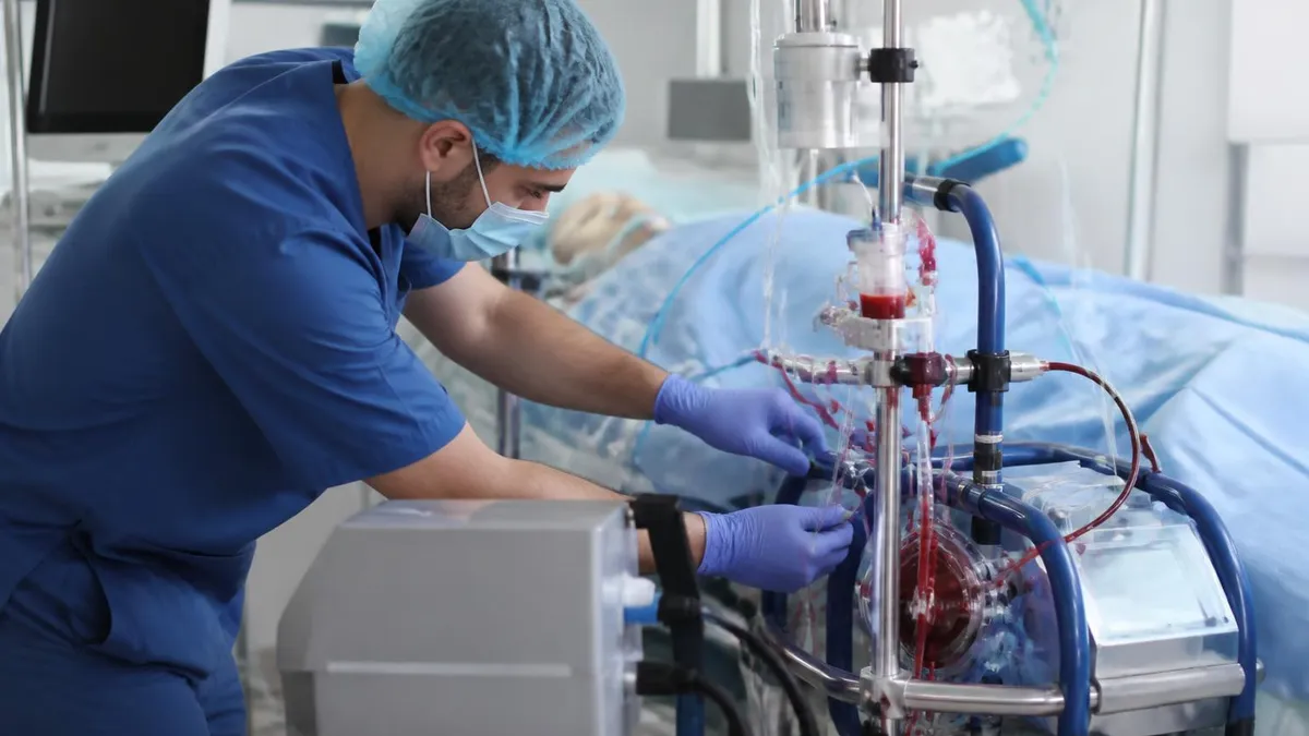
The extracorporeal membrane oxygenation circuit includes a centrifugal or roller pump, a gas-exchange membrane (the oxygenator), a heat exchanger to maintain body temperature, and a complex array of tubing primed with saline or blood products. A dialysis circuit is simpler: a blood pump, a dialyzer cartridge with hollow fibers, and a dialysate delivery system. ECMO circuits must sustain high pressures and flows without hemolysis; dialysis circuits must maintain precise solute gradients and ultrafiltration pressures. For a deeper dive into circuit components, see our guide on ecmo vs dialysis circuit design.
Anticoagulation management is another major differentiator. Because ECMO circuits expose a large surface area of foreign material to blood at high flow rates, continuous heparin infusions titrated to activated clotting time (ACT) or anti-Xa levels are required around the clock. Dialysis patients also receive anticoagulation, but the exposure duration per session is typically three to four hours, allowing for intermittent dosing strategies or even regional citrate anticoagulation that limits systemic effects.
Patient monitoring intensity differs dramatically as well. ECMO patients require continuous bedside nursing in a specialized unit with sweep gas adjustments, oxygenator changes when clot burden builds, and vigilant surveillance for limb ischemia in venoarterial configurations. Dialysis patients on an outpatient basis may attend three sessions per week at a free-standing center with considerably less moment-to-moment oversight. Even critically ill patients receiving continuous renal replacement therapy (CRRT) in the ICU face a monitoring burden that is lower than that of ECMO.
This article explores how extracorporeal membrane oxygenation treatment compares to dialysis across mechanism, patient population, equipment cost, complications, and clinical indications — giving students, nurses, respiratory therapists, and perfusionists the structured knowledge they need to master this distinction on exams and in practice.
ECMO vs Dialysis by the Numbers

Core Mechanisms: How ECMO and Dialysis Work
Blood passes through a membrane oxygenator where oxygen is added and CO₂ is removed. A centrifugal pump replaces or assists the heart's pumping function, supporting patients with severe cardiac or respiratory failure.
Blood flows across a semipermeable hollow-fiber membrane. Diffusion and convection remove uremic toxins, potassium, and excess fluid. The kidneys' filtration role is replaced, but gas exchange and cardiac function remain untouched.
Continuous renal replacement therapy (CRRT) is often run simultaneously on ECMO patients who develop acute kidney injury. CRRT circuits can be integrated into the ECMO circuit or run in parallel, highlighting how these therapies can coexist.
Both ECMO and dialysis require systemic or circuit anticoagulation to prevent clotting on foreign surfaces. ECMO demands continuous heparin infusion with ACT targets of 180–220 seconds; dialysis may use intermittent heparin or citrate.
The extracorporeal membrane oxygenation circuit is substantially more complex than a dialysis circuit, reflecting the higher physiological demands placed on it. At its core, the ECMO circuit consists of a centrifugal pump head (or older roller pump), a polymethylpentene (PMP) membrane oxygenator, a heat exchanger integrated within or adjacent to the oxygenator, and several meters of polyvinyl chloride or silicone tubing. Each component is chosen to minimize thrombogenicity, resist protein adsorption, and maintain gas transfer efficiency over days to weeks of continuous use. The entire circuit is typically coated with heparin or other biocompatible surface treatments to reduce clot formation.
A dialysis circuit, whether used for intermittent hemodialysis or continuous venovenous hemofiltration (CVVH), is designed around a dialyzer cartridge containing thousands of hollow fibers with pores small enough to retain proteins and blood cells while allowing small solutes and water to pass. The blood pump speed is set to 300–400 mL/min, and a separate dialysate pump delivers a buffered electrolyte solution countercurrent to blood flow to maximize concentration gradients. The engineering priorities — tight pore size control, precise transmembrane pressure regulation, and bicarbonate buffering — are entirely different from those of the ECMO circuit.
Oxygenator performance is the central concern in ECMO circuit management. Clinicians monitor pre- and post-membrane pressures to detect fibrin accumulation, inspect the oxygenator for clot streaks under bright light, and measure post-oxygenator PO₂ to confirm adequate gas transfer. When the pressure gradient across the oxygenator rises above 50–60 mmHg or gas transfer deteriorates, the oxygenator must be exchanged — a high-stakes bedside procedure. Dialyzer performance is assessed differently: blood flow resistance, transmembrane pressure, and effluent urea concentration determine adequacy and filter lifetime.
Tubing length and priming volume matter greatly in neonatal ECMO. Neonates undergoing extracorporeal membrane oxygenation in neonates configurations may weigh as little as 2 kg, meaning the ECMO circuit prime volume can exceed the patient's total blood volume. This necessitates priming the circuit with packed red blood cells, fresh frozen plasma, or whole blood rather than crystalloid to prevent severe hemodilution and coagulopathy on initiation. Dialysis circuits for small patients present similar challenges, but the priming volume is lower and the physiological consequences of hemodilution during connection are generally less acute because flows are lower.
Cannula size and placement strategy also diverge significantly. ECMO cannulas are large-bore — typically 15–23 French for drainage and 15–21 French for return — positioned in central vessels such as the jugular vein, femoral vein, or femoral artery depending on the ECMO mode. Dialysis catheters, usually 11–14 French double-lumen lines, are placed in central veins but tolerate much lower flow rates and shorter dwelling times before replacement. ECMO cannulas may remain in place for weeks, introducing infection and thrombosis risks that require specific surveillance protocols distinct from those for dialysis catheters.
Heat exchange is critical in ECMO because blood exposed to the environment in plastic tubing cools rapidly at flows of several liters per minute. ECMO oxygenators incorporate water-jacketed heat exchangers that maintain blood at 35–37°C during normothermic ECMO or allow deliberate cooling to 32–34°C when mild therapeutic hypothermia is desired after cardiac arrest. Dialysis machines also warm dialysate and returning blood, but the thermal mass considerations are less acute given the lower flow rates involved. Understanding these circuit-level differences is essential for any clinician managing critically ill patients on extracorporeal support.
Cost is another stark differentiator. The extracorporeal membrane oxygenation machine price for a complete system — including the console, oxygenator, tubing pack, and cannulas — typically ranges from $30,000 to over $100,000 depending on manufacturer and configuration. Ongoing consumable costs for circuit changes, blood products for priming, and oxygenator replacements add substantially to this figure. A hemodialysis machine costs $15,000–$25,000 for the capital equipment, with per-session consumable costs of $30–$80. The resource intensity of ECMO explains why it remains concentrated in large academic centers rather than distributed across community hospitals.
Venovenous Extracorporeal Membrane Oxygenation, VA-ECMO, and ECMO in COVID
Venovenous extracorporeal membrane oxygenation is used exclusively for respiratory failure when the heart is functioning adequately. Blood is drained from a central vein — commonly the right internal jugular or femoral vein — oxygenated, and returned to a different central vein. Because VV-ECMO does not provide direct cardiac support, patients must maintain sufficient native cardiac output to distribute the oxygenated blood delivered by the circuit. This configuration is the predominant mode used for severe ARDS, including cases seen during the COVID-19 pandemic.
The hallmark of VV-ECMO management is sweep gas titration: increasing oxygen sweep flow raises PO₂, while increasing total sweep gas flow removes more CO₂. Clinicians use serial arterial blood gases to guide these adjustments. Recirculation — where oxygenated blood returning to the venous system is immediately siphoned back into the drainage cannula before reaching the right heart — is a key complication unique to VV configurations, detectable by an unexpectedly high oxygen saturation in the drainage limb and treatable by repositioning cannulas or adjusting flow rates.

ECMO vs Dialysis: Benefits and Limitations Compared
- +ECMO provides life support for both cardiac and pulmonary failure simultaneously — no dialysis equivalent exists
- +VV-ECMO allows complete lung rest, enabling healing from severe ARDS while maintaining oxygenation
- +VA-ECMO can sustain perfusion during cardiac arrest, functioning as a bridge to definitive therapy
- +ECMO can be combined with CRRT for patients with simultaneous cardiac, pulmonary, and renal failure
- +Extracorporeal membrane oxygenation in neonates has dramatically improved survival from congenital diaphragmatic hernia and PPHN
- +Modern ECMO circuits with polymethylpentene oxygenators can safely run for weeks with careful management
- −ECMO carries a 10–30% risk of major bleeding complications due to mandatory anticoagulation and circuit consumption of clotting factors
- −Extracorporeal membrane oxygenation machine price and per-day running costs make it one of the most expensive ICU interventions available
- −Neurological complications including stroke affect 5–15% of ECMO patients, particularly in VA configurations
- −ECMO requires 24/7 specialized bedside nursing and perfusion coverage not available at most hospitals
- −Limb ischemia distal to arterial cannulation occurs in up to 20% of peripheral VA-ECMO patients without distal perfusion cannulas
- −Infection risk from large-bore cannulas dwelling in central vessels increases with duration of support beyond 14 days
Clinical Monitoring Checklist: ECMO vs Dialysis Key Differences
- ✓Confirm ECMO mode (VV vs VA) before interpreting oxygenation parameters — VV does not provide cardiac support
- ✓Check pre- and post-oxygenator pressure gradient every 4 hours to detect fibrin accumulation in the ECMO circuit
- ✓Monitor distal limb perfusion hourly in peripheral VA-ECMO using pulse oximetry on the ipsilateral foot
- ✓Titrate ECMO sweep gas independently of ventilator settings — sweep oxygen controls PaO₂, sweep flow controls PaCO₂
- ✓Distinguish CRRT circuit from ECMO circuit when both are running — never mistake dialysis effluent for ECMO circuit output
- ✓Review anticoagulation targets: ECMO ACT goal typically 180–220 sec vs dialysis heparin protocols targeting lower ACT
- ✓Assess for recirculation in VV-ECMO by comparing drainage limb saturation to systemic venous saturation
- ✓Document ECMO flow in L/min and dialysis blood pump speed in mL/min separately to avoid clinical confusion
- ✓Monitor platelet count and fibrinogen daily — ECMO consumes platelets and promotes hypofibrinogenemia more than dialysis
- ✓Evaluate neurological status at each nursing shift — stroke risk is highest in VA-ECMO and requires early detection
ECMO and Dialysis Can Run Simultaneously in Critically Ill Patients
Up to 50% of adult patients on ECMO develop acute kidney injury severe enough to require renal replacement therapy. CRRT circuits are commonly integrated into the ECMO circuit via a bridge between the drainage and return limbs, allowing both therapies to operate with a single blood pump. This integrated configuration reduces the total extracorporeal volume and the number of venous access sites required, but demands careful monitoring of flows across both systems to avoid circuit collapse or air entrainment.
Extracorporeal membrane oxygenation in neonates occupies a unique and historically significant place in critical care. ECMO was first successfully used in a neonate in 1975 by Robert Bartlett at the University of Michigan, and it revolutionized the treatment of persistent pulmonary hypertension of the newborn (PPHN), meconium aspiration syndrome, congenital diaphragmatic hernia (CDH), and neonatal cardiac anomalies. Before ECMO, mortality rates for severe neonatal respiratory failure exceeded 80%; in experienced centers today, survival rates for neonatal ECMO approach 70–75% for respiratory indications, though CDH remains more challenging with 50–60% survival.
The neonatal ECMO circuit presents unique technical challenges not encountered in adults. Circuit priming volume — typically 300–500 mL in standard circuits — can exceed the neonate's total blood volume of approximately 80 mL/kg. For a 2.5 kg neonate, that represents a total blood volume of only 200 mL, meaning the prime is more than twice the patient's circulating volume.
To prevent catastrophic hemodilution, neonatal circuits are primed with packed red blood cells, albumin, and sodium bicarbonate to achieve near-physiologic hematocrit, pH, and oncotic pressure before cannulation. This stands in sharp contrast to adult ECMO circuits, which are typically primed with crystalloid alone.
Cannula selection for neonates follows strict size-to-vessel ratio guidelines. Neck cannulation via surgical cutdown of the right internal jugular vein and right common carotid artery remains standard for neonatal VV and VA-ECMO respectively, as femoral vessels in neonates are too small for adequate flows. The right common carotid artery is ligated after decannulation in most centers, raising questions about long-term cerebrovascular development that have been studied extensively in ELSO registry follow-up data. Neurodevelopmental outcomes in ECMO survivors are an active research area, with many centers now conducting structured follow-up programs through age 5 and beyond.
Extracorporeal membrane oxygenation for adults follows different cannulation and management paradigms. Adult patients are most commonly cannulated in the femoral vein and femoral artery (for VA-ECMO) or femoral vein and right internal jugular vein (for VV-ECMO) using percutaneous Seldinger technique under ultrasound guidance. The availability of large-bore percutaneous cannulas has made ECMO initiation faster and less dependent on surgical expertise, enabling deployment in cardiac catheterization laboratories and emergency departments for ECPR. Adult ECMO flow targets are weight-based, typically 60–80 mL/kg/min to achieve full cardiopulmonary bypass or 80–100% support of cardiac output.
The extracorporeal membrane oxygenation diagram most commonly shown in educational materials depicts the VV-ECMO configuration: a drainage cannula in the inferior vena cava via the femoral vein, blood passing through the pump and oxygenator, and oxygenated blood returning via the right internal jugular vein into the right atrium. This layout illustrates how blood is oxygenated before entering the right heart and pulmonary circulation, with native lung function supplemented or replaced. VA-ECMO diagrams show the return cannula positioned in the femoral artery, with oxygenated blood injected retrograde into the aorta to support systemic perfusion.
Drug management on ECMO differs substantially from standard ICU pharmacology. The extracorporeal membrane oxygenation circuit sequesters lipophilic, protein-bound drugs in the tubing and oxygenator, reducing plasma concentrations of medications including fentanyl, midazolam, vecuronium, and many antibiotics. Sedation requirements on ECMO are often 2–3 times higher than standard ICU doses to achieve equivalent depth of sedation. Similarly, antibiotic concentrations may fall below minimum inhibitory concentrations for pathogens if standard dosing is used, necessitating therapeutic drug monitoring and dose escalation strategies — an area directly tested in ECMO pharmacology certification exams.
Weaning from ECMO is a structured process distinct from dialysis discontinuation. VV-ECMO weaning involves progressively reducing sweep gas flow to assess native lung recovery — the oxygenator is essentially bypassed by setting sweep to zero while maintaining blood flow, then oxygen saturation and arterial blood gases on minimal ventilator support confirm readiness for decannulation.
VA-ECMO weaning requires simultaneous assessment of hemodynamic stability as flows are reduced, with echocardiographic evaluation of left ventricular function, filling pressures, and mitral regurgitation at each flow reduction step. Dialysis discontinuation simply requires that residual renal function or recovery from AKI supports adequate fluid and solute homeostasis without circuit support.

In ICUs running both ECMO and CRRT simultaneously on the same patient, alarm fatigue and circuit confusion are documented causes of adverse events. Each circuit must be clearly labeled with color-coded tags indicating its function, and nursing handoff checklists should explicitly confirm which pump governs ECMO flow and which governs dialysis. Inadvertent clamping of the ECMO drainage line or incorrect response to a dialysis pressure alarm has resulted in patient harm — institutional protocols should require dual-nurse verification before any circuit intervention.
For clinicians and students preparing for ECMO specialist certification (the ELSO-endorsed CCP or ECP examinations) or critical care board exams, mastering the distinction between ECMO and dialysis is a foundational competency. Exam questions frequently test whether candidates understand that ECMO treats cardiopulmonary failure while dialysis treats renal failure, that the two circuits differ fundamentally in flow rate and component design, and that simultaneous use of both modalities requires specific integration strategies. The extracorporeal membrane oxygenation treatment domain is weighted heavily on pediatric and neonatal critical care boards, where questions about CDH, PPHN, and circuit priming are common.
Understanding the extracorporeal membrane oxygenation procedure from cannulation through decannulation is essential for exam success. Steps include patient selection and consent, anticoagulation loading with heparin (typically 50–100 units/kg IV), cannula placement under imaging or surgical guidance, circuit connection and de-airing, initiation of pump flow with gradual increase to target flow, ventilator rest settings to allow lung recovery, and daily surveillance for complications. Each of these steps generates its own set of testable concepts around hemodynamic management, anticoagulation monitoring, and complication recognition.
Pharmacology questions represent a significant portion of ECMO certification content. Candidates must understand that lipophilic drugs such as fentanyl, sufentanil, midazolam, propofol, and lorazepam are sequestered by ECMO circuits to a much greater degree than hydrophilic drugs such as morphine or vancomycin. They must also know that circuit sequestration is greatest with new circuits and decreases over time as binding sites saturate — meaning sedation requirements may actually decrease after the first 24–48 hours of ECMO without any change in clinical status. This counterintuitive pharmacokinetic behavior is a classic exam question.
Neonatal and pediatric populations generate specific exam content around congenital diagnoses. Extracorporeal membrane oxygenation in neonates with congenital diaphragmatic hernia involves managing pulmonary hypertension in a lung that is structurally hypoplastic, not just functionally impaired — meaning ECMO buys time for pulmonary vascular remodeling but cannot restore normal lung volume. Candidates should know that CDH repair on ECMO remains controversial (some centers prefer pre-ECMO repair, others defer until decannulation), and that right heart function and pulmonary vascular resistance trajectories on ECMO predict decannulation readiness.
Venovenous extracorporeal membrane oxygenation questions on respiratory failure boards emphasize lung-protective ventilation strategy during ECMO support. The goal is ultraprotective ventilation: tidal volumes of 1–3 mL/kg, plateau pressures below 25 cmH₂O, PEEP sufficient to prevent de-recruitment, and FiO₂ reduced to 0.21–0.30 to minimize oxygen toxicity. This is the opposite of what clinicians might reflexively do when a patient's oxygen saturation drops — increasing ventilator support — and is a common misconception tested on exams.
Troubleshooting scenarios are another high-yield exam category. A question might describe an ECMO patient with acutely falling circuit flows, rising drainage line negative pressure, and hemodynamic deterioration — the correct response is to rule out hypovolemia and cannula malposition before increasing pump speed, which can cause hemolysis or cavitation if the drainage line is kinked or the patient is volume-depleted. Similarly, a scenario describing an ECMO patient with sudden loss of pump function requires the candidate to know the appropriate manual cranking procedure and the order of operations for emergency circuit troubleshooting.
Reviewing our comprehensive resources on ecmo vs dialysis circuit architecture will reinforce the component-level knowledge that certification exams test directly. The ability to trace blood flow through the circuit, identify each component by name and function, calculate pressure gradients across the oxygenator, and explain the physiological rationale for each monitoring parameter is the foundation upon which clinical ECMO competency is built — and upon which exam success depends.
Practical preparation for ECMO certification exams should be structured around three core domains: circuit mechanics and troubleshooting, patient population–specific management, and pharmacology. Within circuit mechanics, candidates should be able to sketch the VV and VA-ECMO circuits from memory, label each component, describe the function of the centrifugal pump versus roller pump, and explain why PMP oxygenators have largely replaced silicone membrane oxygenators in modern practice. PMP oxygenators offer lower resistance, better long-term gas transfer, and less plasma leakage than earlier silicone designs — differences with direct clinical and exam implications.
For patient population content, neonatal ECMO dominates the specialty certification landscape. Candidates should memorize the ELSO entry criteria for neonatal respiratory ECMO: oxygenation index (OI) greater than 40 on two consecutive measurements, acute deterioration with OI above 20, or failure to respond to maximal conventional therapy. The oxygenation index formula — (mean airway pressure × FiO₂ × 100) ÷ PaO₂ — must be recalled accurately, as it appears frequently in clinical vignette questions requiring calculation before a management decision can be made.
Pharmacology preparation should include the sequestration rankings of common ICU drugs. In bench studies, fentanyl loses 50–95% of its circuit concentration within the first hour of a new ECMO run. Midazolam shows similarly dramatic sequestration of 40–70%. Vancomycin and other hydrophilic antibiotics show minimal sequestration and can generally be dosed at standard intervals with standard monitoring. However, several beta-lactam antibiotics with intermediate lipophilicity show moderate sequestration — a nuance that is tested in advanced pharmacology practice questions covering the extracorporeal membrane oxygenation treatment impact on drug levels.
Time management during ECMO exams follows the same principles as any high-stakes certification: eliminate obviously wrong answers first, flag questions requiring calculation for second pass, and trust well-rehearsed knowledge over in-the-moment reasoning under pressure. Many ECMO exam questions are scenario-based and require integrating information across two or three domains simultaneously — for example, a neonatal patient on VA-ECMO who develops rising lactate, falling urine output, and loss of pulsatility requires the candidate to recognize low cardiac output state, consider ECMO flow adequacy and left heart distension, and recommend echocardiographic evaluation before escalating interventions.
Hands-on simulation training, where available, dramatically complements cognitive exam preparation. High-fidelity ECMO simulators allow clinicians to practice circuit changes, troubleshoot air entrainment, practice emergency hand-cranking during power failure, and rehearse decannulation procedures in a zero-risk environment. The Extracorporeal Life Support Organization (ELSO) recommends that ECMO specialists maintain competency through both annual didactic review and simulation-based assessment, with specific checkoffs for emergency scenarios. Reviewing ELSO guidelines editions is strongly recommended as a primary reference for exam preparation.
Study groups and peer quizzing accelerate knowledge consolidation for ECMO content. The breadth of material — spanning neonatal through adult populations, respiratory through cardiac indications, circuit mechanics through pharmacokinetics — makes solo studying inefficient. Organizing study sessions around clinical vignettes, with one group member presenting the case and others working through the differential, mirrors the format of exam questions and forces active recall rather than passive review. Use the practice questions in this resource library to benchmark your progress and identify gaps before your exam date.
Finally, staying current with ELSO registry data and published guidelines is essential. The field evolves rapidly: oxygenator technology, cannulation strategies, anticoagulation protocols, and outcome benchmarks all change as registry data accumulates and randomized trials complete. The ELSO Red Book (now in its fifth edition) and the Annals of Thoracic Surgery, Critical Care Medicine, and ASAIO Journal publish the highest-impact ECMO research. Following these sources ensures your exam preparation reflects current evidence rather than outdated conventions — a distinction that certification exam writers specifically test through questions that contrast old versus new practice standards.
ECMO Questions and Answers

ECMO Circuit: How Extracorporeal Membrane Oxygenation Works, Components, and Clinical Use
ECMO Procedure

ECMO Recovery: What to Expect After Extracorporeal Membrane Oxygenation Treatment

ECMO Devices: A Complete Guide to Extracorporeal Membrane Oxygenation Equipment

ECMO vs Bypass: Understanding Extracorporeal Membrane Oxygenation vs Cardiopulmonary Bypass
About the Author

Educational Psychologist & Academic Test Preparation Expert
Columbia University Teachers CollegeDr. Lisa Patel holds a Doctorate in Education from Columbia University Teachers College and has spent 17 years researching standardized test design and academic assessment. She has developed preparation programs for SAT, ACT, GRE, LSAT, UCAT, and numerous professional licensing exams, helping students of all backgrounds achieve their target scores.
Join the Discussion
Connect with other students preparing for this exam. Share tips, ask questions, and get advice from people who have been there.
View discussion (9 replies)