
Difference Between Anatomy and Physiology: A Complete Guide to Understanding Structure vs. Function
❓ Learn the difference between anatomy and physiology, how these sciences work together, and why understanding both is essential for healthcare careers.
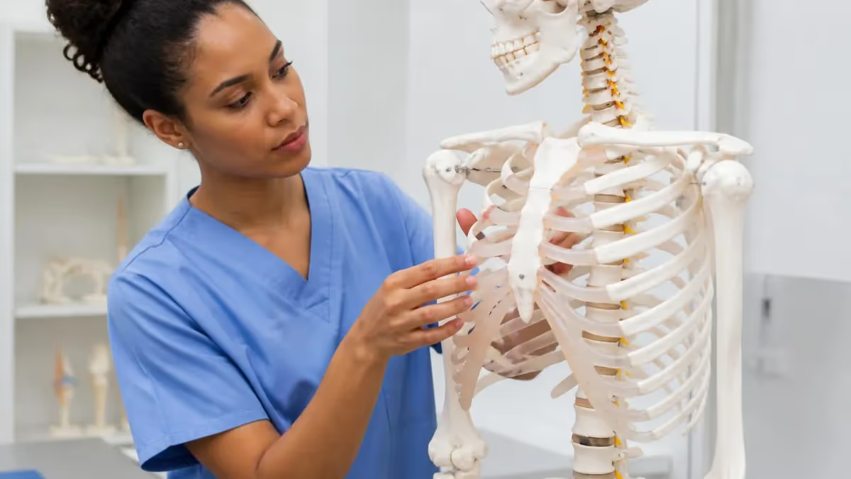
Understanding the difference between anatomy and physiology is one of the first foundational steps for any student entering the health sciences, nursing, or pre-med track. At their core, these two disciplines complement each other perfectly: anatomy focuses on the physical structures of the body — bones, muscles, organs, and tissues — while physiology examines how those structures work, communicate, and sustain life. Together, they form the backbone of modern biomedical science, and separating them conceptually helps students build a stronger, more organized mental framework for studying the human body. Practice with the free Anatomy and Physiology practice test on PracticeTestGeeks (7 questions with answer explanations).
Anatomy, derived from the Greek word anatome meaning "to cut up," is the branch of science dedicated to identifying and describing the form and arrangement of body parts. Anatomists classify structures by location, relationship to neighboring organs, tissue composition, and developmental origin. From gross anatomy — which you can observe with the naked eye — to histology and cellular anatomy visible only under a microscope, the discipline spans every spatial scale of the human body, providing the essential map that all other medical sciences rely upon.
Physiology, by contrast, comes from the Greek physis (nature) and logos (study), and it asks a fundamentally different question: not what is this structure, but what does it do? A physiologist examining the heart does not simply catalog its four chambers — they trace electrical impulses through the sinoatrial node, measure stroke volume, and calculate cardiac output under varying workloads. Physiology is dynamic rather than static, describing processes unfolding in real time across cells, tissues, organs, and organ systems.
The relationship between these two fields is often described as structure determining function. A long bone's hollow shaft reduces weight while preserving strength — that structural feature directly enables efficient locomotion. The alveoli of the lungs have walls only one cell thick, maximizing the diffusion rate of oxygen into capillaries — again, form serving function. Students who grasp this structural-functional relationship early find that memorizing individual facts becomes much easier, because each detail connects logically to a larger mechanical story.
In clinical settings, clinicians draw on both disciplines simultaneously. A radiologist reading an X-ray uses anatomical knowledge to identify structures and physiological knowledge to infer whether those structures are performing normally. A physical therapist designing a rehabilitation program must understand the anatomy of damaged ligaments and the physiology of muscle repair and neural re-education. Emergency physicians interpreting shock understand both the anatomical pathway of blood flow and the physiological cascade of compensatory mechanisms that sustain perfusion when pressure drops.
For students preparing for certification exams, standardized tests, or professional licensing boards, mastering the difference between anatomy and physiology is not merely academic — it directly shapes how exam questions are framed and answered. Questions rarely test one discipline in isolation; they present clinical scenarios that require integrated reasoning across structural and functional domains. Building a clear mental distinction between these two sciences, then learning to blend them fluidly, is the core intellectual skill that separates high-scoring students from average ones.
Whether you are a nursing student, a massage therapy candidate, a pre-med undergraduate, or a curious lifelong learner, this guide will walk you through both disciplines in depth. We will explore definitions, key subdisciplines, the historical development of each field, practical applications in healthcare, common exam strategies, and the specific ways anatomy and physiology interact at every level of biological organization. By the end, you will have a concrete, working understanding of how these two sciences differ and why they belong together.
Free Anatomy and Physiology Study Tools
Interactive Anatomy and Physiology resources on PracticeTestGeeks, free to start:
- Anatomy and Physiology flashcards — review key terms and concepts card by card
- Anatomy and Physiology study guide — topic-by-topic overview of what the exam covers
- Anatomy and Physiology cheat sheet — one-page printable summary for last-minute review
Anatomy and Physiology by the Numbers
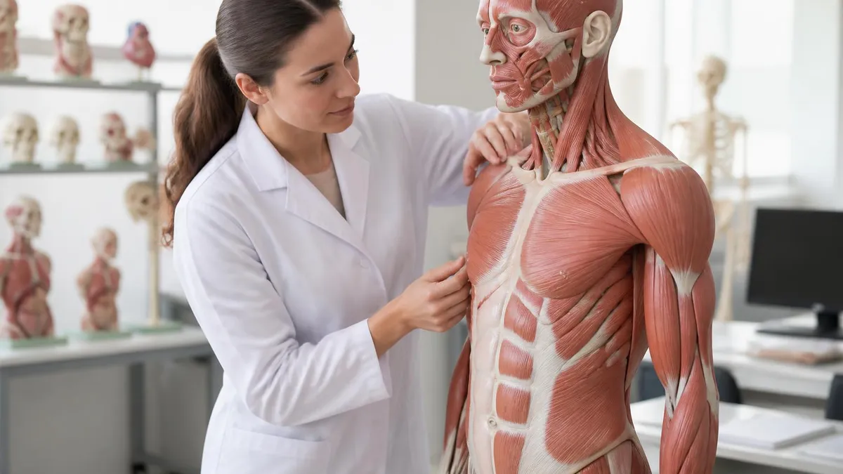
Core Subdisciplines of Anatomy and Physiology
The study of body structures visible to the naked eye, including regional anatomy (studying one area at a time), systemic anatomy (organ systems), and surface anatomy used in clinical examination and imaging interpretation.
Encompasses histology (tissue study) and cytology (cell study). Requires microscopy to visualize structures. Essential for understanding how tissues are organized and how pathological changes appear at the cellular level in disease.
Examines function organ-system by system — cardiovascular, respiratory, renal, endocrine, and more. Each system's physiology links back to its anatomy, explaining how structural design enables the specific regulatory processes needed for homeostasis.
Studies processes occurring within and between individual cells: membrane transport, signal transduction, metabolism, and gene expression. Cell physiology bridges biochemistry and organ-level function, explaining how molecular events produce measurable physical outcomes.
Tracks how structures form from fertilization through birth and maturation. Understanding embryological origin explains congenital anomalies, organ positioning, and why certain pathologies cluster in anatomically related structures.
The principle that structure determines function is so central to biomedical science that it serves as the organizing logic for nearly every anatomy and physiology textbook ever written. When you examine any body structure closely enough, you will find that its physical characteristics — shape, size, material composition, surface texture, and spatial arrangement — are precisely engineered to support the work it must perform. This relationship is not coincidental; it is the outcome of millions of years of evolutionary selection pressuring biological systems toward efficiency.
Consider skeletal muscle as a concrete example. Each muscle fiber is packed with myofibrils, protein filaments of actin and myosin arranged in repeating sarcomere units. This striped, organized architecture is visible under a light microscope and is the direct anatomical basis for the sliding filament theory of contraction. Without understanding that structural arrangement — without knowing where the thick and thin filaments sit relative to each other — the physiology of muscle contraction would be impossible to explain mechanistically. The anatomy does not just accompany the physiology; it generates it.
Neurons provide another compelling illustration. A motor neuron projecting from the spinal cord to a foot muscle in a six-foot adult may have an axon over three feet long. That extraordinary length is an anatomical feature, but it creates an urgent physiological problem: how does an electrical signal travel that distance quickly enough to be useful? The answer is myelination — the Schwann cell wrapping that creates saltatory conduction, allowing impulses to jump from node to node at speeds exceeding 70 meters per second. Anatomy posed the challenge; physiology evolved the solution.
The kidney's nephron demonstrates this relationship at an almost architectural level of elegance. The glomerulus is a knot of capillaries tucked inside a cup-shaped Bowman's capsule — an anatomical arrangement that creates a massive filtration surface area while maintaining a pressure gradient that drives fluid across the membrane. Downstream, the proximal convoluted tubule's brush border of microvilli maximizes reabsorption surface area. The loop of Henle's descending and ascending limbs create a countercurrent multiplier system — a physiological mechanism enabled entirely by the anatomical loop structure. Form and function are inseparable here.
Respiratory anatomy illustrates the same principle at a macroscopic level. The lungs contain approximately 300 million alveoli, each roughly 0.2 millimeters in diameter. Collectively, they create a surface area of about 70 square meters — roughly the size of a tennis court — folded into the volume of two footballs. That anatomical feat of compression directly enables the physiological demand for rapid, massive gas exchange. The diffusion distance across the alveolar-capillary membrane is less than one micrometer, a structural specification that makes oxygen delivery fast enough to support even vigorous aerobic exercise.
Hormonal physiology connects to anatomy through the precise positioning of endocrine glands relative to their target organs and vascular pathways. The adrenal glands sit directly atop the kidneys — not coincidentally, since the hormones they secrete (aldosterone, cortisol, epinephrine) directly regulate renal function and systemic blood pressure. The portal circulation carries hormones from the gut directly to the liver before entering systemic circulation, allowing the liver to metabolize or modify these signals — an anatomical routing that has profound physiological consequences for drug metabolism and nutrient processing.
For students preparing for exams, this structural-functional framework provides a powerful study strategy. Rather than memorizing isolated facts, you can ask: why does this structure look the way it does? What physiological demand drove its evolution? What would fail if this structural feature were absent? These questions transform rote memorization into mechanistic understanding, and that deeper comprehension is precisely what high-stakes exam questions are designed to test. Students who think structurally and functionally simultaneously tend to outperform those who treat anatomy and physiology as separate lists of facts to be memorized independently.
If you want more targeted practice, the anatomy and physiology pre-test page covers the same material with additional questions and explanations.
Anatomy and Physiology Practice Test Questions
Prepare for the Anatomy and Physiology exam with our free practice test modules. Each quiz covers key topics to help you pass on your first try.
Anatomy and Physiology Applied Methods and...
Anatomy and Physiology Exam Questions covering Applied Methods and Techniques. Master Anatomy and Physiology Test concepts for certification prep.
Anatomy and Physiology Assessment and Eval...
Free Anatomy and Physiology Practice Test featuring Assessment and Evaluation. Improve your Anatomy and Physiology Exam score with mock test prep.
Anatomy and Physiology Communication and D...
Anatomy and Physiology Mock Exam on Communication and Documentation. Anatomy and Physiology Study Guide questions to pass on your first try.
Anatomy and Physiology Continuing Educatio...
Anatomy and Physiology Test Prep for Continuing Education Requirements. Practice Anatomy and Physiology Quiz questions and boost your score.
Anatomy and Physiology Core Concepts and P...
Anatomy and Physiology Questions and Answers on Core Concepts and Principles. Free Anatomy and Physiology practice for exam readiness.
Anatomy and Physiology Industry Best Pract...
Anatomy and Physiology Mock Test covering Industry Best Practices. Online Anatomy and Physiology Test practice with instant feedback.
Anatomy and Physiology Musculoskeletal System
Free Anatomy and Physiology Quiz on Musculoskeletal System. Anatomy and Physiology Exam prep questions with detailed explanations.
Anatomy and Physiology Nervous System
Anatomy and Physiology Practice Questions for Nervous System. Build confidence for your Anatomy and Physiology certification exam.
Anatomy and Physiology Professional Standa...
Anatomy and Physiology Test Online for Professional Standards and Ethics. Free practice with instant results and feedback.
Anatomy and Physiology Safety and Compliance
Anatomy and Physiology Study Material on Safety and Compliance. Prepare effectively with real exam-style questions.
Anatomy and Physiology General Concepts
Free Anatomy and Physiology Test covering General Concepts. Practice and track your Anatomy and Physiology exam readiness.
Anatomy and Physiology Integumentary System
Anatomy and Physiology Exam Questions covering Integumentary System. Master Anatomy and Physiology Test concepts for certification prep.
Levels of Biological Organization: Anatomy vs. Physiology Perspective
At the chemical level, anatomy describes the types and arrangements of molecules that make up cellular structures — the phospholipid bilayer of the plasma membrane, the double helix of DNA, the quaternary structure of hemoglobin. Physiology at this level examines how these molecules interact: enzyme kinetics, ATP synthesis, receptor-ligand binding, and second messenger cascades. The structural configuration of a molecule directly determines which reactions it can catalyze and which signals it can transmit.
At the cellular level, anatomy identifies cell types, organelle positions, and membrane specializations — microvilli, cilia, gap junctions, tight junctions. Physiology describes what each specialization does: microvilli increase absorption surface area, cilia move mucus and debris, gap junctions allow rapid electrical coupling between cardiac cells. Understanding the anatomical difference between a ciliated columnar cell and a squamous cell immediately predicts their physiological roles in the respiratory versus capillary walls.
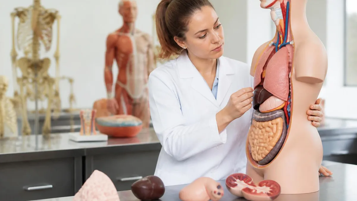
Studying Anatomy vs. Physiology: Different Challenges, Different Rewards
- +Anatomy provides concrete, visual learning anchors — diagrams, models, and cadaver labs make structures tangible and memorable
- +Anatomical knowledge is relatively stable; structures do not change across textbook editions the way physiological models sometimes do
- +Gross anatomy skills translate directly to clinical procedures: suturing, injections, palpation, and imaging interpretation
- +Physiology develops mechanistic reasoning skills that transfer across organ systems and inform clinical problem-solving
- +Physiology explains the 'why' behind symptoms, making pathology and pharmacology courses much easier to understand
- +Integrating both disciplines simultaneously accelerates learning — each reinforces the other, reducing total study time needed
- −Anatomy involves enormous amounts of nomenclature — over 7,500 named structures in Terminologia Anatomica — which demands sustained memorization effort
- −Physiology requires strong math and chemistry foundations; students weak in these areas struggle with osmolarity calculations, Nernst equations, and gas laws
- −Anatomy labs can be emotionally challenging; not all programs offer cadaver access, leaving some students underprepared for clinical settings
- −Physiology concepts are highly abstract — action potentials, countercurrent exchange, and feedback loops require mental modeling skills that take time to develop
- −Both subjects are typically compressed into one or two semesters, creating intense time pressure that favors test-taking strategies over deep comprehension
- −Anatomical variation between individuals means clinical practice sometimes contradicts textbook descriptions, which can confuse students expecting perfect correspondence
Pros and Cons at a Glance
| Pros | Cons |
|---|---|
| Anatomy provides concrete, visual learning anchors — diagrams, models, and cadaver labs make structures tangible and memorable | Physiology requires strong math and chemistry foundations; students weak in these areas struggle with osmolarity calculations, Nernst equations, and gas laws |
| Anatomical knowledge is relatively stable; structures do not change across textbook editions the way physiological models sometimes do | Anatomy labs can be emotionally challenging; not all programs offer cadaver access, leaving some students underprepared for clinical settings |
| Gross anatomy skills translate directly to clinical procedures: suturing, injections, palpation, and imaging interpretation | Physiology concepts are highly abstract — action potentials, countercurrent exchange, and feedback loops require mental modeling skills that take time to develop |
| Physiology develops mechanistic reasoning skills that transfer across organ systems and inform clinical problem-solving | Both subjects are typically compressed into one or two semesters, creating intense time pressure that favors test-taking strategies over deep comprehension |
| Physiology explains the 'why' behind symptoms, making pathology and pharmacology courses much easier to understand | Anatomical variation between individuals means clinical practice sometimes contradicts textbook descriptions, which can confuse students expecting perfect correspondence |
Anatomy and Physiology Exam Prep Checklist
- ✓Map each organ system's anatomy before tackling its physiology — structure first, function second.
- ✓Create a one-page 'structure-function' table for each major organ linking anatomical features to physiological roles.
- ✓Practice labeling unlabeled diagrams from memory, then check against your textbook atlas.
- ✓Write out the steps of each physiological process (e.g., cardiac cycle, action potential) as a numbered sequence without looking at notes.
- ✓Use clinical case vignettes to test whether you can identify which structure is involved based on symptom location and type.
- ✓Review the major body cavities, membranes, and directional terms until you can use them automatically without hesitation.
- ✓Memorize the functional differences between sympathetic and parasympathetic divisions, including target organs and neurotransmitters.
- ✓Practice interpreting histology slides by tissue type — epithelial, connective, muscular, nervous — linking appearance to function.
- ✓Study feedback loops (negative and positive) using real examples: thermoregulation, blood glucose, and childbirth contractions.
- ✓Take timed practice quizzes under exam conditions weekly to track retention and identify gaps before test day.
Structure-Function Thinking Outperforms Rote Memorization
Students who consistently ask 'why does this structure look this way?' and 'what would break if this feature were missing?' retain 40–60% more material over a semester than those who memorize lists in isolation. Every anatomical detail is a physiological clue — treat it that way and exam questions become significantly easier to reason through, even when you encounter unfamiliar phrasing or novel clinical scenarios.
In clinical practice, the boundary between anatomy and physiology dissolves almost completely, because effective diagnosis and treatment require both simultaneously. When a physician hears a systolic murmur through a stethoscope, they immediately invoke anatomical knowledge — which valve is involved, where it sits relative to the chest wall, how its leaflets are arranged — alongside physiological knowledge about pressure gradients, turbulent flow, and the cardiac cycle timing that produces the characteristic sound. Neither discipline alone provides a complete clinical picture.
Diagnostic imaging is perhaps the clearest example of anatomy informing clinical physiology. A computed tomography (CT) scan of the abdomen produces cross-sectional images that require the radiologist to mentally reconstruct three-dimensional anatomy from two-dimensional slices. But the radiologist does not stop at identifying structures — they assess whether those structures are the right size (a physiological indicator of function), whether blood flow patterns appear normal on contrast studies, and whether the tissue density suggests active inflammation, necrosis, or calcification. Anatomy provides the reference; physiology provides the interpretation.
Pharmacology — the science of drugs — is fundamentally applied anatomy and physiology. Every drug target is an anatomical structure (a receptor protein on a specific cell type, an enzyme in a particular tissue, an ion channel in a defined neural pathway), and every drug effect is a physiological outcome (altered membrane potential, changed hormone secretion, modified immune cell activity). Understanding why a beta-blocker slows heart rate requires knowing both the anatomy of cardiac innervation and the physiology of beta-adrenergic receptor signaling in the sinoatrial node.
Physical therapy and occupational therapy rely on integrated anatomical and physiological reasoning at every clinical decision point. A therapist treating a patient with rotator cuff injury must know the exact anatomy of the four muscles and their attachment sites, but they must also understand the physiological principles of tendon healing — the stages of inflammation, proliferation, and remodeling, the mechanical loading thresholds that stimulate collagen synthesis, and the neuromuscular re-education needed to restore coordinated shoulder function. Anatomy defines what is injured; physiology guides how healing proceeds.
Emergency medicine demonstrates the life-or-death stakes of anatomical and physiological integration. A paramedic performing a needle thoracostomy for tension pneumothorax must know the exact anatomical landmark — second intercostal space, midclavicular line — to avoid hitting the neurovascular bundle running under each rib. But they must also understand the physiology of why tension pneumothorax is immediately fatal: progressive air accumulation shifts the mediastinum, compresses the contralateral lung, kinks the great veins, and collapses cardiac output within minutes. Anatomy gives the roadmap; physiology explains the urgency.
Nursing practice integrates these two sciences continuously throughout a shift. Medication administration requires knowing anatomical injection sites (deltoid versus vastus lateralis versus gluteal muscles) and physiological absorption rates from each site. Wound assessment requires recognizing anatomical layers exposed and the physiological stages of wound healing expected at each depth. Patient positioning for respiratory distress requires understanding both the anatomy of dependent lung zones and the physiology of ventilation-perfusion matching that determines which position optimizes oxygenation for a given patient's condition.
For students in allied health programs, understanding the clinical integration of anatomy and physiology transforms abstract coursework into immediately meaningful professional preparation. Every cadaver dissection, every histology slide, every graph of renal clearance versus plasma concentration is a skill being sharpened for patient care. The student who sees anatomy and physiology as two halves of a single clinical language — not two separate courses to be endured — builds the cognitive foundation that carries them from classroom to clinical competency with far less friction along the way.

Most nursing licensing exams (NCLEX-RN, NCLEX-PN), medical board exams (USMLE Step 1), and allied health certification exams integrate anatomy and physiology into clinical application questions rather than testing them as isolated recall. Expect scenario-based questions that describe a patient finding and require you to identify the involved structure AND explain the physiological mechanism. Pure memorization of anatomy or physiology alone is insufficient — integrated understanding is what these exams reward.
Integrating anatomy and physiology into a unified knowledge base is the goal that every health science curriculum aims for, but the path there requires deliberate study strategies tailored to how each discipline is best learned. Anatomy is spatial and visual — it rewards learners who can build three-dimensional mental models and rotate them mentally, who draw structures from memory, who color-code diagrams, and who use physical models or digital anatomy apps to explore spatial relationships from multiple angles. Flashcards work for terminology, but true anatomical mastery requires active visualization and repeated retrieval of structural positions, not just names.
Physiology is sequential and causal — it rewards learners who think in cause-and-effect chains, who trace the logical steps of a process from stimulus to response, and who can identify where a disruption in the chain would produce a specific pathological outcome. The best physiology students write out processes as flowcharts or numbered sequences, then quiz themselves by covering the downstream steps and predicting what comes next. This approach mirrors how exam questions are structured: they describe a disruption early in a physiological process and ask what happens at a later step.
Combining both learning approaches within a single study session produces the strongest retention. A productive session might begin with anatomical review — labeling a heart diagram, identifying the layers of the aortic wall — then transition immediately to physiological application — tracing blood flow through the chambers, calculating pulse pressure from systolic and diastolic values, explaining how aortic valve stenosis alters the pressure-volume loop. Moving fluidly between structure and function within the same study block builds the associative neural pathways that make integrated recall automatic under exam conditions.
Spaced repetition is especially powerful for anatomy and physiology because both subjects contain high volumes of interconnected facts that decay rapidly without review. Students who study intensively before a single exam and then stop reviewing retain far less than students who schedule brief weekly review sessions throughout the semester. Modern spaced repetition software (Anki being the most widely used in medical education) allows students to create cards that test both anatomical identification and physiological explanation, programming optimal review intervals based on individual performance data.
Group study offers particular advantages for anatomy and physiology because teaching a concept to a peer is one of the most effective ways to identify gaps in your own understanding. When you explain the juxtaglomerular apparatus to a classmate, you quickly discover whether you understand both its anatomy (the specialized cells at the afferent arteriole and macula densa) and its physiology (how it regulates renin secretion in response to reduced stretch or sodium delivery). The gaps revealed in group teaching sessions are precisely the gaps most likely to cost points on an exam.
Practice questions are non-negotiable for exam preparation, not because they reveal answers to memorize, but because they train the reasoning process that exams reward. When you encounter a practice question about why a patient with left-sided heart failure develops pulmonary edema, the value is not memorizing that answer — it is practicing the anatomical reasoning (left heart backs up into pulmonary veins) and physiological reasoning (increased hydrostatic pressure exceeds oncotic pressure, driving fluid into alveolar spaces) that produces the answer from first principles.
Students who practice this reasoning process across hundreds of questions before an exam are not just more knowledgeable — they are faster and more confident under time pressure.
For a comprehensive starting point that consolidates anatomical diagrams, physiological summaries, and clinical correlations in one place, many students find that well-organized downloadable resources accelerate their preparation significantly. Building your integrated understanding of anatomy and physiology from structured, reliable materials — and then testing that understanding through rigorous practice — is the most reliable path to exam success in any health sciences program.
As you move through your anatomy and physiology coursework, developing good study habits from the beginning of the semester will save significant stress during exam weeks. The most effective students treat each lecture not as a performance to watch but as an active problem-solving session. They arrive having previewed the topic, they annotate diagrams during class, and they spend fifteen to twenty minutes after each lecture converting notes into questions — flipping every statement into something they must actively retrieve rather than passively recognize.
Understanding anatomical terminology is a high-use early investment. Directional terms (superior, inferior, anterior, posterior, medial, lateral, proximal, distal, superficial, deep), body planes (sagittal, coronal, transverse), and regional terms (brachial, femoral, popliteal, lumbar) appear constantly throughout both anatomy and physiology. Students who internalize these terms in the first two weeks move through subsequent material dramatically faster, because they can parse descriptions of structures and processes without stopping to decode the vocabulary.
Physiology benefits enormously from understanding a small number of foundational principles that recur across every organ system. Fick's law of diffusion explains gas exchange in the lungs, nutrient absorption in the gut, and oxygen delivery to tissues. The Nernst equation underlies action potential generation in neurons, cardiac muscle cells, and skeletal muscle fibers. The Frank-Starling relationship explains cardiac output regulation, but the same length-tension relationship governs skeletal muscle performance. Students who master these cross-cutting principles find that each new organ system requires less new learning — they are applying familiar tools to new anatomical contexts.
Laboratory components of anatomy and physiology courses deserve as much preparation as lectures. Whether your program uses cadaver dissection, virtual anatomy software, histology lab, or a combination, these sessions cement spatial understanding in ways that diagrams alone cannot match. Before each lab, review the structures you expect to identify. During the lab, actively quiz yourself and your partners rather than passively following along. After the lab, sketch what you observed from memory. This three-stage approach (prepare, engage, reconstruct) maximizes what each lab session deposits into long-term memory.
Clinical correlation is one of the most effective study strategies because it gives anatomical and physiological facts an emotional and contextual hook that makes them far more memorable. When you learn about the circle of Willis — the anastomotic ring of arteries at the base of the brain — connecting that anatomy to the physiology of collateral circulation during stroke makes the structure meaningful rather than arbitrary. Every time you learn a new structure or process, spend sixty seconds asking: what disease or injury involves this? What symptom would its dysfunction produce? What treatment targets it?
Time management across a semester-long anatomy and physiology course requires recognizing that these subjects do not lend themselves to cramming. The sheer volume of material — 11 organ systems, hundreds of structures, dozens of physiological processes — means that consistent daily engagement (45-60 minutes minimum) produces far better outcomes than four-hour marathon sessions the night before an exam. Students who build daily review habits in week one of the semester typically enter finals week with genuine confidence rather than desperate catch-up anxiety.
Finally, remember that anatomy and physiology are living sciences, not static encyclopedias. Research continues to refine our understanding of physiological mechanisms, identify new cell types, and revise anatomical classifications. The fundamentals you learn in your coursework are durable and clinically essential, but the habit of curiosity — of asking why structures look as they do and how processes produce their effects — is the intellectual disposition that will serve you throughout an entire career in health science, far beyond any single exam or certification.
Sample Anatomy and Physiology Practice Questions
Try these questions from our free Anatomy and Physiology practice tests. The correct answer and an explanation follow each question.
What is the primary function of the testes in males?
- A. Production of sperm and testosterone
- B. Storage of sperm
- C. Transport of sperm
- D. Production of seminal fluid
Answer: A. Production of sperm and testosterone
The testes are the male gonads responsible for producing sperm (spermatogenesis) and the hormone testosterone, which regulates male secondary sexual characteristics.
What is the primary function of mitochondria in cells?
- A. Protein systhesis
- B. Genetic information storage
- C. Energy production
- D. Detoxification
Answer: C. Energy production
Mitochondria are the "powerhouses" of the cell, generating ATP (adenosine triphosphate) through cellular respiration, which provides energy for cellular functions.
What is the term for the gap between two neurons at a synapse?
- A. Synaptic cleft
- B. Axon hillock
- C. Myelin sheath
- D. Nodes of Ranvier
Answer: A. Synaptic cleft
The synaptic cleft is the narrow fluid-filled space between the presynaptic and postsynaptic membranes where neurotransmitters diffuse.
The tricuspid valve is located between which two structures?
- A. Left atrium and left ventricle
- B. Right ventricle and pulmonary artery
- C. Right atrium and right ventricle
- D. Left ventricle and aorta
Answer: C. Right atrium and right ventricle
The tricuspid valve, named for its three cusps, separates the right atrium from the right ventricle and prevents backflow during contraction.
Anatomy Physiology Questions and Answers
Anatomy and Physiology PDF: Complete Free Download Guide for 2026 Students

Anatomy and Physiology Coloring Workbook: The Complete Guide to Visual Learning, Active Recall, and Mastering Body Systems
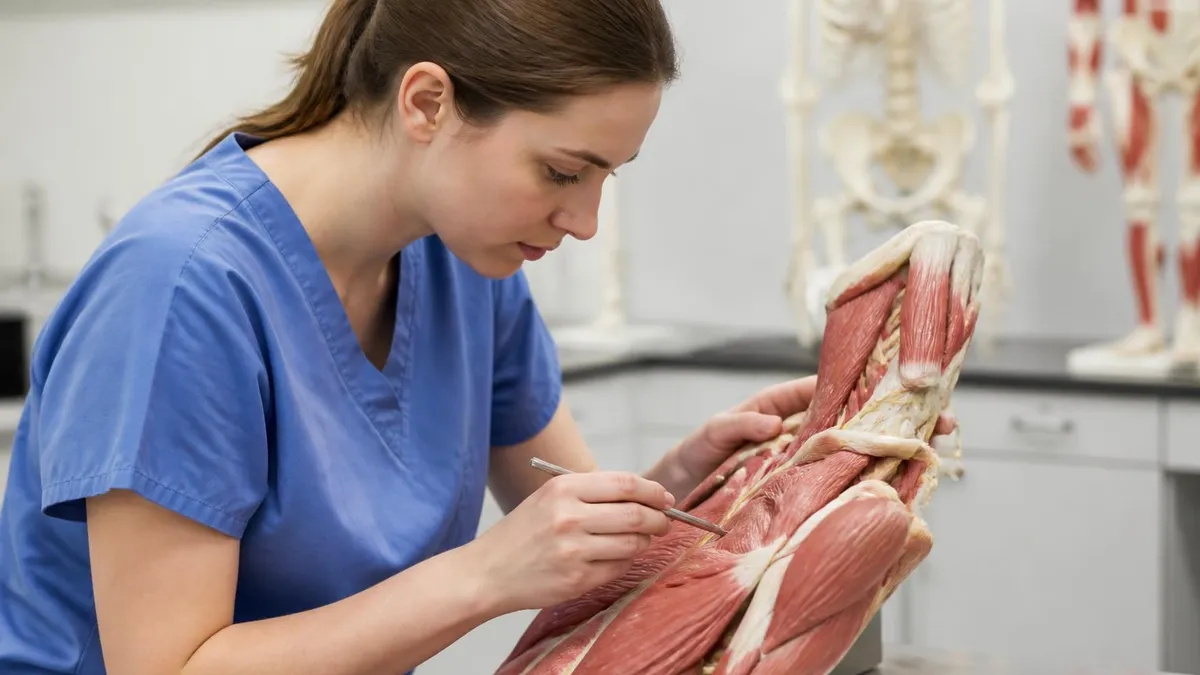
Define Anatomy and Physiology: A Complete Guide to Understanding Human Body Structure and Function
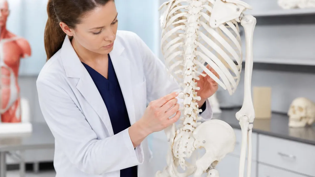
Human Body Anatomy and Physiology: The Complete Guide to Structure, Function, and Integrated Body Systems

Anatomy and Physiology Study Material: Complete Resource Guide
How this page was made: written with AI tools and published by Nathan Beja, founder of Practice Test Geeks, who is responsible for its content. We have not yet matched this exam's facts to an official source, so confirm fees, dates and rules with the exam body. Not reviewed by a subject-matter expert. Editorial policy