
Why Do People Get an MRI? Top Medical Reasons and What to Expect 2026 September
Why do people get MRI scans? Explore the top medical reasons, body parts scanned, what to expect, and how MRI compares to other imaging tests. 🟢
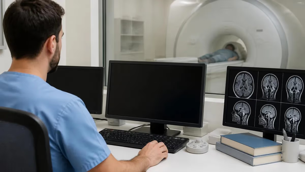
If you have ever wondered why do people get MRI scans so often these days, the short answer is that magnetic resonance imaging produces detail that no other test can match without using ionizing radiation. Physicians order MRIs to confirm a suspected diagnosis, to map out anatomy before surgery, to monitor how a known disease is changing, and to rule out dangerous conditions when symptoms are vague. The combination of soft tissue clarity and safety makes MRI a workhorse of modern medicine.
People are also referred for MRI when an X-ray, ultrasound, or CT scan has already been done but still leaves the clinical picture unclear. For example, an X-ray will quickly show a broken bone, yet it cannot reveal a torn meniscus, a herniated disc, or a small tumor pressing on a nerve. MRI fills that gap. It captures muscles, ligaments, cartilage, the spinal cord, brain tissue, and internal organs with a level of contrast that often changes the treatment plan entirely.
Another reason patients end up inside the scanner is screening. Adults with a strong family history of breast cancer, individuals at risk for hereditary heart disease, and people with prior strokes are often placed on regular MRI surveillance. Early detection through MRI has been linked to better survival rates because lesions can be caught while they are still small and treatable. For many high-risk groups, MRI is now considered the standard of care rather than an optional add-on.
Pain that has not improved with conservative treatment is a leading trigger for an MRI referral. When physical therapy, anti-inflammatory medication, and rest fail to help a sore knee, lower back, or shoulder over six to eight weeks, the next logical step is imaging that can pinpoint the cause. MRI shows whether the pain is coming from a structural problem like a tendon tear, a bone bruise, or a nerve impingement, and it guides the decision between continued therapy, injection, or surgery.
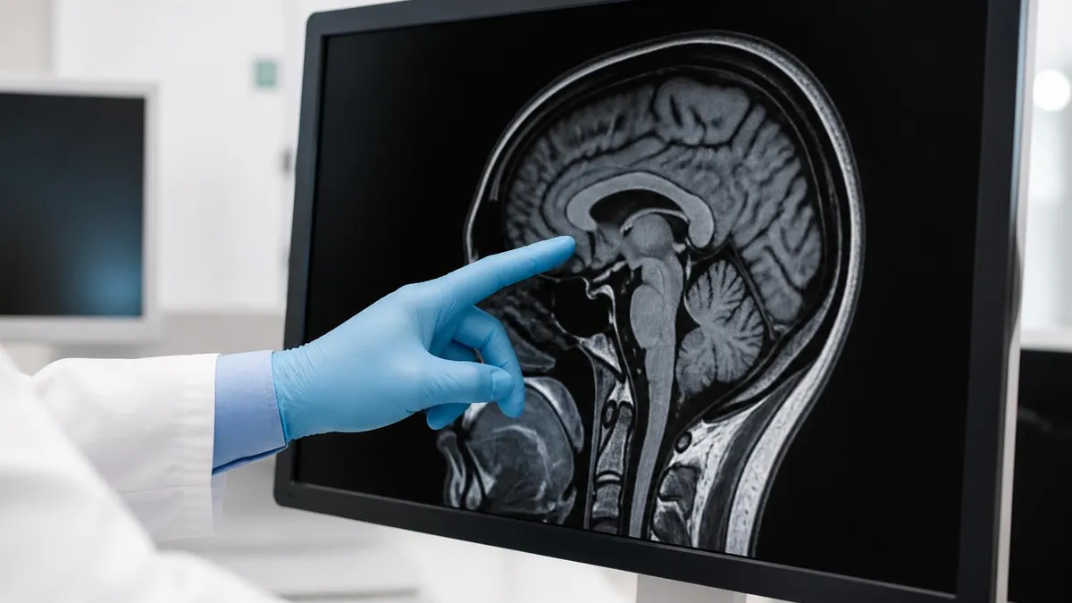
Neurological symptoms are another common reason. Patients with persistent headaches, sudden vision changes, numbness, weakness, seizures, or memory problems are routinely scheduled for a brain MRI. The scan can detect strokes, multiple sclerosis plaques, tumors, aneurysms, and inflammation that a CT might miss entirely.
Because the brain and spinal cord are made almost entirely of soft tissue, MRI is the imaging modality of choice for nearly every neurological complaint that lasts more than a few days. Doctors also use it to monitor known conditions over time. For a broader perspective on how this technology evolved into a routine diagnostic, the history of MRI shows how quickly it became a clinical standard.
Finally, MRI is requested when cancer is suspected or being staged. It helps oncologists see exactly how far a tumor extends, whether lymph nodes are involved, and how the patient is responding to treatment. Surgeons use MRI maps to plan incisions that protect critical structures, and radiation oncologists use them to design beam paths that spare healthy tissue. Across nearly every specialty, MRI has become the test that turns guesswork into precision medicine, which explains why millions of scans are performed each year.
Understanding why your provider has ordered an MRI can ease anxiety and help you prepare. Whether the goal is to confirm a diagnosis, screen for disease, or monitor a treatment, knowing the purpose puts you in control. This guide walks through the most common reasons for MRI, what happens during the exam, who should and should not have one, and how MRI fits alongside other imaging tests in everyday clinical practice across the United States.
MRI in the U.S. by the Numbers
Top Medical Reasons for an MRI
Torn ligaments, ruptured tendons, cartilage damage, and muscle tears are invisible on X-rays. MRI reveals them clearly, which is why orthopedic surgeons rely on it before recommending repair surgery or extended physical therapy.
Headaches, seizures, weakness, vision loss, and unexplained dizziness often lead to a brain or spine MRI. The scan detects strokes, MS lesions, tumors, aneurysms, and pinched nerves that influence treatment decisions.
MRI maps the exact size and location of tumors in the breast, prostate, liver, brain, and other organs. It helps oncologists plan surgery, choose radiation fields, and track how the cancer responds to therapy.
Cardiac MRI measures heart muscle function, scarring, and blood flow without radiation. It is used to investigate cardiomyopathy, congenital defects, and inflammation when echocardiograms cannot give a full answer.
Patients with multiple sclerosis, Crohn's disease, liver disease, or known tumors return for periodic MRIs to see whether their condition is stable, improving, or worsening so treatment can be adjusted in time.
Different body regions are scanned for very different reasons, and understanding the most common targets helps explain why your doctor may order one type of MRI over another. The brain is the single most imaged area because it harbors so many conditions that mimic each other. Patients with persistent headaches, memory loss, slurred speech, or weakness typically receive a brain MRI to rule out stroke, tumor, multiple sclerosis, or hemorrhage. Many of these conditions look identical on initial exam, so imaging is essential.
The spine ranks second in scan volume. Lower back pain affects roughly 80 percent of American adults at some point, and when conservative treatment fails, MRI shows exactly which disc is herniated, which nerve is being compressed, and whether the spinal cord is involved. Cervical spine MRI is ordered for neck pain with arm tingling, while thoracic MRI is used less often but is crucial in cases of unexplained mid-back pain, trauma, or suspected tumors. Each level has its own typical findings.
The knee is the most scanned joint in the country. Athletes, weekend warriors, and aging adults all develop meniscus tears, ACL injuries, cartilage breakdown, and patellar tendinitis that MRI captures in fine detail. The shoulder follows closely, where rotator cuff tears, labral injuries, and impingement syndromes drive most referrals. Hip MRIs are increasingly common as physicians recognize labral tears and femoroacetabular impingement as causes of pain once attributed to vague soft tissue strain or arthritis alone.
Abdominal and pelvic MRIs are ordered for conditions that ultrasound or CT cannot fully clarify. Liver lesions, pancreatic masses, kidney tumors, adrenal nodules, and gynecologic problems such as fibroids, endometriosis, and ovarian cysts are all best characterized with MRI. For men, prostate MRI has become a standard tool before biopsy, helping urologists target suspicious areas and avoid unnecessary procedures. The lack of radiation makes pelvic MRI especially attractive for younger patients who may need repeat imaging.
Cardiac MRI is a specialized but growing application. Cardiologists order it to measure ejection fraction with precision, detect myocardial scarring from prior heart attacks, evaluate suspected myocarditis, and assess congenital heart disease in both children and adults. Unlike echocardiography, cardiac MRI provides tissue characterization, which means it can distinguish inflamed muscle from healthy muscle and old scar from acute injury. This level of detail directly changes medication choices and surgical planning.
Breast MRI is reserved for high-risk screening and for clarifying findings from mammography or ultrasound. Women with BRCA gene mutations, a strong family history, or dense breast tissue are often placed on annual breast MRI surveillance. The scan can find cancers years before they become palpable, which dramatically improves treatment outcomes. The technology continues to improve, and as machines become more accessible patients have more options at MRI imaging centers throughout the country.
Less obvious but equally important are MRIs of the inner ear, the orbits behind the eyes, the temporomandibular joint, the pituitary gland, and the peripheral nerves. Each of these specialized exams answers a specific clinical question that no other test can address as reliably. The diversity of body parts that benefit from MRI is one of the main reasons the technology has become indispensable to modern diagnosis across virtually every medical specialty practiced in the United States today.
MRI Practice Test Questions
Prepare for the MRI - Magnetic Resonance Imaging exam with our free practice test modules. Each quiz covers key topics to help you pass on your first try.
MRI Knowledge
MRI Exam Questions covering Knowledge. Master MRI Test concepts for certification prep.
MRI Physics
Free MRI Practice Test featuring Physics. Improve your MRI Exam score with mock test prep.
MRI Anatomy and Pathology
MRI Test Prep for MRI Anatomy and Pathology. Practice MRI Quiz questions and boost your score.
MRI Anatomy and Positioning
MRI Questions and Answers on MRI Anatomy and Positioning. Free MRI practice for exam readiness.
MRI Contrast Agents
Free MRI Quiz on MRI Contrast Agents. MRI Exam prep questions with detailed explanations.
MRI Patient Care and Positioning
MRI Practice Questions for MRI Patient Care and Positioning. Build confidence for your MRI certification exam.
MRI vs CT vs X-ray vs Ultrasound
MRI and CT both produce detailed cross-sectional images, but they answer different questions. CT uses X-ray beams and is fast, making it the first choice in trauma, stroke alerts, and acute chest pain when speed matters most. It excels at showing bones, bleeding, and lung tissue within seconds.
MRI takes longer but uses no ionizing radiation and shows soft tissue with far more contrast. It is preferred for evaluating the brain, spinal cord, ligaments, cartilage, and pelvic organs. When a CT shows something ambiguous, MRI is often ordered as the follow-up to characterize the finding more precisely before treatment decisions are made.
Benefits and Drawbacks of Getting an MRI
- +No ionizing radiation, making it safer for repeat scans
- +Unmatched soft tissue detail for muscles, nerves, and organs
- +Multiple imaging planes from a single exam session
- +Excellent for detecting early-stage tumors and lesions
- +Useful in patients of all ages, including children
- +Contrast agents are generally well tolerated
- +Helps avoid exploratory surgery by clarifying diagnoses
- −Scans take 30 to 60 minutes, requiring patients to lie still
- −Loud knocking and humming noises during operation
- −Closed-bore design can trigger claustrophobia
- −Not safe for some implants, pacemakers, or metallic foreign bodies
- −Significantly more expensive than X-ray or ultrasound
- −Limited availability in rural areas can mean long waits
How to Prepare for Your MRI Scan
- ✓Tell your doctor about any pacemaker, defibrillator, or implanted device before scheduling
- ✓Disclose previous surgeries, especially those involving metal clips or plates
- ✓Mention any history of welding, metal work, or shrapnel injury to your eyes
- ✓Bring a list of current medications and known allergies, especially to gadolinium contrast
- ✓Wear loose, comfortable clothing without metal zippers, snaps, or underwire
- ✓Remove all jewelry, watches, hair pins, and piercings before entering the scanner room
- ✓Inform staff if you are pregnant or might be pregnant before the exam begins
- ✓Eat and drink normally unless your physician gives specific fasting instructions
- ✓Ask about sedation or open MRI options if you experience claustrophobia
- ✓Arrive 15 to 30 minutes early to complete safety screening paperwork
Even small metal objects can become dangerous projectiles inside an MRI suite
The MRI magnet is always on, even when no scan is in progress. Loose keys, oxygen tanks, and unscreened implants have caused serious injuries and deaths. Honest answers during the screening questionnaire are the single most important safety step you can take as a patient preparing for an MRI.
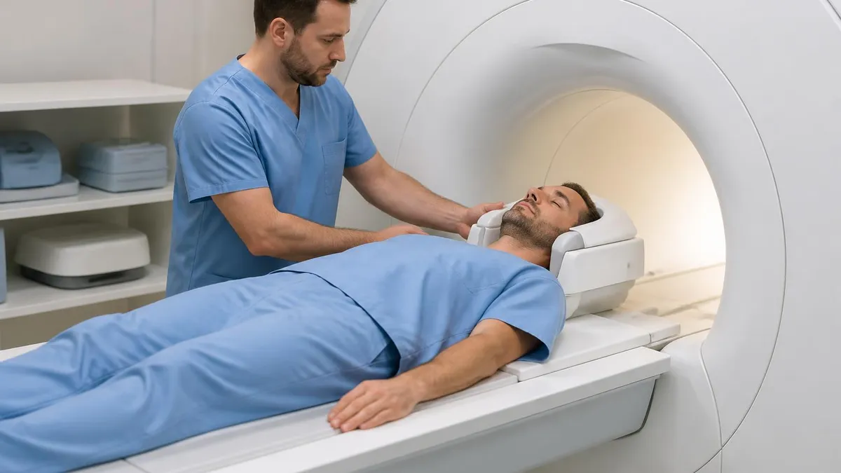
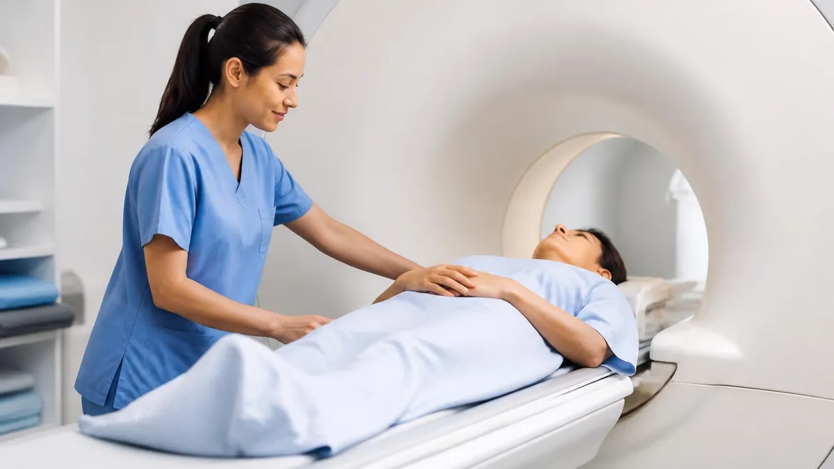
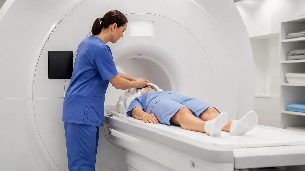
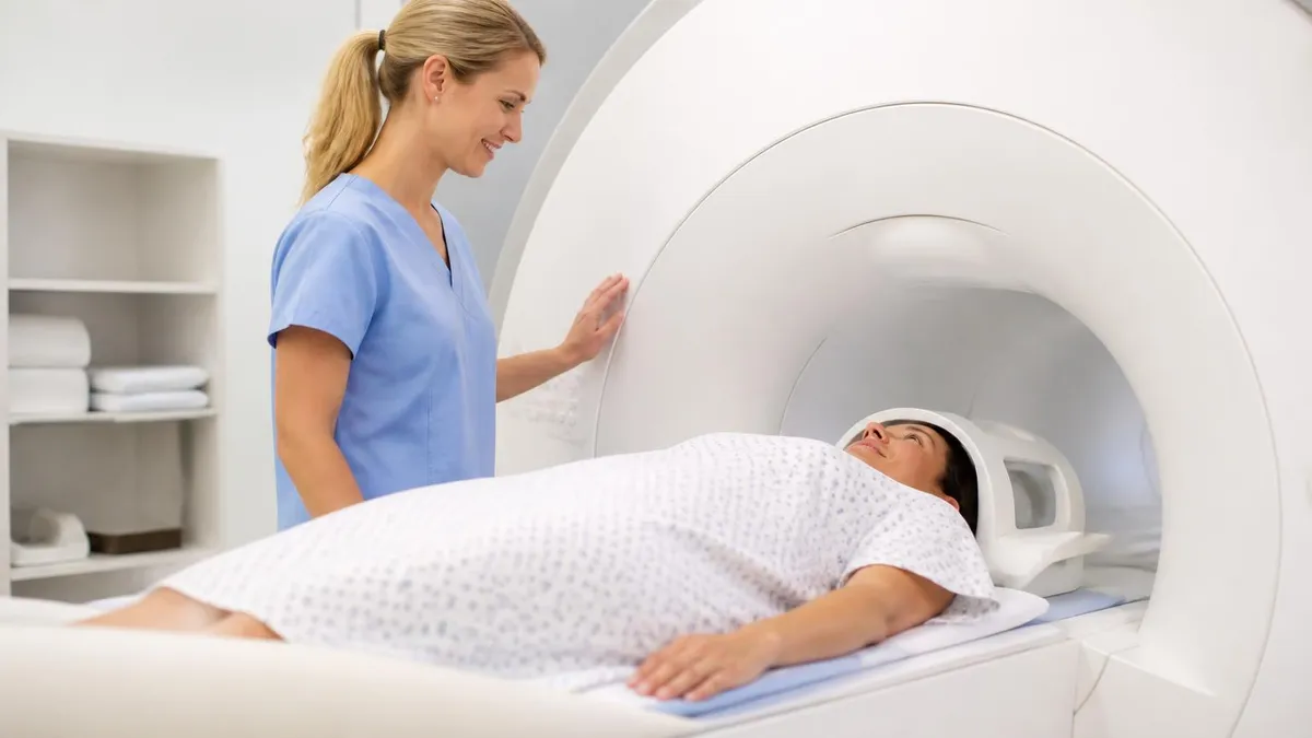
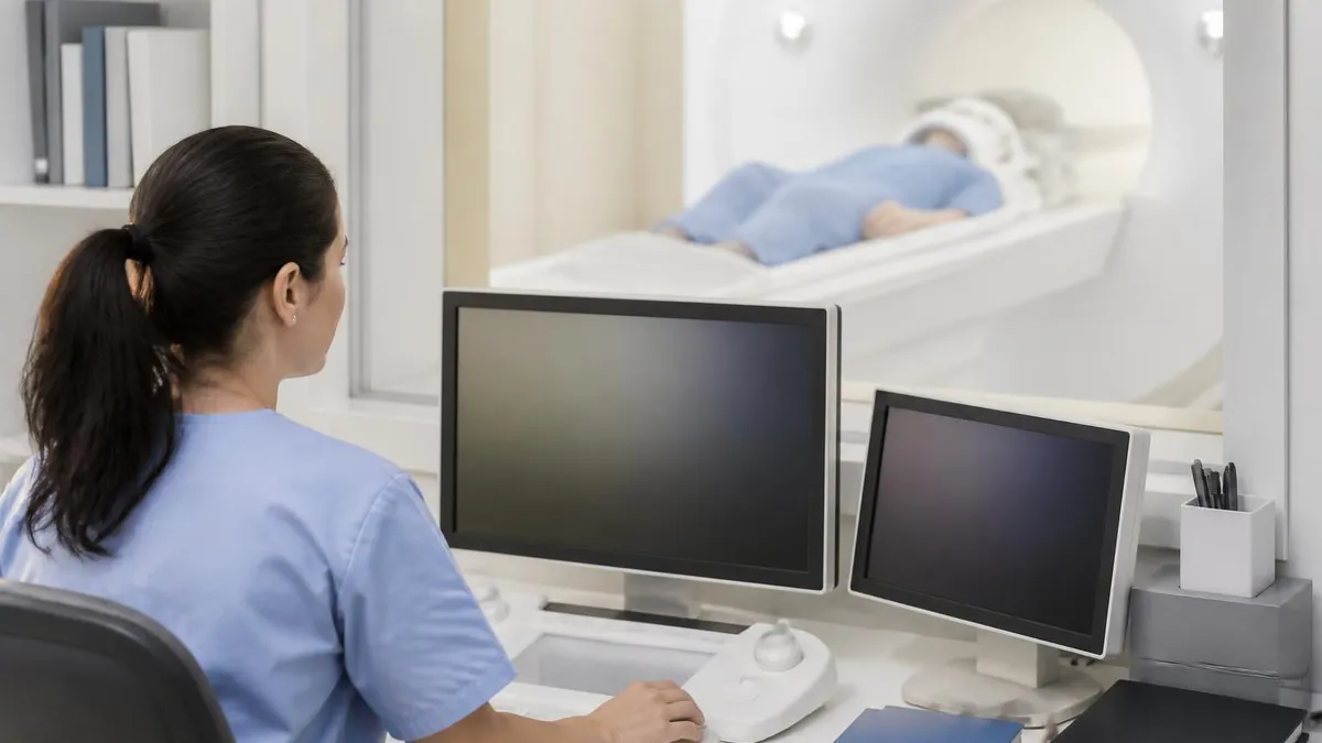
Once you have checked in and changed into a gown, a technologist will walk you through the safety screening one more time before leading you into the scanner room. The MRI itself looks like a large doughnut or tube. You will lie on a padded table that slides into the bore of the magnet. Pillows, blankets, and cushions are usually offered, and you will be given foam earplugs or headphones because the machine generates loud knocking sounds during each pulse sequence as the gradients turn on and off.
The technologist communicates with you through an intercom from the adjacent control room. You will be told when each sequence is about to start and roughly how long it will last. Some sequences are short, only 30 seconds, while others last several minutes. Holding still is essential because even small movements can blur the images and force the scan to be repeated. Most patients find that breathing slowly and closing their eyes makes the time pass much more quickly than expected during the exam.
If the scan requires contrast, an IV line will be placed in your arm before you go into the scanner. Partway through the exam, the technologist will inject a small amount of gadolinium-based contrast through that line. You may feel a brief cool sensation as the dye enters your vein, but most people notice nothing at all. The contrast helps highlight blood vessels, tumors, and areas of inflammation. To learn how it is used in detail, review MRI with and without contrast.
Some MRI exams require specialized positioning. A shoulder MRI may have your arm rotated outward in a coil, while a wrist scan often has you lying face down with your arm extended overhead in the so-called superman position. Cardiac MRI requires you to hold your breath repeatedly during imaging. The technologist will explain each step and let you practice the breath holds before the actual sequences begin. Comfort during these positions is important because the body part being scanned must remain perfectly still throughout each sequence.
For patients with claustrophobia, several strategies can make the exam tolerable. Many centers now offer wide-bore scanners that have a larger opening than traditional MRI tubes. Open MRI machines exist as well, although image quality is sometimes slightly lower. Mild oral sedation prescribed by your physician, mirrored goggles that show video or scenery, and bringing a trusted family member into the room are all options. Communicating your concerns ahead of time gives the staff time to plan and ensure a comfortable, successful exam.
The total time in the scanner depends on what is being imaged and whether contrast is used. A simple brain MRI without contrast may take 20 minutes, while a multi-region cancer staging scan with contrast can take well over an hour. The technologist will give you an accurate estimate when you check in. During the exam you can usually squeeze a call ball that alerts the staff if you need to stop. Knowing this safety feature is available gives many patients peace of mind throughout the scan.
After the last sequence finishes, the table slides out and the technologist helps you up. If you received contrast, the IV will be removed and a small bandage applied. You can resume normal activities immediately in most cases. Driving home, returning to work, and eating normally are all fine. The technologist does not interpret images. That task belongs to the radiologist, who will review the scan in detail and send a written report to the ordering physician, typically within 24 to 48 hours of the exam.
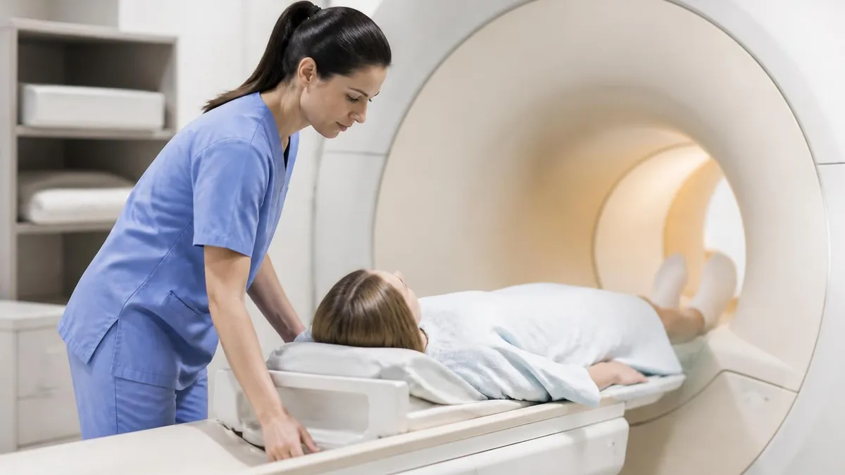
Patients with certain pacemakers, cochlear implants, aneurysm clips, or metallic eye fragments may not be candidates for MRI. Always disclose every implant, surgery, and occupational metal exposure. Some devices are MRI-conditional, meaning they can be scanned only under specific protocols.
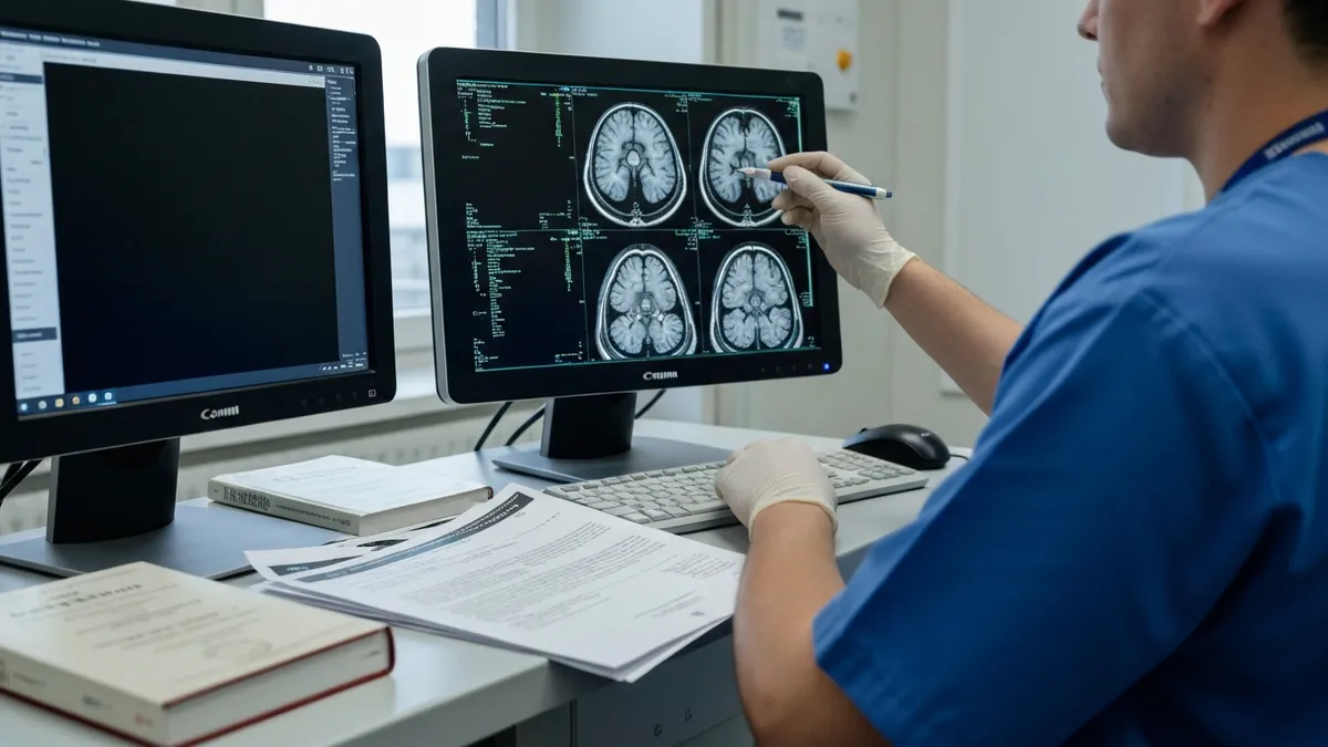
After the MRI is complete, the next step is interpretation. A board-certified radiologist analyzes every image, comparing different sequences and any prior imaging you have had. The radiologist looks for abnormalities, measures lesions, and notes incidental findings. A detailed report is then sent to the physician who ordered the exam. Most reports are completed within one to two business days, though stat scans for emergencies are read within minutes. Patients can typically access the report and images through a patient portal as well.
Your ordering physician will discuss the results with you, usually at a follow-up appointment. If the scan confirms the suspected diagnosis, treatment can move forward quickly. If something unexpected appears, additional testing may be recommended. This could include a biopsy, a different imaging study, a referral to a specialist, or simply a repeat MRI in a few months to monitor the area. Decisions are based on the clinical picture, not the image alone, which is why the conversation with your doctor matters as much as the report itself.
Incidental findings are common and often nothing to worry about. A small cyst on the liver, a benign meningioma seen on a brain MRI, or a tiny disc bulge in the lumbar spine may show up without causing any symptoms. Radiologists routinely flag these findings so that your doctor can decide whether they need monitoring. Most do not require treatment, but transparency is important. Your physician can put any incidental finding in context, comparing it to your history and current symptoms to determine the right next step.
If contrast was used, you may be asked to drink extra water for the remainder of the day to help your kidneys clear the gadolinium. Patients with normal kidney function clear it quickly with no issues. Those with severe kidney disease are typically not given gadolinium in the first place, since the risk of a rare but serious condition called nephrogenic systemic fibrosis must be carefully weighed. Your physician and radiologist work together to choose the safest contrast protocol for your individual situation.
Insurance and billing questions often come up after the exam. MRIs are expensive, and prior authorization is required by most insurance plans before the scan is performed. If the authorization was approved, your out-of-pocket cost will depend on your deductible and copay. Outpatient imaging centers usually charge less than hospital-based MRI departments for the same study, so it can be worth asking about pricing differences before scheduling, especially if you have a high-deductible plan or are paying out of pocket.
Many patients want to understand more about what the abbreviation actually means and where the field is heading. Reviewing what MRI medical abbreviation stands for can give helpful context. Knowing that magnetic resonance imaging refers to the way hydrogen atoms in your body respond to a powerful magnetic field helps demystify the process. It also explains why MRI is so different from radiation-based tests and why preparation, screening, and contrast protocols are central to every exam performed.
Finally, keep your imaging records. Ask the facility for a CD or digital copy of your MRI before you leave. If you ever change doctors, move to a new state, or seek a second opinion, having the actual images rather than just a report can speed up care significantly. Comparison with prior scans is one of the most important tools a radiologist has, and providing old studies often prevents unnecessary repeat imaging. Treat your MRI records as part of your personal medical history and store them somewhere safe and easy to find.
Preparing for an MRI is mostly about communication and comfort. Confirm the appointment time and location the day before, and reread any instructions sent by the imaging center. Plan to arrive at least 15 to 30 minutes early so that paperwork, screening forms, and a brief consultation with the technologist can be completed without rushing. If you wear contact lenses, makeup with metallic shimmer, or hair products with metallic flakes, plan to remove them. Even cosmetic items can sometimes interfere with image quality.
Choose clothing that is easy to change out of and free from metal. Many centers provide gowns, but wearing simple cotton t-shirts and elastic-waist pants is also acceptable in many cases. Leave valuables at home. Most facilities offer lockers for personal items, but minimizing what you bring reduces stress and the risk of losing something during the visit. If you take regular medications, bring a written list with dosages so that the technologist or radiologist can review it during the screening process before the exam.
Eat a light meal beforehand unless you have been told to fast. A balanced snack can prevent dizziness and help you remain still during the scan. Stay well hydrated, especially if contrast will be used, since good hydration supports kidney function. Skip caffeine if it makes you fidgety, but otherwise follow your normal routine. If you take anti-anxiety medication for the exam, arrange for someone to drive you home afterward, as you should not operate a vehicle while feeling drowsy from sedation of any kind.
Mental preparation matters as much as physical preparation. Many patients feel anxious before an MRI, especially if they have never had one. Watching a short video of an MRI procedure online or asking the technologist to walk you through the equipment can take much of the mystery out of the process. Practicing slow, controlled breathing for a few days before the appointment can also help you stay calm in the scanner. Music delivered through MRI-safe headphones is offered at most modern imaging centers as well.
If you have implants of any kind, gather documentation before your appointment. Pacemaker cards, surgical implant records, and any letters from the manufacturer noting MRI safety status are extremely helpful. Some implants are labeled MRI-conditional, meaning they can be scanned under specific magnet strengths and protocols. The screening process is far smoother when this paperwork is available. When in doubt, call the imaging center several days before your scan so that they can verify compatibility and adjust the protocol if needed before you arrive.
Plan for the unexpected. While MRI is generally a smooth process, occasionally scans need to be repeated, additional sequences may be added, or a contrast injection may be requested mid-exam. Building extra time into your schedule prevents stress if any of these happen. Bring a book or download a podcast for the waiting area. Knowing the report is not usually available immediately also helps manage expectations. Asking when results will be ready and how you will be notified gives you a clear timeline for follow-up after the appointment ends.
Last, take advantage of the resources around you. Imaging center staff perform thousands of MRIs each year and have heard every concern. They can answer questions about claustrophobia, contrast, pricing, and what to expect. Your physician can also clarify why the scan was ordered and what answer it is meant to provide. Going into the exam informed and relaxed makes for better images and a better experience overall. That preparation is part of why MRI continues to deliver the precise diagnostic information patients and doctors increasingly rely on.
MRI Questions and Answers
The History of MRI: From Discovery to Modern Medicine
MRI With and Without Contrast: How It Works, What to Expect
MRI Medical Abbreviation: What MRI Stands For and Why It Matters
MRI Imaging Centers: Complete Guide to Independent Outpatient MRI Facilities
St Jude Pacemaker MRI Compatibility List: A Complete Safety and Scanning Guide
About the Author

Medical Laboratory Scientist & Clinical Certification Expert
Johns Hopkins UniversityDr. Sandra Kim holds a PhD in Clinical Laboratory Science from Johns Hopkins University and is certified as a Medical Technologist (MT) and Medical Laboratory Scientist (MLS) through ASCP. With 16 years of clinical laboratory experience spanning hematology, microbiology, and molecular diagnostics, she prepares candidates for ASCP board exams, MLT, MLS, and specialist certification tests.
Join the Discussion
Connect with other students preparing for this exam. Share tips, ask questions, and get advice from people who have been there.
View discussion (8 replies)