
Can You Get an MRI with a Copper IUD? Complete Safety Guide 2026 September
Can you get an MRI with a copper IUD? ✅ Learn what's safe, what to tell your technologist, and how to prepare for your scan.

If you have a copper IUD and your doctor has ordered an MRI, you are almost certainly asking: can you get an MRI with a copper IUD? The short answer is yes — copper IUDs are considered MRI-conditional and, in the vast majority of cases, safe to scan around at field strengths of 1.5 Tesla and 3 Tesla.
Copper is a non-ferromagnetic metal, meaning it is not attracted to the powerful magnetic field inside the MRI bore the way iron or steel would be. Extensive testing by device manufacturers and independent researchers has confirmed that copper IUDs do not migrate, heat dangerously, or malfunction during standard MRI procedures.
Understanding why copper IUDs are generally safe requires a brief look at MRI physics. The scanner uses a static magnetic field, rapidly switching gradient fields, and radiofrequency (RF) pulses to generate images. Ferromagnetic implants — those containing iron — can experience significant torque and translational forces.
Copper, however, is paramagnetic at room temperature, which means its interaction with the magnetic field is negligibly weak. The primary concern with any conductive metal in an MRI is RF heating, but copper IUDs are so small that the induced current and associated heat rise remain well within accepted safety limits established by the International Electrotechnical Commission (IEC) and the American College of Radiology (ACR).
The most common copper IUDs used in the United States include the Paragard (TCu380A). Paragard has been tested at 3T and has received MRI-conditional labeling, which means it can be scanned under specific conditions without known risks to the patient. The ACR's Committee on MRI Safety classifies Paragard as MRI conditional, not MRI unsafe. This distinction matters enormously: MRI unsafe devices must never enter the scanner room, while MRI conditional devices can be scanned when the stated conditions are met. Knowing your device brand and model helps your MRI technologist verify safety before the scan begins.
Hormonal IUDs like Mirena, Kyleena, Liletta, and Skyla contain a small amount of barium sulfate for X-ray visibility, but their frames are made from polyethylene — a non-metallic plastic — with no copper component. These devices have their own MRI-conditional labeling and are also generally considered safe for MRI. However, this article focuses specifically on copper IUDs, since patients often worry that copper's electrical conductivity might pose unique risks. In clinical practice, both copper and hormonal IUDs are routinely scanned without removal, and radiologists rarely cite the IUD as a contraindication.
It is always your responsibility — and your care team's — to disclose all implants before an MRI. Most facilities require patients to complete a detailed metal screening questionnaire. Listing your IUD type, brand, and approximate insertion date gives the MRI team the information they need to verify the device's MRI safety status in resources like the ACR MRI Safety Manual or MRIsafety.com. Never assume the technologist already knows about your IUD; proactive disclosure prevents avoidable scan delays and ensures the right safety checks are performed before you enter the scanner room.
For patients interested in mri with iud and other advanced MRI protocols, understanding how your implant interacts with different scan sequences is increasingly important. Some specialized sequences, particularly those using high specific absorption rate (SAR) pulses or very fast switching gradients, may require additional safety review even for MRI-conditional devices. Your radiologist or MRI physicist can calculate whether the planned sequence falls within the conditions specified by your IUD manufacturer, ensuring both image quality and patient safety are optimized.
Finally, it is worth noting that removal of a copper IUD specifically to allow an MRI is almost never medically necessary and is strongly discouraged unless your physician has a compelling clinical reason. IUD insertion and removal carry small but real risks including infection, uterine perforation, and failed reinsertion. The safety profile of copper IUDs in MRI environments is well-established, and the overwhelming consensus among radiologists, gynecologists, and medical physicists is that the scan can and should proceed with the device in place.
MRI and Copper IUD Safety by the Numbers
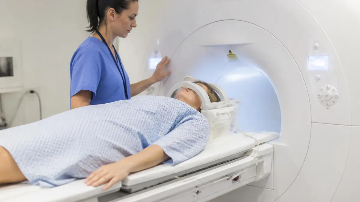
Step-by-Step: Getting an MRI Safely with a Copper IUD
Confirm Your IUD Brand and Model
Notify the Ordering Physician
Complete the MRI Safety Screening Form Honestly
Speak Directly with the MRI Technologist
Proceed with Scan — No Removal Needed
Follow Up If You Notice Any Changes
The physics underlying MRI safety for metallic implants centers on three distinct interactions: static magnetic field forces, gradient-induced electrical currents, and radiofrequency heating. For copper IUDs, the static field is the least concerning of the three. Copper's magnetic susceptibility is approximately −9.63 × 10⁻⁶ SI units, placing it firmly in the diamagnetic category — it is actually very slightly repelled rather than attracted by magnetic fields.
This means the net translational force (the tendency to pull the device toward the center of the magnet) is vanishingly small compared to the gravitational force already acting on the device inside your body. No clinically meaningful movement results.
Gradient magnetic fields switch on and off thousands of times per second during an MRI scan. When a conductive loop — even a small one — is placed in a time-varying magnetic field, Faraday's law of induction dictates that a current will be induced in that loop. Copper IUDs do form a partial conductive loop.
However, studies measuring induced currents in Paragard-type devices have found that the resulting forces are orders of magnitude smaller than those generated by body movement or normal uterine contractions. The ACR and device manufacturers have confirmed that no clinically significant torque or vibration occurs from gradient interactions at approved field strengths.
Radiofrequency heating is the most technically nuanced concern. RF pulses at the Larmor frequency (approximately 64 MHz at 1.5T and 128 MHz at 3T) deposit energy into conductive structures, potentially raising their temperature. The specific absorption rate (SAR) limit for the whole body is set at 4 W/kg by the IEC, with more conservative limits for localized tissue.
Because the Paragard IUD is small — the crossbar is only 32 mm wide and the vertical stem is 36 mm long — it does not create significant antenna-like resonance at clinical MRI frequencies. Multiple phantom and in-vivo studies have recorded temperature increases of less than 1°C at the device location during standard clinical sequences at 3T, well below the 2°C threshold generally considered the upper limit for safe tissue heating.
One nuance worth understanding is that SAR varies with sequence type. T1-weighted spin echo sequences with many rapid RF pulses deliver more SAR than gradient echo or single-shot sequences. Most clinical MRI protocols are designed to operate in IEC normal operating mode (SAR ≤ 2 W/kg whole body), which produces even lower heating than the maximum allowed exposure. If your referring physician requests a specialized protocol with high SAR — such as certain cardiac or breast MRI sequences — the MRI physicist can calculate the expected temperature rise at the IUD location and determine whether additional precautions are needed.
It is also important to understand MRI labeling terminology, because the terms used on device package inserts have precise regulatory meanings. "MR Safe" means the device poses no known hazards in all MRI environments — typically reserved for entirely non-metallic items like plastic catheters. "MR Conditional" means the device is safe only when specific conditions are met, such as maximum field strength, maximum SAR, and minimum gradient slew rate. "MR Unsafe" means the device must never enter the MRI environment.
Copper IUDs fall into the MR Conditional category, and the conditions stated in Paragard's labeling — 3T or less, normal operating mode SAR — are met by virtually all routine clinical MRI examinations in US hospitals and imaging centers.
Field strength above 3T is used in research settings, and a small number of 7T whole-body MRI systems are now entering clinical use in specialized academic centers. If you are scheduled for a 7T MRI, you should specifically ask whether your copper IUD has been tested at that field strength. As of the current literature, Paragard's MRI-conditional labeling extends to 3T; testing at 7T is limited and not yet incorporated into the official product labeling. Research participants who use 7T systems are typically screened and counseled individually by the site's MRI safety officer.
Overall, the preponderance of scientific evidence — spanning bench testing, computer simulation, phantom experiments, and clinical case series — supports the conclusion that copper IUDs present no meaningful safety hazard during MRI examinations performed at 1.5T or 3T under standard clinical conditions. Radiologists, gynecologists, and MRI safety experts broadly agree: the benefit of performing a needed diagnostic scan vastly outweighs the negligible and theoretical risks associated with scanning a copper IUD.
For a fuller set of questions on this topic, see our mri abdomen and pelvis cpt code resource.
For a fuller set of questions on this topic, see our open mri near me resource.
IUD Types and MRI Compatibility: Copper vs. Hormonal vs. Unknown Devices
The Paragard TCu380A is the only copper IUD approved by the FDA for use in the United States. Its frame is made of polyethylene with copper wire wound around the vertical stem and copper sleeves on the horizontal arms. Because copper is non-ferromagnetic, Paragard has been tested and labeled MRI conditional at field strengths up to 3 Tesla. The temperature rise at the device during standard clinical sequences at 3T has measured less than 1°C in multiple published studies, and no significant torque or displacement forces have been detected.
Patients with Paragard can undergo MRI of any body part — including the pelvis — without removal of the device. Pelvic MRI with a copper IUD in place may show a small artifact (a void or distortion) in the uterine image near the device location, which is generally not clinically problematic. Radiologists are trained to recognize IUD artifacts and account for them in their interpretation. If precise imaging of the uterine cavity itself is the clinical goal, your radiologist may recommend a specific sequence or imaging plane that minimizes the artifact.
Pros and Cons of Scanning with a Copper IUD In Place vs. Removal
- +No procedural risk — avoids the small but real risks of IUD removal and reinsertion (infection, perforation, expulsion)
- +Copper is non-ferromagnetic, so translational and torque forces are negligibly small at 1.5T and 3T
- +Paragard carries MRI-conditional labeling approved at field strengths used in virtually all routine clinical MRI
- +Temperature rise at the device during standard sequences measures less than 1°C — far below the safety threshold
- +Scan can proceed without delay — no need to schedule a separate removal appointment before imaging
- +Avoids gap in contraceptive coverage that could result if IUD is removed and reinsertion is delayed
- −Copper IUD creates a small imaging artifact in the uterus that may slightly obscure nearby structures on pelvic MRI
- −Device must be disclosed on the safety screening form; patients who forget may cause scan delays
- −Safety labeling only extends to 3T; patients undergoing 7T research MRI require individual evaluation
- −High-SAR specialized sequences may require additional physicist review before proceeding
- −Patients with unknown or very old IUD models cannot rely on standard conditional labeling without further investigation
- −Some facilities may have overly conservative local policies and may unnecessarily delay or refuse scanning — requiring escalation to the radiologist
Pre-MRI Checklist for Patients with a Copper IUD
- ✓Locate your IUD insertion card or call your gynecologist's office to confirm the exact brand and model of your device.
- ✓Notify the doctor ordering your MRI about your copper IUD so it can be noted on the imaging referral.
- ✓Call the imaging facility before your appointment to alert staff that you have a copper IUD.
- ✓Complete the MRI safety screening questionnaire fully and accurately — check yes for IUD and write in the device name.
- ✓Tell the MRI technologist about your IUD in person before entering the scanner room.
- ✓Ask the technologist to verify your device's MRI-conditional status in the ACR safety database or MRIsafety.com.
- ✓Confirm that the planned scan will be performed at 3T or lower field strength, which is within Paragard's labeled conditions.
- ✓Ask whether any sequences in your protocol use high SAR settings that may require additional physicist review.
- ✓Remove all external metal (jewelry, hair clips, belt buckles) as you normally would for any MRI appointment.
- ✓After the scan, contact your gynecologist if you notice unusual cramping, spotting, or cannot feel your IUD strings.
The ACR Classifies Paragard as MRI Conditional — Not MRI Unsafe
Many patients and even some non-radiology healthcare providers mistakenly assume that any metal implant is a contraindication to MRI. In fact, the American College of Radiology explicitly classifies the Paragard copper IUD as MRI conditional at 3T or less, meaning it can be safely scanned under standard clinical conditions. Removal before MRI is almost never medically necessary and is actively discouraged due to procedural risks.
When you arrive at the MRI facility, the interaction with the MRI technologist is your most important safety checkpoint. Technologists are trained in MRI safety and have access to up-to-date databases that list the MRI status of thousands of implants and devices. Tell them you have a copper IUD and give them the brand name — Paragard, for example — as soon as you meet them in the screening area. If you have the insertion card with you, bring it; it often contains the model number, lot number, and manufacturer information that makes the safety lookup faster and more precise.
The technologist will search MRIsafety.com (maintained by Dr. Frank Shellock) or the ACR's implant safety resources, both of which are considered authoritative references in clinical MRI safety. The listing for the Paragard TCu380A confirms MRI conditional status at 1.5T and 3T and provides the specific conditions under which scanning is approved. These conditions align with normal clinical operating modes, so the technologist will almost always be able to confirm quickly that your scan can proceed without modification.
In some cases, the technologist may need to consult the supervising radiologist or an on-site MRI safety officer — particularly if your IUD is an unfamiliar model or if the planned scan involves unusual parameters. This consultation is a normal part of the safety workflow and should not alarm you. It typically takes only a few minutes and results in written documentation in your medical record confirming that the safety review was performed. Do not feel pressured to decline the consultation or rush the process; this extra step exists to protect you.
If you are having a pelvic MRI specifically — for example, to evaluate the uterus, ovaries, or pelvic floor — tell the technologist that your IUD is in the pelvis so they can anticipate the artifact it will produce on imaging. The technologist and radiologist will choose imaging sequences and orientations that minimize artifact interference while still capturing the diagnostic information your physician needs. Common strategies include using sequences with shorter echo times, adjusting the imaging plane to move the artifact away from the structure of interest, or employing artifact-reduction techniques such as view-angle tilting.
Patients sometimes ask whether the MRI can dislodge or displace their IUD. This is a common concern, but the evidence strongly says no. Multiple studies, including in-vivo imaging sessions where the uterus was imaged directly during and after MRI, have shown no IUD displacement attributable to the scan. The uterine muscle and the anchoring mechanism of the IUD's arms are more than sufficient to resist the trivial magnetic forces involved. Post-MRI IUD string checks — the same self-check gynecologists recommend monthly — are a reasonable precaution but are not required by any clinical guideline specifically because of MRI exposure.
Anxiety about being in the MRI scanner is real and common. If you experience claustrophobia, discuss this with your physician before the appointment; mild anxiolytic medication or open-bore MRI options may be available. The presence of your IUD does not affect anxiety management options. Patients with IUDs can use all standard comfort measures: headphones with music, mirror attachments, prism glasses, or sedation when clinically indicated. The goal is a successful scan that gives your physician the information needed to guide your care.
After your MRI, the images will be interpreted by a radiologist who will note the presence of the IUD artifact in their report and distinguish it from any pathological findings. If the radiologist has any uncertainty about whether a finding near the IUD is real or artifactual, they may recommend additional imaging — such as ultrasound — to clarify. This is standard radiological practice and does not indicate a safety problem with your IUD or the MRI itself.
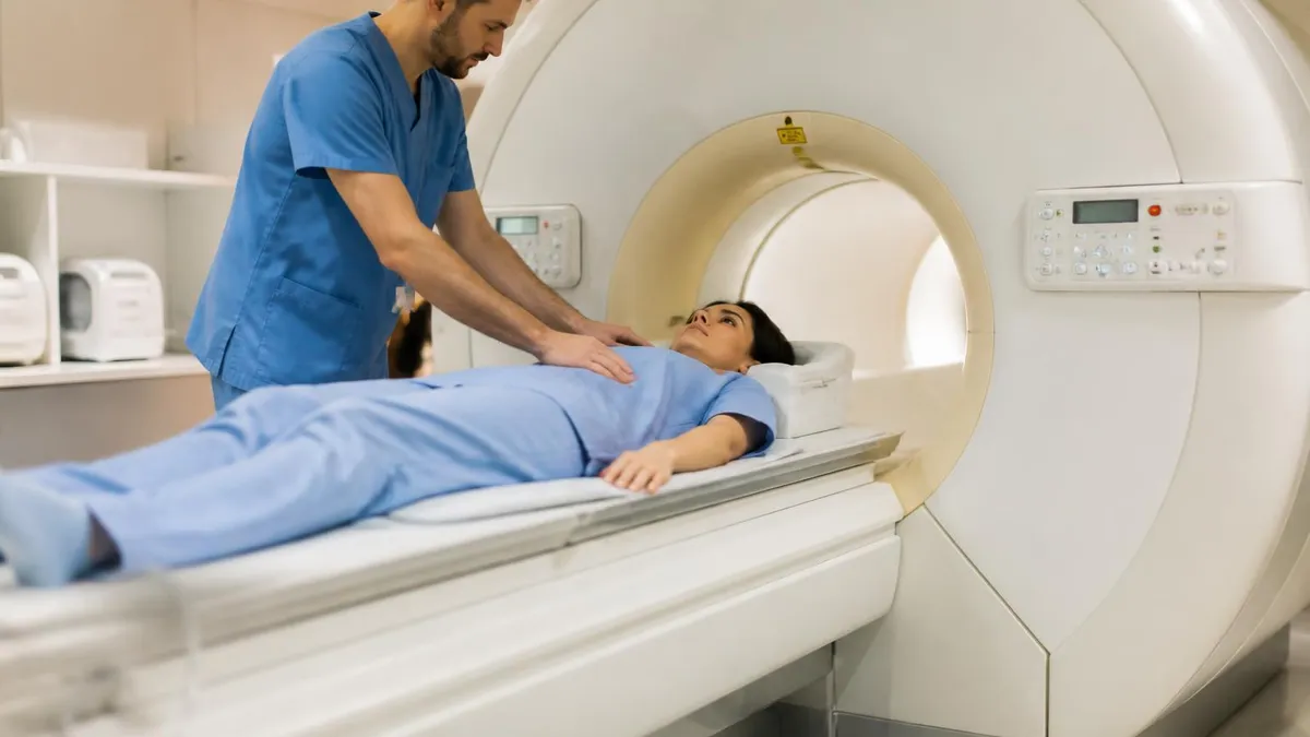
Paragard's MRI-conditional labeling currently covers field strengths up to 3 Tesla. If you are referred to a 7T research or clinical MRI — now available at select academic centers — inform the site's MRI safety officer about your copper IUD and request device-specific evaluation before the scan. Similarly, patients who do not know the brand or model of their IUD and cannot obtain medical records should not proceed to MRI without a case-by-case review by a qualified MRI physicist or safety officer.
One area where patients sometimes receive conflicting information is from facility staff who are unfamiliar with current MRI safety standards. It is not uncommon for a patient to be told — incorrectly — that their IUD must be removed before an MRI. This outdated guidance dates to an era before comprehensive MRI safety testing of IUDs was available and before MRI-conditional labeling became standard practice. If you encounter resistance at an imaging facility, you are within your rights to ask to speak with the supervising radiologist, who can review the manufacturer's labeling and ACR guidance and make an authoritative decision.
The ACR's manual on MRI safety, updated regularly, is the gold standard reference for clinical decision-making in the United States. It explicitly lists the Paragard TCu380A as MRI conditional and provides the testing conditions under which it was evaluated. Radiologists who have completed residency training or MRI safety continuing education will be familiar with this guidance. If you feel that a facility is refusing your scan based on outdated or incorrect information, your ordering physician can intervene by communicating directly with the radiologist on duty.
It is also worth noting that the European Society of Urogenital Radiology (ESUR) and the Royal Australian and New Zealand College of Radiologists (RANZCR) publish similar guidance affirming the MRI safety of copper IUDs under standard clinical conditions. If you have had an IUD inserted outside the United States and are unsure of its MRI status, checking these international resources — or the global implant safety database at MRIsafety.com — is a reasonable approach. Most copper IUDs sold internationally share the same non-ferromagnetic copper-and-polyethylene construction as Paragard and have similar safety profiles.
Pregnancy and IUD interaction is occasionally raised as a concern in the context of MRI. MRI during the first trimester is generally avoided when possible due to limited data on fetal safety, but this is a separate concern from IUD safety.
Copper IUDs are highly effective (greater than 99%) at preventing pregnancy, so the scenario of being pregnant with a copper IUD in place — while possible in rare cases of device failure — is uncommon. If there is any possibility of pregnancy, a pregnancy test should be performed before pelvic MRI regardless of IUD status, in accordance with standard pre-MRI screening protocols.
Gadolinium-based contrast agents, sometimes used to enhance MRI images, are an entirely separate consideration from IUD safety. If your MRI order includes contrast, discuss any kidney disease, allergies, or prior contrast reactions with your radiologist before the scan. The presence of a copper IUD has no bearing on the decision to use or avoid gadolinium contrast. These are independent safety considerations governed by different sets of guidelines and risks.
Patients preparing for the ARRT MRI registry examination should be familiar with the classification system for implants — MR Safe, MR Conditional, and MR Unsafe — and should understand the specific physical interactions (static field torque, gradient-induced forces, RF heating) that determine a device's classification. IUD safety is a frequently tested topic in MRI registry preparation, and understanding the reasoning behind copper IUD's MRI-conditional status will help candidates answer related questions confidently on the registry exam.
In summary, the answer to whether you can get an MRI with a copper IUD is a clear and well-supported yes — with the straightforward conditions that you disclose the device, verify its MRI-conditional status with the technologist, and confirm the scan will be performed at 3T or less under standard clinical SAR limits. The evidence base supporting this conclusion spans decades of device testing, international safety guidelines, and real-world clinical experience across millions of patients.
For MRI technologists and students studying for the ARRT MRI registry, questions about implant safety — including IUDs — represent an important knowledge domain. The registry tests candidates on their ability to assess patient safety, identify contraindications, and apply the ACR's MRI safety framework in clinical scenarios. Understanding the difference between MR Safe, MR Conditional, and MR Unsafe devices is foundational, and copper IUDs are a textbook example of an MR Conditional device with a well-characterized safety profile that allows scanning under defined conditions.
Technologists should be prepared to look up any unfamiliar device in authoritative safety databases before allowing a patient to enter the scanner room. The two most widely used resources in US clinical practice are MRIsafety.com and the ACR's implant safety guide. Both are searchable by device name, manufacturer, and model number. When a device listing confirms MR Conditional status, the technologist documents the lookup result in the patient's pre-scan screening record. This documentation protects both the patient and the facility and creates an auditable safety trail.
For facilities conducting pelvic MRI, it is helpful to establish a standard protocol note for patients with IUDs. This note should specify the device type, the database reference used for safety verification, the field strength of the exam, and the relevant SAR level of the protocol.
Many academic radiology departments and large imaging networks have created internal reference guides that list the most common IUDs with their MRI status pre-verified, which speeds the screening process for patients who can confirm their device brand. Standardizing this workflow reduces variability and the chance that a busy technologist might skip the verification step under time pressure.
From a patient education standpoint, gynecologists and primary care providers play a critical role in preparing patients for MRI. A brief counseling message at the time of IUD insertion — explaining that the device is MRI conditional, encouraging the patient to keep their insertion card, and advising them to always disclose the IUD on medical screening forms — can prevent confusion years later when a patient is referred for MRI. Some gynecologists now routinely provide patients with a wallet card listing the device name, model, and MRI conditional status, similar to the cards given to patients with cardiac pacemakers.
Artifact management is a practical skill that becomes relevant when the area being imaged is close to the copper IUD. The copper creates a susceptibility artifact — a localized distortion of the magnetic field — that shows up as a void or blooming region on the MRI image. This artifact is largest on gradient echo sequences with long echo times and smallest on fast spin echo sequences with short echo times. Radiologists interpreting pelvic MRI routinely account for this artifact by reviewing multiple sequences and imaging planes. In practice, the artifact almost never prevents a diagnostic-quality examination from being completed.
Technologists and radiologists who regularly perform pelvic MRI develop familiarity with the characteristic appearance of IUD artifacts for each device type. Copper IUDs tend to produce larger susceptibility artifacts than hormonal IUDs due to copper's higher magnetic susceptibility compared to the materials used in hormonal device frames. Knowing which device is present helps the radiologist predict the size and shape of the artifact and distinguish it confidently from pathological findings such as fibroids, polyps, or adenomyosis that might otherwise be confused with device-related artifact on a single sequence.
Students and technologists who want to deepen their understanding of implant safety physics will benefit from reviewing the ASTM F2052 standard for measurement of magnetically induced displacement force, the ASTM F2213 standard for magnetically induced torque, and the ASTM F2182 standard for measurement of RF-induced heating.
These are the test standards that device manufacturers use to characterize implants before applying for MRI-conditional labeling. Familiarity with these standards helps explain why certain devices are safe at 1.5T but not 3T, or why normal SAR mode is safe while first-level controlled mode requires additional review — distinctions that will appear on the MRI registry examination.
MRI Questions and Answers
DWI MRI: Diffusion-Weighted Imaging Explained (2026 Guide)

MRI Lumbar Spine Without Contrast: Complete Guide to the Scan, What It Shows, and How to Prepare
MRI History: From Nuclear Resonance to Modern Scanners

MRI of Cervical Spine: Complete Guide to Imaging, Anatomy, Pathology, and Patient Preparation

Where to Get an MRI: Facilities, Costs, and How to Choose
About the Author

Medical Laboratory Scientist & Clinical Certification Expert
Johns Hopkins UniversityDr. Sandra Kim holds a PhD in Clinical Laboratory Science from Johns Hopkins University and is certified as a Medical Technologist (MT) and Medical Laboratory Scientist (MLS) through ASCP. With 16 years of clinical laboratory experience spanning hematology, microbiology, and molecular diagnostics, she prepares candidates for ASCP board exams, MLT, MLS, and specialist certification tests.
Join the Discussion
Connect with other students preparing for this exam. Share tips, ask questions, and get advice from people who have been there.
View discussion (8 replies)