
MRI While Pregnant: Safety, Risks, Trimester Guidelines, and What to Expect
🗨️ Is MRI while pregnant safe? Learn trimester-by-trimester guidelines, real risks, gadolinium concerns, and what radiologists recommend for pregnant patients.
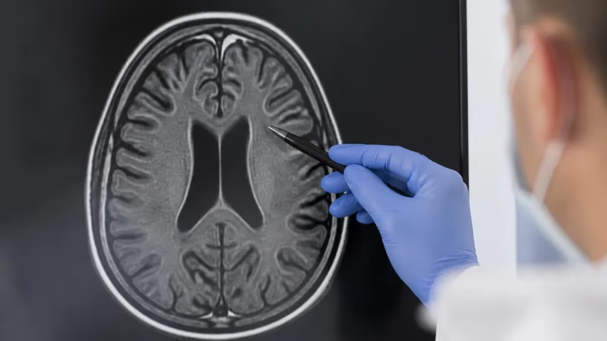
MRI while pregnant is one of the most common and anxiety-inducing questions that expectant mothers face when their physician orders advanced imaging. Unlike X-ray or CT scans, MRI does not use ionizing radiation, which means it avoids one of the primary concerns associated with imaging during pregnancy. Instead, MRI relies on powerful magnetic fields and radiofrequency pulses to generate detailed soft-tissue images, making it an attractive diagnostic tool when clinical necessity outweighs the theoretical risks. Understanding the distinction between these imaging modalities helps patients and providers make well-informed decisions during each trimester.
Despite the absence of ionizing radiation, MRI during pregnancy is not considered entirely risk-free, particularly during the first trimester. The biological effects of strong magnetic fields on a developing embryo or fetus are not fully understood, and no large-scale randomized controlled trials on humans have been conducted due to obvious ethical constraints. Most of the safety data comes from epidemiological studies, animal research, and retrospective reviews of patients who underwent MRI before their pregnancies were confirmed. These studies have been largely reassuring, but the absence of evidence is not the same as evidence of absence.
The American College of Radiology (ACR) and the Society for Maternal-Fetal Medicine both state that MRI can be performed at any gestational age when the diagnostic information is clinically necessary and when the benefit to the mother outweighs any potential fetal risk. Critically, the ACR recommends that MRI should not be withheld if it is the best diagnostic option available. Providers are encouraged to document their clinical rationale when ordering MRI for pregnant patients, especially during the first trimester when organogenesis is occurring.
One of the most important considerations in prenatal MRI is whether gadolinium-based contrast agents (GBCAs) will be used. Gadolinium crosses the placenta and enters fetal circulation, and while it is eventually cleared through fetal urine into the amniotic fluid, it can recirculate before being fully eliminated. Animal studies have linked high-dose gadolinium exposure to teratogenic effects, and a large Canadian cohort study published in JAMA in 2016 found associations between first-trimester gadolinium use and rheumatological, inflammatory, and infiltrative skin conditions in children, although absolute risks remained very low.
Radiologists and MRI technologists play a central role in the safety discussion. Before scanning a pregnant patient, the imaging team should confirm gestational age, review the clinical indication, assess whether non-contrast MRI would suffice, and ensure the patient has provided informed consent that reflects an accurate understanding of known and unknown risks. Optimizing scan parameters to minimize specific absorption rate (SAR) and acoustic noise is also standard practice. When in doubt, consultation between the ordering physician, radiologist, and maternal-fetal medicine specialist is the safest pathway.
For patients preparing for MRI or studying for registry exams, understanding how MRI interacts with pregnancy is a core competency. Exam questions frequently test knowledge of FDA pregnancy categories for contrast agents, ACR guidelines, magnetic field strength thresholds, and the biological mechanisms behind potential fetal risks. Reviewing these concepts in the context of clinical scenarios will help both technologists and radiologists answer questions with confidence. Studying the nuances of mri while pregnant guidelines alongside anatomical protocols deepens overall exam readiness.
This article walks through the complete clinical picture — from first-trimester precautions to third-trimester considerations, contrast agent policies, noise mitigation strategies, and what patients should expect on the day of their scan. Whether you are a patient trying to understand your options, a student preparing for the ARRT MRI registry, or a technologist refreshing your protocols, this comprehensive guide gives you the evidence-based information you need to navigate prenatal MRI safely and confidently.
MRI During Pregnancy by the Numbers
Trimester-by-Trimester MRI Guidelines
First Trimester (Weeks 1–13): Proceed With Caution
Second Trimester (Weeks 14–27): Generally Considered Safe
Third Trimester (Weeks 28–40): Logistical and Physiological Challenges
Postpartum: Gadolinium and Breastfeeding Considerations
Emergency Situations: MRI Supersedes Trimester Concerns
Gadolinium-based contrast agents (GBCAs) represent the most significant and well-documented risk associated with MRI during pregnancy. All GBCAs are classified as FDA Pregnancy Category C, meaning that animal studies have demonstrated adverse fetal effects at high doses, but adequate human studies do not exist to fully characterize the risk. In clinical practice, gadolinium is reserved for situations where the non-contrast MRI is diagnostically insufficient and the clinical question cannot be answered by alternative imaging methods. The decision to administer gadolinium to a pregnant patient should always involve a senior radiologist and, ideally, the patient's obstetrician or maternal-fetal medicine specialist.
The mechanism of concern with gadolinium involves its ability to cross the placenta. Once in fetal circulation, gadolinium is filtered by the fetal kidneys and excreted into amniotic fluid. However, unlike adults who excrete gadolinium efficiently through adult kidneys, the fetus swallows amniotic fluid, re-absorbs gadolinium, and re-excretes it — creating a prolonged recirculation loop. This extended tissue exposure window is the primary reason why gadolinium carries more theoretical risk in fetuses than in adults. The half-life of gadolinium in fetal tissue is not well established in humans, but animal models suggest it is significantly longer than in adults.
The landmark 2016 cohort study by Ray et al., published in JAMA, analyzed over 1.4 million births in Ontario, Canada. Among the 397 women who received gadolinium MRI during pregnancy, first-trimester exposure was associated with a statistically significant increase in the risk of rheumatological, inflammatory, and infiltrative skin conditions in children at one to four years of age.
The absolute risk increase was small — approximately 4 additional cases per 1,000 children exposed — but the finding reinforced the position that gadolinium should only be used in pregnancy when clearly essential. Stillbirth and neonatal death rates did not differ significantly from the unexposed group, which was reassuring.
Not all GBCAs carry the same risk profile. Linear GBCAs are known to deposit gadolinium in tissues including the brain, bone, and skin at higher rates than macrocyclic GBCAs. Because of this, the FDA and the European Medicines Agency have restricted the use of certain linear agents. When gadolinium is deemed necessary during pregnancy, macrocyclic agents such as gadobutrol (Gadavist) and gadoteridol (ProHance) are preferred because of their greater thermodynamic and kinetic stability and lower tendency for transmetallation — the process by which gadolinium is released from its chelating ligand.
Patients who receive gadolinium during pregnancy should be counseled before the scan about the known and unknown risks. Informed consent documentation is not legally required in all states, but it represents best practice and protects both the provider and the patient. The consent discussion should cover the indication for gadolinium, why non-contrast imaging was insufficient, the class of agent being used, and the known findings from the Ray et al. study. Patients should have the opportunity to ask questions and, when the situation is non-emergent, to consult with their OB or MFM before making a decision.
For MRI technologists and radiologists preparing for registry examinations, gadolinium pharmacology and pregnancy safety are high-yield topics. Questions often focus on the mechanism of placental transfer, FDA classification, the preference for macrocyclic agents, and the clinical threshold for gadolinium use during each trimester. Understanding the Ray et al. study findings and their limitations is important for nuanced exam answers that reflect real clinical practice rather than oversimplified rules. Connecting this knowledge to broader contrast safety frameworks strengthens retention and application during the exam.
Beyond gadolinium, some providers also consider the potential effects of radiofrequency heating on fetal tissue. The specific absorption rate (SAR) measures the rate at which RF energy is absorbed by body tissue, and elevated SAR has theoretical potential to raise fetal core temperature. Most modern 1.5T and 3T scanners have built-in SAR monitoring systems that automatically limit RF deposition. Technologists should select sequences with the lowest SAR consistent with diagnostic image quality, use body-coil limitations wisely, and document any SAR-related parameter adjustments made during a prenatal scan. These practices reflect both technical competence and patient-centered care.
MRI Practice Test Questions
Prepare for the MRI - Magnetic Resonance Imaging exam with our free practice test modules. Each quiz covers key topics to help you pass on your first try.
MRI Knowledge
MRI Exam Questions covering Knowledge. Master MRI Test concepts for certification prep.
MRI Physics
Free MRI Practice Test featuring Physics. Improve your MRI Exam score with mock test prep.
MRI Anatomy and Pathology
MRI Test Prep for MRI Anatomy and Pathology. Practice MRI Quiz questions and boost your score.
MRI Anatomy and Positioning
MRI Questions and Answers on MRI Anatomy and Positioning. Free MRI practice for exam readiness.
MRI Contrast Agents
Free MRI Quiz on MRI Contrast Agents. MRI Exam prep questions with detailed explanations.
MRI Patient Care and Positioning
MRI Practice Questions for MRI Patient Care and Positioning. Build confidence for your MRI certification exam.
MRI Safety Protocols by Clinical Indication
When a pregnant patient presents with acute neurological symptoms — sudden severe headache, focal neurological deficits, vision changes, or altered consciousness — MRI of the brain is frequently the imaging modality of choice because it avoids the ionizing radiation of CT while providing superior soft-tissue contrast. Conditions such as intracranial hemorrhage, venous sinus thrombosis, posterior reversible encephalopathy syndrome (PRES), and acute ischemic stroke all require prompt and accurate diagnosis that MRI can provide more definitively than any alternative modality.
In these emergencies, the gestational trimester becomes secondary to maternal stabilization. PRES is disproportionately common in pregnant women due to the hypertensive disorders of pregnancy, and MRI with DWI and FLAIR sequences can diagnose it without contrast in most cases. When gadolinium is considered necessary — such as when CNS vasculitis or an intracranial tumor is suspected — the clinical team should document the indication clearly and select a macrocyclic GBCA at the lowest effective dose. Rapid scan completion also minimizes total RF energy deposition.
MRI vs. Other Imaging Modalities During Pregnancy
- +No ionizing radiation — eliminates the primary embryologic risk associated with X-ray and CT
- +Superior soft-tissue contrast compared to ultrasound for complex diagnoses
- +Multi-planar imaging capability allows comprehensive anatomical assessment in a single session
- +No known cumulative dose effect — multiple MRI scans do not compound biological risk the way radiation does
- +Fetal MRI can characterize anomalies beyond the resolution of obstetric ultrasound
- +Preferred modality for appendicitis in pregnancy when ultrasound is inconclusive per ACR guidelines
- −Theoretical risk of RF heating and SAR-related fetal temperature elevation, especially at higher field strengths
- −Gadolinium contrast crosses the placenta and has uncertain long-term developmental effects
- −Loud acoustic noise during scanning — up to 110 dB — poses theoretical risk to fetal auditory development
- −First-trimester safety data is limited due to ethical restrictions on prospective human trials
- −Patient positioning in the third trimester is challenging and may compromise image quality
- −Longer scan times compared to CT increase patient discomfort and fetal motion artifact risk
Pre-Scan Checklist for Pregnant Patients Undergoing MRI
- ✓Confirm gestational age with the ordering physician and document it in the scan request before scheduling.
- ✓Verify the clinical indication and ensure it meets the threshold of medical necessity for prenatal MRI.
- ✓Complete a standard MRI safety screening form with specific pregnancy questions, including trimester and due date.
- ✓Assess whether gadolinium contrast is required or whether a non-contrast protocol can answer the clinical question.
- ✓If gadolinium is planned, obtain and document informed consent covering known fetal risks and the clinical rationale.
- ✓Select sequences with the lowest specific absorption rate (SAR) consistent with diagnostic image quality.
- ✓Prepare acoustic hearing protection — earmuffs or earplugs — to reduce fetal exposure to gradient noise.
- ✓Position the patient with a left lateral wedge in the second and third trimester to prevent aortocaval compression.
- ✓Monitor maternal vital signs before, during, and after the scan, especially in late pregnancy.
- ✓After the scan, document all scan parameters, SAR levels, and contrast administered in the patient's medical record.
ACR Position: MRI Should Not Be Withheld When Clinically Necessary
The American College of Radiology states explicitly that MRI during pregnancy should not be withheld if it is the best available imaging option for the clinical question at hand. Delaying a necessary MRI to wait for a safer trimester can itself result in harm through diagnostic delay. The decision framework centers on whether the benefit of the diagnostic information to the mother and fetus outweighs the theoretical — and largely unquantified — risk of MRI exposure at any given gestational age.
The clinical evidence base for MRI safety during pregnancy has grown substantially over the past two decades, though significant knowledge gaps remain.
The most frequently cited concern — teratogenicity from magnetic field exposure — has not been demonstrated in human populations at the field strengths currently used in clinical practice (1.5T and 3T). A 2016 systematic review published in Radiology examined outcomes from multiple cohort studies and found no statistically significant association between prenatal MRI exposure and adverse perinatal outcomes including congenital malformations, stillbirth, or neonatal death when gadolinium was not administered. These findings have been replicated in smaller studies with similar conclusions.
Animal studies using much higher field strengths (up to 9.4T) and prolonged exposure durations have documented effects including delayed ossification and reduced birth weight in rodent models. However, direct extrapolation from rodent data to human clinical MRI at 1.5T or 3T is methodologically problematic. The field strengths used in animal studies often far exceed those of clinical systems, and the duration and intermittent nature of clinical MRI scans differ substantially from the chronic whole-body exposures used in controlled animal experiments. Nonetheless, these findings justify the precautionary principle, particularly during the first trimester when embryogenesis is most active.
Acoustic noise is an underappreciated aspect of MRI safety in pregnancy. Gradient switching during MRI generates noise levels between 82 and 118 dB depending on the sequence and scanner model. Prolonged exposure to noise exceeding 85 dB is associated with sensorineural hearing loss in adults and animals. The fetal ear begins developing around week 20 of gestation and is functional by approximately week 25.
For MRI scans performed after 24 weeks, acoustic shielding from amniotic fluid reduces fetal noise exposure by approximately 30 dB, bringing most clinical MRI sequences below the threshold of concern for fetal auditory damage. That said, use of maternal earplugs is standard practice and provides additional attenuation.
Thermal effects from RF energy deposition represent another theoretical risk. The specific absorption rate (SAR) determines the rate at which tissue absorbs RF energy and converts it to heat. Fetal thermoregulation depends entirely on maternal thermoregulation — the fetus has no independent mechanism to dissipate heat. A core temperature rise of 1.5°C above baseline has been associated with teratogenic effects in animal models.
Modern MRI scanners continuously monitor whole-body and local SAR and automatically restrict imaging parameters to prevent unsafe heating. In practice, the temperature increase observed in fetal tissue during standard clinical MRI is well below the 1.5°C threshold, especially for sequences optimized for SAR reduction.
Magnetic field gradient switching generates time-varying magnetic fields (dB/dt) that can theoretically induce electrical currents in biological tissue — a phenomenon known as peripheral nerve stimulation. This effect, while detectable in adult patients as tingling or fluttering sensations, has no established direct correlate in fetal tissue because the developing nervous system has different electrical properties. Current IEC standards limit dB/dt values to prevent peripheral nerve stimulation in adult patients, and these limits are applied uniformly regardless of whether the patient is pregnant. Whether these limits adequately protect fetal neural tissue remains an open research question.
For students preparing for the ARRT MRI registry examination, the safety evidence framework is directly testable. Questions may present a clinical vignette of a pregnant patient needing emergent neuroimaging and ask which imaging modality is preferred, which contrast class is safest, or which trimester carries the highest theoretical risk. Understanding the hierarchy of concerns — ionizing radiation is eliminated, gadolinium is the primary modifiable risk, thermal and acoustic effects are mitigated by modern scanner safety systems — helps structure answers in a logical, evidence-based way. Connecting these safety principles to specific scanner protocols reinforces both clinical competence and exam performance.
The intersection of MRI physics, patient safety, and obstetric medicine is one of the richest topics in the imaging sciences. A technologist or radiologist who understands not just the protocols but the biological rationale behind each precaution is better equipped to answer patient questions, adapt protocols to unexpected clinical situations, and advocate for evidence-based practice in their department. Registry exams reward depth of understanding precisely because the real clinical world requires it — especially in high-stakes situations involving vulnerable patients like pregnant women and their developing fetuses.
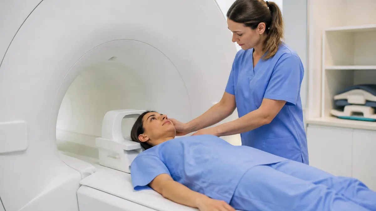
Gadolinium-based contrast agents should never be administered to a pregnant patient in the first trimester without explicit approval from a senior or attending radiologist and, when time permits, consultation with the patient's obstetrician or maternal-fetal medicine specialist. The first trimester represents the highest-risk window for organogenesis, and gadolinium recirculation through amniotic fluid prolongs fetal tissue exposure. Document clinical necessity thoroughly and use the lowest effective dose of a macrocyclic agent if gadolinium cannot be avoided.
For patients scheduled for MRI while pregnant, the day-of-scan experience deserves careful attention to both clinical safety and patient comfort. Arriving at the MRI suite, patients will be asked to complete or re-confirm their MRI safety screening questionnaire. The technologist will review the answers specifically for pregnancy details, including gestational age, whether they have received any gadolinium contrast during this pregnancy, and whether they have any implanted devices such as intrauterine devices (IUDs), which are generally MRI-safe but should be documented. The technologist may also ask about any prior adverse reactions to contrast media if gadolinium is planned.
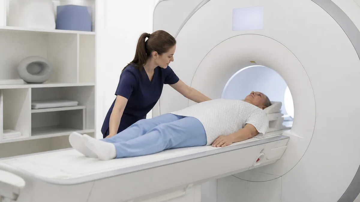
Once inside the scanner room, positioning is tailored to gestational stage. First-trimester patients typically tolerate the standard supine position without difficulty. Second-trimester patients may begin to experience mild discomfort from prolonged supine positioning, and the technologist should offer a small cushion under the knees to reduce lumbar strain.
Third-trimester patients should almost universally be positioned with a left lateral wedge or rolled blanket under the right hip to achieve 10–15 degrees of left lateral tilt, preventing the gravid uterus from compressing the inferior vena cava and reducing venous return. A call button should always be placed in the patient's hand so she can signal distress immediately.
Acoustic noise management is a standard part of prenatal MRI preparation. All patients undergoing MRI receive hearing protection, but for pregnant patients this step is particularly emphasized. Single-use foam earplugs inserted correctly can reduce noise exposure by 25–33 dB, bringing most gradient-intensive sequences below the occupational threshold for sustained noise exposure. Some centers also provide headphones that play music or audio instructions, which serve dual purposes: noise attenuation and patient distraction to reduce anxiety during the scan. Patients with significant claustrophobia may require anxiolytic premedication; for pregnant patients, the choice of anxiolytic should always be discussed with the obstetrician.
Scan duration is an important variable to communicate to pregnant patients before they enter the magnet. A typical non-contrast brain MRI takes 30–45 minutes. An abdominal or pelvic MRI may take 45–75 minutes depending on the clinical question and whether breath-holding sequences are required. For pregnant patients in the third trimester, extended scan durations can cause backache, hip discomfort, and anxiety from sustained confinement. Technologists should communicate expected duration before the scan, offer structured rest intervals if the protocol permits, and check in verbally with the patient at regular intervals during sequences to monitor for distress.
If gadolinium contrast is administered, the patient should be monitored for at least 30 minutes post-injection in most facilities, consistent with standard contrast safety protocols. The risk of acute adverse reactions to gadolinium in pregnant patients is not higher than in the general population, but any acute reaction — urticaria, bronchospasm, hypotension — requires the same aggressive management regardless of pregnancy status.
Epinephrine, antihistamines, and corticosteroids are all administered when clinically indicated even in pregnancy, because the risk of an untreated anaphylactic reaction to both the mother and fetus far exceeds any theoretical medication risk. Post-scan hydration is encouraged to facilitate renal clearance of contrast.
Follow-up communication between the imaging team and the ordering provider is essential after any prenatal MRI. The radiology report should document the specific sequences performed, whether contrast was used and the type and dose, SAR levels if elevated protocols were employed, and any technical limitations related to fetal motion or patient positioning.
This documentation serves both clinical and medico-legal purposes and ensures that the full imaging history is available to the obstetric team managing the patient's ongoing care. The radiologist's impression should explicitly address the clinical question that prompted the scan, with a clear statement of findings and their clinical implications.
Patients frequently ask whether their baby will be affected by the loud noise during the MRI. This is a valid concern that deserves a thoughtful, evidence-based response. As outlined earlier, amniotic fluid provides approximately 30 dB of acoustic shielding, and maternal soft tissue provides additional attenuation.
The frequencies at which gradient coils generate peak noise (1–3 kHz) are in the speech range, and while this is audible to a second- or third-trimester fetus, exposure during a single clinical scan is unlikely to cause permanent auditory damage. Reassuring patients with accurate information reduces anxiety and improves scan compliance, both of which contribute to diagnostic image quality. Technologists should be trained to deliver this information consistently and compassionately.
Preparing for registry examinations on MRI safety topics requires more than memorizing guidelines — it requires understanding the clinical reasoning behind each recommendation so you can apply it in novel scenarios on exam day. The ACR Manual on Contrast Media and the ACR-SPR Practice Parameter for the Safe and Optimal Performance of Fetal MRI are foundational documents that every MRI technologist and radiologist should review.
These documents are freely available online and represent the authoritative US clinical standards for prenatal imaging. Exam questions are often written to test whether candidates understand the principles behind the guidelines, not just the rules themselves.
When studying MRI safety in pregnancy, organize your knowledge around four key dimensions: (1) field strength and thermal effects, (2) gadolinium and placental transfer, (3) acoustic noise and fetal auditory development, and (4) patient positioning and cardiovascular safety in late pregnancy. Each dimension has its own set of mechanisms, risk thresholds, and mitigation strategies. Creating a comparison matrix across these dimensions helps consolidate the material and makes it easier to retrieve under exam pressure. Flash cards covering the SAR limits, FDA contrast categories, dB thresholds, and trimester-specific guidelines are particularly useful for high-yield fact retention.
Practice questions are essential for translating knowledge into exam performance. The best questions for this topic are clinical vignettes that require you to integrate multiple concepts simultaneously — for example, a scenario involving a 28-week pregnant patient with suspected appendicitis who has a peanut allergy and a documented adverse reaction to gadolinium in a previous scan. Working through this type of multi-variable scenario requires you to prioritize which risks are most immediately dangerous, which protocols are most appropriate, and what steps you would take in what sequence. Vignette-based practice far outperforms rote memorization for registry-style questions.
Beyond exam preparation, building clinical expertise in prenatal MRI is increasingly valuable for practicing technologists. As MRI scanner availability increases and radiation-reduction initiatives in obstetric imaging gain momentum, the volume of prenatal MRI referrals is expected to grow. Technologists who are comfortable with the unique positioning, communication, and protocol optimization challenges of prenatal MRI are valuable members of any imaging team. Pursuing continuing education specifically in MRI safety and obstetric imaging, attending relevant sessions at AHRA or SMRT meetings, and reviewing published case reports involving prenatal MRI complications all contribute to ongoing professional development.
Communication with patients is as important as technical competence. Pregnant patients are often anxious about any medical procedure, and MRI — with its loud noise, enclosed space, and association with serious diagnoses — can be particularly frightening. Technologists who take time to explain what the machine does, why it is ordered, what the noise is and why it occurs, and what specific steps are being taken to protect the baby report better patient cooperation and higher-quality scans.
Scripting reassuring language — without minimizing legitimate risks — is a skill that can be developed with training and peer feedback. Patient-centered communication is now explicitly included in ARRT continuing competency frameworks for a reason.
The broader principle underlying all MRI safety guidance in pregnancy is proportionality: the risk of the imaging procedure must be weighed against the risk of not imaging, and that calculation must be made transparently and collaboratively with the patient and her care team. There is no universal answer that applies to all pregnant patients in all clinical situations.
The first-trimester patient with a mild headache and no neurological signs is a very different scenario than the 32-week patient with sudden onset left-sided weakness. Developing the clinical judgment to differentiate these scenarios, apply the appropriate guidelines, and communicate the rationale clearly is the ultimate goal of both clinical training and registry examination preparation.
As MRI technology continues to evolve — with faster scan times, lower SAR sequences, and improved motion correction algorithms — the risk-benefit calculation for prenatal MRI will continue to shift in favor of scanning when clinically indicated. Compressed sensing techniques and simultaneous multi-slice acquisition can reduce scan times by 50% or more compared to conventional protocols, which is particularly beneficial for pregnant patients who are uncomfortable in the supine position.
Ultra-low-SAR sequences designed specifically for fetal and prenatal imaging are an active area of development at major academic centers. Staying current with these advances through peer-reviewed literature and professional organization resources is part of the commitment to evidence-based prenatal imaging practice.
MRI Questions and Answers

MRI of Cervical Spine: Complete Guide to Imaging, Anatomy, Pathology, and Patient Preparation

Knee MRI Images: A Complete Guide to Reading, Understanding, and Interpreting Knee Scans

Ace Your Exam with MRI Practice Test 2026 Prep

MRI Tech School 2026 — Programs, Requirements, and What to Expect
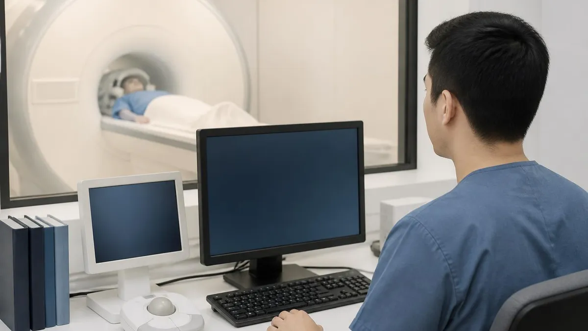
MRI Procedure: What to Expect Before, During and After Your Scan
About the Author

Medical Laboratory Scientist & Clinical Certification Expert
Johns Hopkins UniversityDr. Sandra Kim holds a PhD in Clinical Laboratory Science from Johns Hopkins University and is certified as a Medical Technologist (MT) and Medical Laboratory Scientist (MLS) through ASCP. With 16 years of clinical laboratory experience spanning hematology, microbiology, and molecular diagnostics, she prepares candidates for ASCP board exams, MLT, MLS, and specialist certification tests.
Join the Discussion
Connect with other students preparing for this exam. Share tips, ask questions, and get advice from people who have been there.
View discussion (8 replies)