
Why Would a Doctor Order an MRI of the Abdomen? Reasons, CPT Codes, and Open MRI Options
🎯 Why would a doctor order an MRI of the abdomen? Learn the reasons, CPT codes, open MRI machine options, prep steps, and costs before your scan.
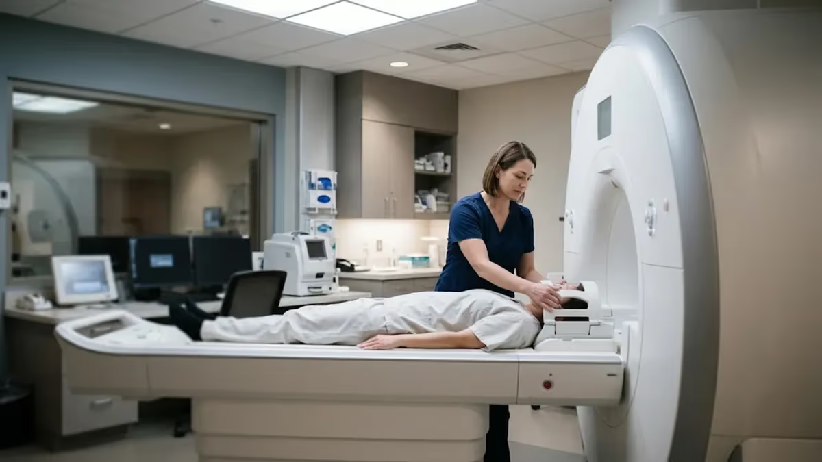
Why would a doctor order an MRI of the abdomen? Usually because ultrasound or CT left a question unanswered. MRI uses magnets and radio waves, not radiation, to image the liver, pancreas, kidneys, spleen, and bile ducts. Patients who worry about tight spaces often ask about an open MRI machine, and some abdominal studies can be done on wide-bore or open systems, though image quality varies. This guide explains the reasons, codes, and preparation. Practice with the free MRI - Magnetic Resonance Imaging practice test on PracticeTestGeeks (11 questions with answer explanations).
Doctors have several imaging tools, and each suits different questions. Ultrasound is quick and inexpensive, CT excels at emergencies and bone, and MRI wins on soft-tissue detail. Learning how these differ clarifies many mri scan uses across the body. For the abdomen, MRI is often the problem solver: it can tell a harmless cyst from a suspicious mass, map bile ducts without a catheter, and track chronic disease without repeated radiation exposure.
Common triggers include persistent upper abdominal pain, abnormal liver blood tests, unexplained weight loss, jaundice, or a lesion spotted incidentally on another scan. A physician may also order the study to stage a known cancer, monitor Crohn disease, or evaluate a kidney mass when CT contrast is unsafe. Each reason follows the same logic: when a clearer, safer, or more specific picture will change treatment, MRI becomes worth the longer time and higher cost.
The order itself carries a billing code. Abdominal MRI is reported with CPT 74181 for a scan without contrast, 74182 with contrast, and 74183 for without followed by with contrast. Knowing the code helps when you call your insurer about prior authorization, which most plans require for advanced imaging. Your doctor's office usually submits the request, but confirming approval before the appointment can prevent a surprise bill weeks later.
Many patients scan the abdomen together with the pelvis, especially when symptoms span both regions. A pelvic MRI CPT code is separate: 72195 without contrast, 72196 with contrast, and 72197 for without then with contrast. Gynecologic searches often phrase this as MRI pelvis w wo gyn CPT, which points to 72197. Combined studies bill each region separately, so ask the scheduler exactly which codes the radiologist intends to use.
Comfort matters too. Standard tubes are about 60 centimeters wide, while wide-bore scanners reach 70 centimeters, and a true open design leaves the sides unenclosed. Open systems usually run at lower field strength, so some detailed abdominal sequences take longer or look grainier. A radiologist can advise whether your clinical question tolerates that trade-off, or whether a wide-bore closed scanner, perhaps with mild sedation, offers the best balance of comfort and image quality.
This article walks through the main reasons for abdominal MRI, what the scan detects, how open and closed options compare, how to find locations, and how to prepare. You will also find checklists, practice resources, and answers to frequent questions. Use it to talk with your doctor confidently, not to self-diagnose. Only a clinician who knows your history can decide whether this test is the right next step for you.
Free MRI - Magnetic Resonance Imaging Study Tools
Interactive MRI - Magnetic Resonance Imaging resources on PracticeTestGeeks, free to start:
- Free MRI Knowledge Questions and Answers — 11 questions with instant scoring and answer explanations
- MRI - Magnetic Resonance Imaging flashcards — review key terms and concepts card by card
- MRI - Magnetic Resonance Imaging study guide — topic-by-topic overview of what the exam covers
- MRI - Magnetic Resonance Imaging cheat sheet — one-page printable summary for last-minute review
Abdominal MRI by the Numbers
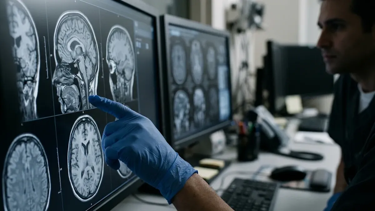
Main Reasons a Doctor Orders an Abdominal MRI
Abnormal liver enzymes, suspected tumors, fatty liver grading, cirrhosis follow-up, and bile duct blockages are leading reasons. MRI characterizes liver lesions precisely, and MRCP sequences show the bile ducts without needles or catheters, often avoiding more invasive procedures.
Unexplained upper abdominal pain, pancreatitis follow-up, cysts, or suspected masses often lead to MRI. It shows the pancreatic duct and cyst features well, helping doctors decide between surveillance, biopsy, or surgery for findings that other scans leave unclear.
When ultrasound or CT finds a kidney or adrenal nodule, MRI can distinguish fat-containing benign growths from suspicious ones. It is also useful for patients who cannot safely receive iodine-based CT contrast because of allergy or reduced kidney function.
Crohn disease and related conditions need repeated imaging over years. MR enterography shows bowel wall thickening, fistulas, and abscesses without radiation, which matters for younger patients who may need many scans across a lifetime of disease monitoring.
Doctors use MRI to stage known cancers, check for spread to the liver, and judge treatment response. Its contrast between tumor and healthy tissue helps surgeons plan operations and lets oncologists compare scans accurately over several months of therapy.
What an Abdominal MRI Can Show That Other Scans Miss
The core strength of MRI is soft-tissue contrast. Different tissues hold water and fat differently, and the scanner converts those differences into sharply separated gray tones. In the abdomen, this means a radiologist can often tell fluid from solid tissue, fat from scar, and blood from tumor. CT shows many of these structures but with less contrast, and ultrasound can be limited by bowel gas or body habitus.
Liver imaging is the classic example. A spot seen on CT may be a simple cyst, a hemangioma, a regenerative nodule, or cancer. MRI sequences with and without gadolinium contrast show how the spot fills and clears, a pattern radiologists use with systems such as LI-RADS in at-risk patients. That can spare many people a biopsy, or confirm quickly that a biopsy is truly needed.
Bile duct and pancreatic imaging benefits from a technique called MRCP, short for magnetic resonance cholangiopancreatography. It uses heavily fluid-weighted images so that bile and pancreatic fluid appear bright, outlining stones, strictures, and cysts. Compared with ERCP, an endoscopic procedure that carries a small risk of pancreatitis, MRCP is entirely noninvasive. Doctors often use it first to decide whether a therapeutic endoscopy is warranted at all.
Kidneys and adrenal glands also image well. Adrenal nodules are common, and many are benign adenomas containing microscopic fat. Chemical-shift MRI sequences detect that fat, which can reassure patients without surgery. Kidney masses can be assessed for enhancement, a feature suggesting solid tumor rather than cyst. Because gadolinium does not carry the same iodine allergy concerns, MRI offers an alternative for patients who cannot receive CT contrast safely.
Blood vessels and organs can be viewed in motion-friendly ways as well. Magnetic resonance angiography can show the aorta, renal arteries, and portal vein without catheter placement, and it can detect clots or narrowing. Doctors may order it for suspected renal artery stenosis, portal vein thrombosis, or before liver surgery. Sequences differ, so the exact protocol is chosen by the radiologist according to the clinical question on the order.
MRI also has limits. It is slower than CT, more sensitive to patient motion and breathing, and less effective for tiny calcifications or kidney stones. Gas in the bowel can distort images. For acute trauma or suspected appendicitis in adults, CT often remains the first choice because speed matters. Doctors weigh these trade-offs, which is why you may have an ultrasound or CT before an MRI is ever ordered.
Finally, remember that the scan answers a specific question. The order should state the symptom or finding, such as characterize liver lesion or evaluate pancreatic cyst. That wording guides which sequences are run and whether contrast is used. If your order simply says abdominal pain, ask your doctor what they hope to learn. Clear clinical questions produce better protocols, shorter scans, and reports that are more useful to everyone.
Open MRI Machine vs Closed Bore for Abdominal Scans
An open MRI machine has magnets above and below the patient with open sides, which eases claustrophobia and accommodates larger bodies. Many are low or mid field, roughly 0.2 to 1.2 tesla. That lower strength reduces signal, so images can be less sharp, and scans often run longer. For simple questions, such as a large mass or joint-style imaging, results can be perfectly adequate.
Abdominal imaging is more demanding. Organs move with breathing, bowel, and heartbeat, and diagnostic detail depends on fast, high-signal sequences. Some open systems cannot perform advanced liver or MRCP protocols well. If your doctor suspects a small lesion, ask whether the facility's open scanner is approved for abdominal work, or whether a wide-bore alternative would produce a more reliable diagnostic answer.
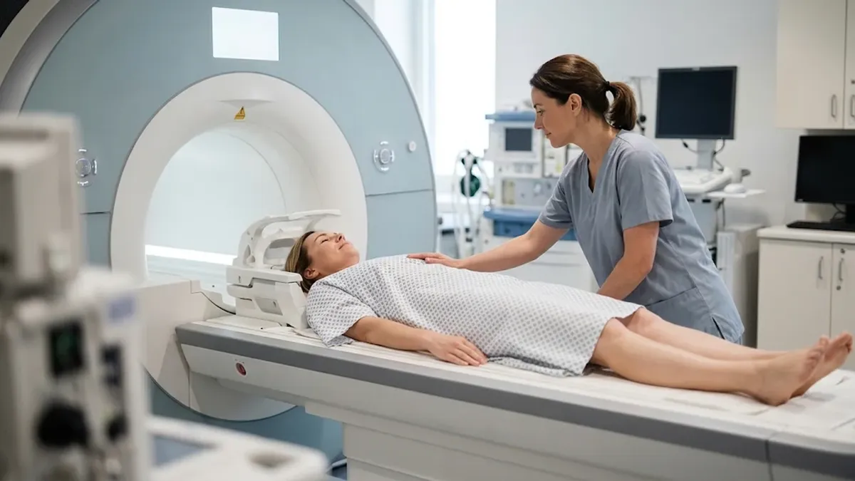
Is an Open MRI Machine Right for Your Abdominal Scan?
- +Open sides reduce claustrophobia and panic for many anxious patients
- +Accommodates larger body sizes that may not fit standard tubes
- +Easier for children to be accompanied by a parent during imaging
- +Often quieter than some high-field closed scanners
- +Can allow positions that are more comfortable for painful conditions
- +Useful for simple questions such as follow-up of a known large mass
- −Lower field strength usually means lower image detail
- −Scans often take longer, increasing motion blur from breathing
- −Some advanced liver, MRCP, and enterography protocols may be unavailable
- −Fewer facilities offer open systems, so travel may be required
- −Radiologists may recommend repeating the scan on a closed scanner
- −Small lesions can be harder to characterize with confidence
Pros and Cons at a Glance
| Pros | Cons |
|---|---|
| Open sides reduce claustrophobia and panic for many anxious patients | Lower field strength usually means lower image detail |
| Accommodates larger body sizes that may not fit standard tubes | Scans often take longer, increasing motion blur from breathing |
| Easier for children to be accompanied by a parent during imaging | Some advanced liver, MRCP, and enterography protocols may be unavailable |
| Often quieter than some high-field closed scanners | Fewer facilities offer open systems, so travel may be required |
| Can allow positions that are more comfortable for painful conditions | Radiologists may recommend repeating the scan on a closed scanner |
| Useful for simple questions such as follow-up of a known large mass | Small lesions can be harder to characterize with confidence |
Checklist: Prepare for Your Abdominal MRI Order
- ✓Ask your doctor what question the abdominal MRI should answer.
- ✓Confirm the CPT code with the scheduler and your insurer.
- ✓Obtain prior authorization before booking the appointment.
- ✓Tell staff about implants, pacemakers, clips, or metal fragments.
- ✓Report kidney disease, pregnancy, or prior contrast reactions.
- ✓Follow fasting instructions, commonly four to six hours.
- ✓Ask whether an open or wide-bore scanner is appropriate.
- ✓Arrange a driver if you will take anti-anxiety medication.
- ✓Wear comfortable clothing without metal zippers or fasteners.
- ✓Practice breath-holding for twenty seconds before the appointment.
Ask for the Clinical Question, Not Just the Scan
The most useful thing you can ask is what your doctor hopes to learn. A clear question determines the scanner, the contrast decision, and the CPT code, which saves time and money. Ask it before you book, not after.
Costs, Insurance, and CPT Codes for Abdominal and Pelvic MRI
Cost is one of the biggest surprises patients face. Prices for an abdominal MRI vary widely across the United States, from several hundred dollars at a negotiated cash rate at a freestanding center to several thousand dollars billed by a hospital outpatient department. Your actual cost depends on your deductible, coinsurance, and whether the facility is in network. Always request a written estimate before scheduling anything.
CPT codes drive billing. For the abdomen, 74181 covers a scan without contrast, 74182 covers contrast only, and 74183 covers an exam without contrast followed by contrast. The with and without combination takes longer and costs more. Doctors choose it for lesion characterization. Ask whether contrast is truly needed for your question, because a noncontrast study may be adequate for some follow-up indications.
A pelvic MRI CPT code works the same way. Code 72195 is without contrast, 72196 is with contrast, and 72197 is without followed by with contrast. People searching MRI pelvis w wo gyn CPT are usually looking for 72197, commonly used for evaluating fibroids, adenomyosis, endometriosis, or ovarian masses. If your doctor orders both regions, the abdomen and pelvis each carry their own code, and charges add together.
Prior authorization is the next hurdle. Most commercial plans and Medicare Advantage plans require approval for advanced imaging, often through a separate benefits manager. The ordering office submits clinical notes, and approval is tied to a specific code and facility. If the facility or code changes later, for example from 74181 to 74183, you may need new approval. Verify this before arriving for the appointment.
Out-of-pocket planning helps. Ask whether the center offers a self-pay discount, since some independent imaging sites advertise flat prices well below hospital charges. Compare in-network facilities by calling and asking for the total price including the radiologist's reading fee, which is sometimes billed separately. Health savings and flexible spending accounts can pay for scans, and many centers offer payment plans for patients with high deductibles.
Watch out for hidden extras. Contrast agent charges, sedation, and professional reading fees may appear on separate bills. If you receive an unexpected charge, request an itemized statement and compare the codes against your original estimate. Billing errors are common, and appeals frequently succeed when the code on the claim does not match the authorization. Keep your approval number and every written estimate in a single folder.
Remember that cheaper is not always better if the scan must be repeated. A low-field open scanner may cost less per visit but could produce nondiagnostic images, leading to a second scan on a better machine. Discuss this with your doctor so you choose the right equipment the first time. A single, well-protocoled exam usually costs less overall than two attempts at a bargain price.
Gadolinium contrast requires attention to kidney function, especially if you have severe kidney disease or are on dialysis. Tell the staff about pregnancy, allergies, and prior reactions. Never assume a facility has your records; confirm them before the appointment.
Open Bore MRI Locations and Finding the Closest Open MRI to Your Location
Many people type closest open MRI to my location into a search engine and choose the first result. A better approach is to start with a referral list from your doctor, then narrow by equipment. Not every center advertising open imaging performs abdominal work on that unit. Ask directly whether the scanner is used for liver, pancreas, or MRCP studies, or only for joints and spine.
Open bore MRI locations are less common than standard centers, especially in smaller cities. Large hospitals, academic medical centers, and regional imaging networks are the likeliest to offer open or wide-bore equipment. Rural patients may need to travel an hour or more. When distance is a concern, ask whether a wide-bore scanner at a nearer facility could meet your comfort needs without sacrificing diagnostic quality.
Check accreditation and staffing. The American College of Radiology accredits MRI units after reviewing physics tests and sample images, and many insurers prefer accredited sites. Ask whether a board-certified radiologist interprets your images, and whether subspecialty readers handle body MRI. For complicated findings such as liver tumors, a fellowship-trained body radiologist can add meaningful accuracy, even when scanner hardware seems similar across competing centers.
Use official tools for your search. Your insurer's provider directory lists in-network facilities and sometimes shows cost estimates. The ACR facility search lets you confirm accreditation. Call at least two locations and compare the answers. Write down the field strength, bore diameter, expected scan duration, price estimate, and whether the center can provide sedation or a support person in the room. These details make comparison simple.
Scheduling details matter more than most patients expect. Abdominal MRI usually needs a fasting window, so a morning slot is often easiest. Ask how long you should plan to stay, since check-in, intravenous placement, and recovery can double the time on the table. If you are anxious, request the first appointment of the day, when schedules are least likely to run late and waiting-room time stays short.
If your first choice cannot accommodate you, do not abandon the scan. Tell your doctor which barrier you faced, whether claustrophobia, body size, or distance. Referral offices keep lists of centers that handle such cases, including sites with larger bore sizes or anesthesia support. Many patients complete an excellent abdominal MRI after a single phone call that redirected them from an unsuitable open unit.
Finally, bring a family member if allowed, and ask to see the scanner before the appointment. Many centers will show you the room, explain the sounds, and let you practice lying in position. That preview often eases fear dramatically. Combined with a well-chosen machine and clear instructions, it turns a stressful appointment into a manageable one and improves the chance of getting clean, diagnostic images.
Practical Tips for a Smooth Abdominal MRI Experience
Preparation begins days before the scan. Read every instruction from the facility, including fasting rules, medication guidance, and arrival time. Most centers ask you to avoid solid food for four to six hours so the stomach and bowel are quiet and the gallbladder is full. Drink water unless told otherwise, and take regular medications with small sips unless your doctor instructs you differently.
Dress for comfort and safety. Wear loose clothing without metal, such as sweatpants and a plain cotton shirt, or expect to change into a gown. Leave jewelry, hairpins, watches, and credit cards in a locker, since magnets can damage cards and metal can heat or distort images. Remove medication patches that contain metal backing, and ask staff about tattoos or permanent makeup containing iron pigment.
Breath-holding is the skill that most improves abdominal images. The technologist will ask you to inhale, exhale, and hold for roughly fifteen to twenty-five seconds, repeated many times. Practice at home by holding a comfortable, relaxed breath, not a forced deep one. Consistent breath-holds at the same point in your breathing cycle keep organs in the same position from image to image.
Manage anxiety proactively. Tell the technologist if you feel nervous, because they can talk you through each sequence, adjust the room lighting, or provide a call button and headphones. Some people close their eyes before entering the bore and keep them closed. Focus on slow breathing and counting. If you take prescribed anti-anxiety medication, arrange transportation, since you cannot drive afterward and the facility may refuse to proceed otherwise.
Stay as still as possible between breath-holds. Motion is the most common reason an image must be repeated, adding minutes and sometimes forcing a second visit. Get comfortable on the table before the scan begins, ask for cushions under your knees, and tell staff immediately about any discomfort. The loud knocking and buzzing is normal and harmless, and earplugs or headphones protect your hearing.
After the scan, you can usually resume normal activity immediately unless you received sedation. If you received gadolinium, drink fluids and eat normally. Results typically reach your doctor within one to three business days, though urgent cases may be faster. A radiologist writes the report, and your ordering physician explains it. Ask for a copy of the images on disc or portal for future comparison.
Finally, keep good records. Save the CPT code, date, facility, and scanner type, because future doctors may want to compare studies made on similar equipment. If you change providers, request the images, not just the written report. Comparing old and new scans is one of the most powerful uses of MRI, and having your history ready can shorten diagnosis for any later symptoms that develop.
Sample MRI - Magnetic Resonance Imaging Practice Questions
Try these questions from our free MRI - Magnetic Resonance Imaging practice tests. The correct answer and an explanation follow each question.
A 2D spin-echo scan uses TR = 500 ms, a 256 (phase) x 256 (frequency) matrix, and 2 NEX. What is the approximate scan time?
- A. 2 minutes 8 seconds
- B. 4 minutes 16 seconds
- C. 8 minutes 32 seconds
- D. 1 minute 4 seconds
Answer: B. 4 minutes 16 seconds
2D scan time = TR x number of phase-encoding steps x NEX. Here that is 0.5 s x 256 x 2 = 256 s, or about 4 minutes 16 seconds. The frequency matrix does not affect scan time.
Parallel imaging techniques such as SENSE or GRAPPA shorten scan time mainly by:
- A. Increasing the TR
- B. Skipping some phase-encoding lines and using multi-element coil sensitivity data to fill them in
- C. Reducing the number of frequency-encoding samples
- D. Raising the main magnetic field strength
Answer: B. Skipping some phase-encoding lines and using multi-element coil sensitivity data to fill them in
Parallel imaging undersamples k-space in the phase-encoding direction by an acceleration factor (R). It then uses the spatial sensitivity profiles of a multi-channel phased-array coil to unfold the aliased image (SENSE) or rebuild the missing lines (GRAPPA). The trade-off is lower SNR, roughly by the square root of R multiplied by the coil geometry (g) factor.
When imaging the lumbar spine, which position best opens the intervertebral foramina and reduces lumbar lordosis for improved visualization?
- A. Prone with legs extended
- B. Supine with legs elevated on a cushion (knees and hips flexed)
- C. Lateral decubitus
- D. Prone with pillow under abdomen
Answer: B. Supine with legs elevated on a cushion (knees and hips flexed)
Flexing the hips and knees while supine flattens lumbar lordosis, opens intervertebral foramina, and improves disc and nerve root visualization.
Which sequence uses both magnitude and phase data from a high-resolution 3D gradient echo acquisition to highlight hemosiderin, deoxyhemoglobin, and calcification?
- A. Diffusion tensor imaging
- B. Double inversion recovery
- C. Arterial spin labeling
- D. Susceptibility-weighted imaging (SWI)
Answer: D. Susceptibility-weighted imaging (SWI)
SWI combines magnitude and filtered phase images to greatly increase sensitivity to susceptibility differences such as blood products and calcium.
Take the full MRI - Magnetic Resonance Imaging practice test
MRI Questions and Answers

CT vs MRI Imaging: How They Differ, When Each Is Used, and What to Expect

MRI Prep: Complete Patient Guide to Preparing for Your MRI Scan

Travel MRI Tech 2026: Salary, Requirements & ARRT MRI Exam
Dangers of MRI Machines: Safety Risks, Side Effects, Pelvic MRI CPT Codes, and Open Bore Options
GE 1.5 MRI Machines: Models, Pelvic MRI CPT Codes, and Open Bore Options
Written and edited by Nathan Beja, founder of Practice Test Geeks, with AI assistance. Exam facts are checked against the official sources linked below. Editorial policy
Sources & references
Exam facts on this page (fees, format, dates, passing scores) are checked against these official sources; the date is our last check. Editorial policy
- assets-us-01.kc-usercontent.com — Renewal, score validity, eligibility, exam format, retake policy, test dates, time limit · verified Nov 2026
- arrt.org — Fees, passing score, score release, exam information · verified Nov 2026
- home.pearsonvue.com — Test delivery · verified Nov 2026