
Dangers of MRI Machines: Safety Risks, Side Effects, Pelvic MRI CPT Codes, and Open Bore Options
🔎 Dangers of MRI machines explained: projectile risks, gadolinium, heat, noise, plus pelvic MRI CPT code basics and how to find open bore scanners.
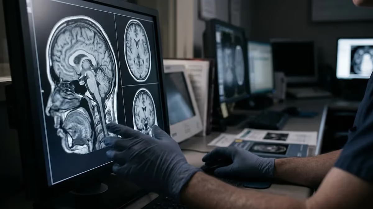
The dangers of MRI machines are real but narrow, and almost all of them are preventable with good screening. Magnetic resonance imaging uses a powerful static magnet, rapidly switching gradient coils, and radiofrequency pulses instead of ionizing radiation, so it carries no X-ray dose. That makes it one of the safest ways to see inside the body. The risks come from the magnet itself, implanted devices, contrast agents, and human error in the scanner room.
A typical clinical scanner runs at 1.5 or 3 tesla. Earth's magnetic field is roughly 50 microtesla, so a 3T magnet is about 60,000 times stronger. That field is always on, even when no scan is running, and it never switches off during a power outage. A ferromagnetic object carried into the room can accelerate toward the bore at lethal speed. This single fact drives nearly every safety rule technologists follow every day.
Beyond the magnet, patients ask about heating, noise, and contrast dye. Radiofrequency energy warms tissue slightly, and the scanner limits this through specific absorption rate calculations. Gradient coils produce loud knocking sounds that can exceed 100 decibels without protection. Gadolinium-based contrast carries a rare kidney-related risk. None of these hazards is trivial, yet serious injuries are uncommon because facilities follow the American College of Radiology safety framework and screen every patient and visitor before entry.
This guide walks through each hazard in plain language and then connects safety to practical topics people search for, including the pelvic MRI CPT code used for billing, how to find open bore MRI locations, and what to do when you want the closest open MRI to my location. For a broader look at mri safety and side effects compared with CT, see our companion article on how the two modalities differ.
If you are a patient, you will learn what to tell the technologist, which implants matter, and how to handle anxiety inside the bore. If you are an MRI technologist student, you will see how Zone I through IV access control works and why screening forms exist. Either way, the goal is the same: a scan that produces a clear diagnostic image without harm to anyone in or near the room.
Real numbers help put risk in perspective. Tens of millions of MRI exams are performed in the United States each year, and serious adverse events remain rare compared with that volume. Most reported incidents involve burns from skin contact with cables, missed metal in clothing, or unscreened implants. In other words, most harm traces back to a process failure rather than to the technology itself, which is why education and strict checklists matter so much.
Throughout this article, you will find a stat summary, a breakdown of the main hazards, side-by-side comparisons of scanner designs, a screening checklist, and a full FAQ. Use the quick links to jump to the section you need. Nothing here replaces advice from your radiologist, referring physician, or the facility's MRI safety officer, but it should give you the vocabulary to ask smart questions and to feel prepared on scan day.
MRI Safety by the Numbers
The Main Dangers of MRI Machines at a Glance
Ferromagnetic objects such as oxygen tanks, scissors, and wheelchairs are pulled toward the magnet with great force. This is the most dangerous hazard in the room and the reason strict zone access control and metal screening exist for every person.
Pacemakers, defibrillators, cochlear implants, clips, and neurostimulators may move, heat, or malfunction. Devices labeled MR Conditional are safe only within defined limits, so staff verify the exact model and scan settings before any exam begins.
Radiofrequency pulses warm tissue, and conductive loops, cables, or skin-to-skin contact can concentrate heat into a burn. Padding, straight lead routing, and prompt reporting of warmth or tingling keep this risk very low in practice.
Gradient coils create knocking and buzzing that can exceed 100 decibels. Without earplugs or headphones, the noise can cause temporary or even permanent hearing damage, which is why protection is mandatory for every patient and staff member.
Gadolinium agents can rarely cause allergic-like reactions, and some older agents were linked to nephrogenic systemic fibrosis in severe kidney disease. Screening, agent selection, and reserving contrast for cases where it changes care reduce this risk.
Projectile Hazards, Implants, and the Four MRI Safety Zones
The projectile effect is the most dramatic of all the dangers of MRI machines. Ferromagnetic items such as oxygen cylinders, scissors, wheelchairs, floor buffers, and IV poles are pulled toward the magnet's isocenter with force that rises sharply as they get closer. In 2001, a six-year-old boy died at a New York hospital when an oxygen tank flew into the bore. That incident reshaped national guidance and remains a standard teaching case in technologist programs.
To control that risk, the ACR divides the MRI suite into four zones. Zone I is the public area. Zone II is where patients are screened and interviewed. Zone III is restricted to trained personnel and is where the magnet's field becomes a hazard. Zone IV is the scanner room itself. Anyone crossing from Zone II into Zone III must be cleared, and doors are locked or supervised so unscreened people never wander toward the magnet.
Implanted devices are the second major concern. Older pacemakers, defibrillators, cochlear implants, aneurysm clips, and neurostimulators can move, heat, malfunction, or be damaged by the field. Many modern devices are labeled MR Conditional, meaning they are safe only under defined field strengths, gradient limits, and scan conditions. Staff verify the exact make and model against manufacturer instructions before scheduling. If a device cannot be confirmed, the scan is usually postponed or switched to CT.
Metal that you cannot see also matters. Metal fragments in the eye from grinding or welding can migrate and cause blindness, so facilities often ask workers with that history to get orbit radiographs first. Shrapnel, bullets, and some surgical hardware require review. Hair products, cosmetics, and tattoo inks may contain metallic particles that warm up during a scan. Transdermal patches with foil backing can burn skin. Honest, detailed answers on the screening form are a patient's best protection.
Terminology matters on exam day and on registry tests. MR Safe means an item poses no known hazard in any MR environment, such as a plastic cup. MR Conditional means it is safe under specific conditions. MR Unsafe means it poses a known hazard, such as a steel oxygen tank. Items carry standardized green, yellow, and red icons, and technologists must never assume an object is safe simply because it looks nonmetallic or is small.
Emergencies inside the scanner need a special plan. If a patient codes, staff do not bring a crash cart into Zone IV. They stop the scan, remove the patient from the room, and begin resuscitation outside the field. A quench, the rapid venting of cryogenic helium to collapse the field, is reserved for situations where someone is pinned by the magnet and cannot be freed otherwise. Quenching is costly and must be done carefully because escaping gas can displace oxygen.
Training is the best defense. Annual safety education for all staff, including nurses, anesthesiologists, transport staff, and housekeeping, reduces accidents more than any technology. Many hospitals add ferromagnetic detection systems at the Zone III door as a second layer. Patients can help by removing jewelry, hearing aids, credit cards, phones, and belts before entering. Visitors who accompany a child or relative must also be screened, since the magnet affects everyone equally, regardless of why they are there.
Pelvic MRI CPT Code and Open Bore MRI Locations Explained
The pelvic MRI CPT code depends on contrast use. CPT 72195 describes MRI of the pelvis without contrast. CPT 72196 covers the pelvis with contrast only. CPT 72197 covers the pelvis without contrast followed by contrast and further sequences, which is the combination most people mean when they search for the MRI pelvis w wo gyn CPT. Gynecologic indications include fibroids, adenomyosis, endometriosis, ovarian masses, and staging of cervical or endometrial cancer.
Because the codes describe the study and not the diagnosis, insurers rely on your ICD-10 diagnosis to judge medical necessity. Prior authorization is common, especially for 72197. The contrast agent itself is typically billed separately under a HCPCS supply code, and fees vary widely between hospital outpatient departments and freestanding imaging centers. Ask for the exact CPT code before the appointment, request a self-pay estimate if uninsured, and compare facilities, since the same code can differ in price several-fold.
Open Bore MRI vs Closed Bore MRI: Which Is Better for You?
- +Open bore designs greatly reduce the enclosed feeling for claustrophobic patients
- +Side access lets a parent or companion stay in contact during the scan
- +Larger patients often fit more comfortably than in a standard tube
- +Lower sedation needs can reduce anesthesia-related risks
- +Easier positioning helps people with pain, limited mobility, or kyphosis
- +Many open units are in outpatient centers with lower prices than hospitals
- −Lower field strength can mean longer scans and reduced detail
- −Small lesions may be harder to see than on 1.5T or 3T systems
- −Not every exam protocol, such as advanced pelvic or neuro studies, is available
- −Fewer open bore MRI locations exist, so travel may be longer
- −Magnetic hazards and implant rules are exactly the same as closed systems
- −Motion blur is more likely if long sequences are hard to hold still for
Pelvic MRI Safety Screening Checklist: What to Do Before You Scan
- ✓Complete the MRI screening form honestly and in full.
- ✓Bring your implant or pacemaker identification card.
- ✓List every past surgery, including approximate dates and hospitals.
- ✓Tell staff about shrapnel, bullets, or metal work injuries.
- ✓Report your IUD brand and any other implanted devices.
- ✓Remove jewelry, watches, hairpins, hearing aids, and piercings.
- ✓Leave phones, credit cards, and keys outside Zone III.
- ✓Disclose pregnancy, possible pregnancy, and kidney disease.
- ✓Ask for the pelvic MRI CPT code and verify insurance authorization.
- ✓Request earplugs and agree on a squeeze-ball signal before the scan.
Most MRI Harm Is a Screening Failure, Not a Machine Failure
The magnet cannot be turned off, so every serious MRI accident traces back to something that entered Zone IV when it should not have. A complete screening form, a verified implant, and a checked pocket are the three cheapest and strongest protections available.
Contrast, Heat, and Noise: Lesser-Known Dangers of MRI Machines
Gadolinium-based contrast agents are the most common drug-related concern. They are given by IV to highlight inflammation, tumors, and blood vessels. Most patients feel nothing, though some report nausea, headache, or a cool sensation. Mild reactions occur in a small fraction of doses, and severe allergic-like reactions are roughly one in ten thousand or fewer. Facilities keep emergency medications nearby, and patients are asked to remain on site briefly after injection when feasible.
Nephrogenic systemic fibrosis, or NSF, is the serious complication historically linked to certain linear gadolinium agents in people with severe kidney impairment. It causes skin thickening and can affect joints and organs. The ACR classifies agents into groups, and Group II macrocyclic agents carry very low NSF risk. Kidney function screening, often with an eGFR below 30 as the trigger for caution, remains standard for at-risk patients such as those with dialysis needs, diabetes, or advanced age.
Studies since 2014 have shown that small amounts of gadolinium can remain in the brain, bones, and skin, even in people with normal kidneys. In 2018 the FDA added a class warning and required a medication guide. No consistent harm has been proven from this retention in patients with normal kidney function, but radiologists now use contrast only when it changes management and favor macrocyclic agents. Patients who need many follow-up scans should ask whether non-contrast protocols are enough.
Radiofrequency pulses deposit energy, which tissue absorbs as heat. Scanners calculate SAR, measured in watts per kilogram, and stay within limits; in normal operating mode the whole-body limit is 2 W/kg. Burns happen when conductive loops form, such as skin-to-skin contact between the thighs or crossed ankles, or when cables touch the patient. Technologists use foam pads to separate limbs and route leads straight. Tell staff immediately if you feel warmth, tingling, or burning during the scan.
Fast switching gradient fields can induce small currents in nerves, leading to peripheral nerve stimulation. Patients describe tapping or twitching in the arms, legs, or torso. It is uncomfortable but not dangerous, and scanners have limits to keep stimulation below painful thresholds. Squeeze the call ball if it happens and the technologist can adjust parameters. Fast sequences such as echo planar imaging are the most likely to cause this sensation, and it typically stops the moment the sequence ends.
Gradient coils vibrate against the magnet housing, producing knocking, buzzing, and chirping that can exceed 110 decibels in some sequences. That is comparable to a rock concert and can cause temporary threshold shifts or permanent hearing damage without protection. Earplugs, headphones, or both are mandatory and are especially important for children, anesthetized patients, and people unable to report discomfort. Quiet sequences exist on newer systems and can cut noise substantially for sensitive patients.
Sedation introduces its own dangers. Children and anxious patients may need sedation, and monitoring must be done with MR Conditional equipment, because standard monitors and pumps can be pulled by the field or fail. Respiratory depression is the main concern, and a trained clinician must watch pulse oximetry and capnography continuously. Ask whether the facility has an anesthesia team and an MR-compatible ventilator before booking a sedated scan, particularly at a freestanding outpatient center.
The magnet is on around the clock. Do not carry phones, keys, tools, oxygen tanks, or medical equipment into Zone IV, and never assume a device is safe without written confirmation of its MR status. If an object becomes stuck to the magnet, stop and call for trained help.
Claustrophobia, Pregnancy, and Special Populations: Reducing MRI Risk
Anxiety is the most common reason people struggle with MRI. Estimates suggest that a meaningful minority, often cited between 2 and 15 percent, cannot finish a standard scan without help. Panic is not physically dangerous, but a patient who moves ruins the images and may need to repeat the exam. Breathing exercises, a mirror or prism glasses, a cooling fan, music, and a companion's hand on the ankle all help.
Options exist for those who still cannot tolerate a tube. Wide-bore and open bore systems reduce the enclosed feeling. Feet-first positioning keeps the head outside the bore for many pelvic and lower body studies. Short-acting oral anxiolytics can be prescribed by your doctor before the appointment. If you plan to take one, arrange a ride home, because you cannot safely drive afterward, and tell the facility so staff can monitor you.
MRI without contrast is generally considered acceptable in pregnancy when the information is needed, and it is the preferred imaging option for many conditions because it avoids radiation. No harm to the fetus has been demonstrated at clinical field strengths. Gadolinium, however, crosses the placenta and is avoided unless the benefit clearly outweighs the risk. Pregnant staff members may work in the MRI department but many policies advise them not to remain in the scanner room during scanning.
Pelvic imaging brings its own safety questions. Many women with intrauterine devices ask about metal. Most modern copper and hormonal IUDs are MR Conditional or MR Safe, but you should always report the brand. The exam coded as MRI pelvis w wo gyn CPT 72197 uses contrast, so renal function, allergy history, and pregnancy status are reviewed first. Glucagon may be given to reduce bowel motion, and bladder instructions vary by protocol.
Children need age-appropriate preparation. Child life specialists, toy scanners, and movies shown through MR-safe goggles reduce the need for sedation. Older adults often have more implants, such as joint replacements, stents, and cardiac devices, along with reduced kidney function that affects contrast decisions. Hip and knee replacements are usually safe to scan, though they can distort images near the metal. A complete surgical history, including approximate dates and hospitals, speeds clearance.
Patients with cardiac devices need a coordinated plan. An MR Conditional pacemaker is typically programmed into a special MRI mode before the scan, monitored throughout by a nurse or cardiac technician, and then reprogrammed afterward. Some centers also scan patients with older or abandoned leads under strict protocols after risk-benefit review. This workflow takes time and staff, so schedule early and bring your device identification card, which lists the manufacturer, model, and implant date.
Workers around MRI systems face different exposures, including repeated movement through strong fields, which can cause transient dizziness, a metallic taste, or visual flashes called magnetophosphenes. These effects typically fade quickly. Technologists should move slowly near the bore, follow guidelines on occupational exposure, and avoid carrying sensitive implants into Zone IV. Employers must provide hearing protection, ongoing training, and incident reporting systems so that near-misses lead to process improvements instead of repeated mistakes.
Practical Tips for a Safe MRI Scan and Smarter Scheduling
Prepare early. Read the screening form when it arrives, and gather device cards, operative reports, and a medication list. If you have any implant, call the facility days ahead rather than on scan day, because verifying a device can take hours. Wear comfortable clothing without metal, such as cotton sweats, or plan to change into a gown. Leave jewelry, watches, and wallets at home, and bring glasses only if the technologist confirms they are metal-free.
Ask about the exam plan. Confirm which body part is being imaged, whether contrast will be used, and how long the scan will take. A pelvic exam may run 30 to 45 minutes, while multi-sequence gynecologic protocols can approach an hour. Knowing the time helps you plan for breathing and bladder comfort. If instructions include fasting or limited fluid intake, follow them exactly, since certain abdominal and pelvic protocols depend on an empty stomach or a particular bladder state.
Handle insurance before arriving. Ask your doctor's office for the pelvic MRI CPT code, usually 72195, 72196, or 72197, and call your insurer to verify coverage and prior authorization. Request the facility's cash price if your deductible is high. Many freestanding centers charge far less than hospital outpatient departments for the same code, so a quick comparison of two or three options can save hundreds of dollars without affecting the quality of the images.
Use proximity wisely. Searching for the closest open MRI to my location is useful, but nearest is not always best. Weigh field strength, radiologist expertise, accreditation, and wait time. An open scanner five minutes away that cannot answer your clinical question may force a repeat scan elsewhere. Call ahead, ask whether a particular scanner suits your body size and exam, and make sure your referring physician knows where the results will be sent.
During the scan, communicate. You will be given a squeeze ball or call button; use it for pain, heat, panic, or dizziness. Stay still, breathe normally unless told otherwise, and follow breath-hold instructions precisely, since motion is a leading cause of repeated sequences. Use the earplugs even if the room seems quiet at first. If you are claustrophobic, close your eyes before entering the bore and keep them closed until the exam is over.
After the scan, watch for delayed symptoms. If you received contrast, drink fluids unless restricted, and report hives, swelling at the injection site, or breathing trouble promptly. Burns can appear hours later as blisters or red patches; call the facility if you notice any. ACR guidance says breastfeeding mothers generally do not need to interrupt nursing after a macrocyclic gadolinium agent, though some choose to pump and discard milk for 12 to 24 hours as a precaution.
Finally, students preparing for the ARRT MRI exam should practice safety scenarios as thoroughly as physics. Questions often test zone definitions, MR Conditional labeling, quench procedures, SAR limits, and contrast screening. Pair written study with timed practice questions, review each missed answer, and explain the concepts aloud. For patients and future technologists alike, the core rule is simple: when in doubt, stop, ask, and verify before anyone enters Zone IV.
MRI Questions and Answers

CT vs MRI Imaging: How They Differ, When Each Is Used, and What to Expect

MRI Prep: Complete Patient Guide to Preparing for Your MRI Scan

MRI Preparation Guide: How to Prep for Your Scan Step by Step

Travel MRI Tech 2026: Salary, Requirements & ARRT MRI Exam

MRI Internal Auditory Canal: Complete Guide to IAC Imaging, Anatomy, Indications, and Interpretation
How this page was made: written with AI tools and published by Nathan Beja, founder of Practice Test Geeks, who is responsible for its content. Exam facts are checked against the official sources listed below. Not reviewed by a subject-matter expert. Editorial policy
Sources & references
Exam facts on this page (fees, format, dates, passing scores) are checked against these official sources; the date is our last check. Editorial policy
- arrt.org — Passing score, score release, fees, exam information · verified Oct 2026
- assets-us-01.kc-usercontent.com — Renewal, score validity, test delivery, eligibility, exam format, test dates, time limit · verified Oct 2026