
CT vs MRI Imaging: How They Differ, When Each Is Used, and What to Expect
CT MRI imaging explained: 💯 how each scan works, when doctors choose one over the other, preparation tips, and what technologists need to know.
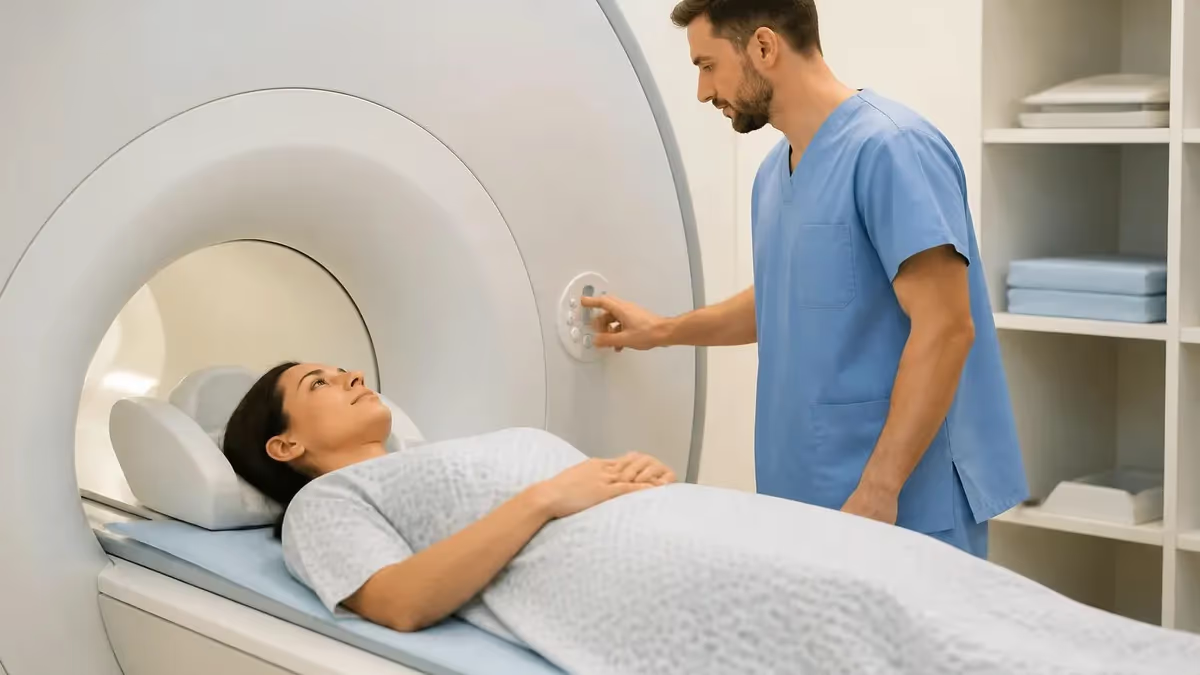
Understanding ct mri imaging is essential for patients, students, and practicing technologists alike. Both computed tomography (CT) and magnetic resonance imaging (MRI) are cross-sectional imaging modalities that allow clinicians to see inside the body without surgery, yet they rely on entirely different physical principles, produce distinct types of contrast, and excel in different clinical scenarios.
Choosing the right modality can mean the difference between detecting a subtle lesion early and missing it entirely, which is why every imaging professional must be fluent in the strengths and limitations of each technology. For a detailed breakdown of one of the most frequently ordered MRI exams, see our guide on ct mri imaging of the cervical spine.
CT scanning uses ionizing radiation — specifically a rotating X-ray tube and detector array — to acquire hundreds of thin axial slices that a computer reconstructs into two-dimensional and three-dimensional images. The speed of CT is one of its greatest assets: a modern 64-slice or 256-slice CT scanner can image the entire chest, abdomen, and pelvis in a single breath-hold of five to fifteen seconds. This makes CT the go-to modality in emergency departments where time is critical, particularly for suspected pulmonary embolism, aortic dissection, or acute hemorrhagic stroke where rapid triage is life-saving.
MRI, by contrast, uses a powerful static magnetic field combined with radiofrequency pulses to excite hydrogen protons in the body's water and fat molecules. When those protons return to equilibrium, they emit signals that the scanner's receiver coils capture and the system converts into images.
Because MRI does not use ionizing radiation, it is considered safer for repeated imaging and for vulnerable populations such as pregnant patients (after the first trimester with appropriate clinical justification) and children who require multiple follow-up scans throughout their lives. The trade-off is acquisition time: even a routine brain MRI typically runs twenty to forty-five minutes.
The tissue contrast achievable with MRI vastly exceeds what CT offers for soft-tissue structures. MRI can differentiate between gray matter and white matter in the brain, detect cartilage tears in joints, visualize ligament integrity, and characterize liver lesions based on their signal behavior across multiple pulse sequences — tasks that CT simply cannot perform with comparable accuracy. This is why MRI dominates in neurological imaging, musculoskeletal evaluation, and oncologic staging when soft-tissue detail determines treatment planning.
CT, on the other hand, remains unmatched for imaging cortical bone detail, detecting calcifications, evaluating the lungs (where MRI suffers from respiratory motion and low proton density in air-filled spaces), and guiding interventional procedures where metal instruments would be incompatible with the MRI environment. CT angiography has also become a powerful tool for evaluating coronary artery disease, peripheral arterial disease, and cerebrovascular anatomy, often rivaling or replacing conventional catheter angiography in many clinical situations.
For MRI technologists preparing for board examinations and credentialing, a thorough understanding of how CT and MRI compare is not merely academic — it is a testable domain that appears consistently on the American Registry of Radiologic Technologists (ARRT) MRI examination and the American Registry of Magnetic Resonance Imaging Technologists (ARMRIT) exam. Knowing when a radiologist or ordering physician might prefer one modality over the other, understanding the physics behind each, and being able to counsel patients on what to expect all fall within the professional scope of the registered MRI technologist.
This article provides a comprehensive, exam-focused guide to CT and MRI imaging: the underlying physics, clinical applications, patient preparation, safety considerations, and study strategies to help you master this topic whether you are a student entering the field or an experienced tech refreshing your knowledge before registry renewal.
CT and MRI Imaging by the Numbers
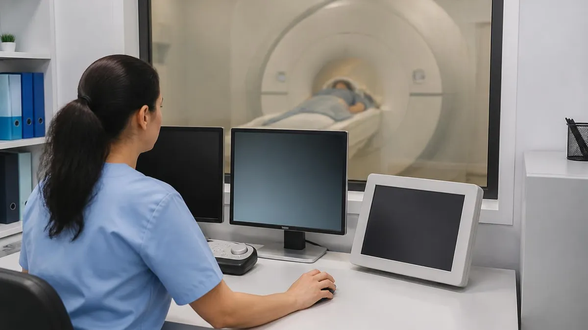
Core Physics Principles: CT vs. MRI
CT measures how much X-ray radiation different tissues absorb (attenuate). Dense structures like cortical bone appear bright (high Hounsfield units); air appears black. Image contrast depends entirely on tissue density differences, making CT excellent for bone and calcifications.
MRI exploits the magnetic properties of hydrogen protons. T1 relaxation reflects how quickly protons realign with the main field; T2 relaxation reflects how quickly they lose phase coherence. Varying pulse sequence parameters (TR and TE) produces dramatically different tissue contrasts from the same patient.
Higher magnetic field strength (3T vs. 1.5T) improves MRI signal-to-noise ratio, enabling thinner slices and finer anatomical detail. CT spatial resolution is generally higher for bone structures, but MRI wins for soft-tissue contrast resolution across nearly every anatomical region.
CT uses iodinated contrast agents injected intravenously to highlight vascular structures and increase lesion conspicuity. MRI uses gadolinium-based contrast agents (GBCAs), which shorten T1 relaxation times and cause enhancing tissues to appear bright on T1-weighted post-contrast sequences.
CT delivers ionizing radiation doses typically ranging from 2 to 20 millisieverts depending on body part and protocol. MRI delivers zero ionizing radiation, making it preferable for pediatric patients, pregnant patients (when clinically justified), and anyone requiring frequent imaging surveillance.
The clinical applications of CT and MRI imaging overlap in some regions but diverge sharply in others, and understanding these distinctions is fundamental to practicing competent diagnostic imaging. In the brain and central nervous system, MRI is the gold standard for virtually all non-emergency indications.
Demyelinating diseases such as multiple sclerosis, brain tumors, posterior fossa lesions, and subtle cortical dysplasias are far better visualized on MRI than CT because the magnetic resonance signal can differentiate between tissue types at a level of contrast that X-ray attenuation simply cannot match. The posterior fossa and brainstem — regions prone to streak artifacts on CT from the dense surrounding temporal bones — are imaged with exceptional clarity on MRI.
In acute stroke management, the two modalities serve complementary and sequential roles. CT without contrast is the first scan obtained in any suspected stroke patient because it is fast, universally available, and immediately distinguishes hemorrhagic from ischemic stroke — a distinction that determines whether thrombolytic therapy can be administered safely.
Within minutes of CT ruling out hemorrhage, CT angiography maps the cerebral vasculature to locate the occlusion, and CT perfusion can estimate the ischemic penumbra. MRI diffusion-weighted imaging (DWI), however, detects acute ischemia within minutes of onset with far greater sensitivity than CT, which is why comprehensive stroke centers proceed to MRI when the clinical picture warrants it and time permits.
For musculoskeletal imaging, MRI dominates in nearly all soft-tissue applications. Meniscal tears, anterior cruciate ligament (ACL) ruptures, rotator cuff pathology, labral tears, and bone marrow edema patterns (which indicate stress reactions, contusions, and early avascular necrosis) are indications that drive the vast majority of outpatient MRI orders.
CT, by contrast, is preferred for evaluating complex fracture patterns, particularly in the pelvis, calcaneus, tibial plateau, and spine, where precise three-dimensional reconstruction guides surgical planning. CT is also the modality of choice for assessing fracture healing and the integrity of orthopedic hardware, since MRI is often severely degraded by metal artifact from implants.
Abdominal and pelvic imaging represents perhaps the most nuanced arena for modality selection. CT of the abdomen and pelvis with intravenous and oral contrast remains the workhorse of emergency abdominal imaging, diagnosing appendicitis, bowel obstruction, diverticulitis, mesenteric ischemia, and solid organ trauma with high accuracy and remarkable speed.
However, for liver lesion characterization, MRI with hepatobiliary contrast agents such as gadoxetate disodium (Eovist) has become the definitive technique, differentiating benign hepatocellular adenomas and focal nodular hyperplasia from malignant hepatocellular carcinoma with a level of specificity that CT cannot approach. MRCP (magnetic resonance cholangiopancreatography) has largely replaced diagnostic ERCP for evaluating the bile ducts and pancreatic duct because it is non-invasive and does not carry the risk of procedure-related pancreatitis.
Cardiac imaging represents one of the fastest-growing application areas for both modalities. Cardiac CT, specifically coronary CT angiography (CCTA), has revolutionized the non-invasive evaluation of coronary artery disease. Modern high-resolution scanners can acquire images of the coronary arteries in a single heartbeat, providing detailed visualization of plaque burden, stenosis severity, and coronary anatomy that guides decisions about catheterization. Cardiac MRI, on the other hand, is the reference standard for myocardial viability assessment, cardiomyopathy characterization, and congenital heart disease evaluation, providing functional data — ejection fraction, wall motion, myocardial strain — alongside detailed tissue characterization through late gadolinium enhancement sequences.
Pulmonary imaging illustrates one area where CT holds a decisive advantage. The lungs present a particular challenge for MRI because air contains very few hydrogen protons to generate signal, and respiratory motion during the longer MRI acquisition windows causes significant image degradation. High-resolution CT (HRCT) of the chest is the definitive technique for evaluating interstitial lung disease, bronchiectasis, emphysema, and small pulmonary nodules.
CT is also the primary tool for lung cancer screening in high-risk populations, following the USPSTF guidelines recommending annual low-dose CT for adults aged 50 to 80 who meet smoking history criteria. While emerging MRI techniques using hyperpolarized gas contrast agents and ultrashort echo time (UTE) sequences are advancing pulmonary MRI, these remain largely research tools and are not yet in routine clinical deployment.
For technologists, understanding these application-specific strengths helps in multiple ways: it enables more meaningful patient education conversations, supports appropriate clinical communication with ordering providers, and directly informs the exam preparation and protocol selection decisions that define expert-level MRI practice. Registry examination questions frequently present clinical scenarios and ask which modality or sequence is most appropriate, rewarding candidates who have internalized the clinical reasoning behind imaging choices rather than simply memorizing isolated facts.
MRI Practice Test Questions
Prepare for the MRI - Magnetic Resonance Imaging exam with our free practice test modules. Each quiz covers key topics to help you pass on your first try.
MRI Knowledge
MRI Exam Questions covering Knowledge. Master MRI Test concepts for certification prep.
MRI Physics
Free MRI Practice Test featuring Physics. Improve your MRI Exam score with mock test prep.
MRI Anatomy and Pathology
MRI Test Prep for MRI Anatomy and Pathology. Practice MRI Quiz questions and boost your score.
MRI Anatomy and Positioning
MRI Questions and Answers on MRI Anatomy and Positioning. Free MRI practice for exam readiness.
MRI Contrast Agents
Free MRI Quiz on MRI Contrast Agents. MRI Exam prep questions with detailed explanations.
MRI Patient Care and Positioning
MRI Practice Questions for MRI Patient Care and Positioning. Build confidence for your MRI certification exam.
MRI and CT Scan Types: Protocols and Sequences Explained
MRI protocols are built from multiple pulse sequences, each weighted to highlight different tissue properties. T1-weighted sequences (short TR, short TE) make fat appear bright and fluid appear dark, making them ideal for anatomy and post-contrast enhancement. T2-weighted sequences (long TR, long TE) make fluid bright and fat intermediate, which is excellent for detecting edema, inflammation, and most pathological processes. FLAIR (Fluid-Attenuated Inversion Recovery) suppresses cerebrospinal fluid signal to reveal periventricular lesions that would otherwise blend with bright CSF on standard T2 images.
Diffusion-weighted imaging (DWI) detects restriction of water molecule movement, making acute stroke ischemia and highly cellular tumors appear bright within minutes of onset. Gradient echo (GRE) sequences are sensitive to magnetic susceptibility effects, making them the preferred choice for detecting hemorrhage, calcium, and iron deposition. STIR (Short Tau Inversion Recovery) suppresses fat signal uniformly and is widely used in musculoskeletal MRI to reveal bone marrow edema. Technologists must understand how changing TR, TE, and flip angle shifts image contrast to troubleshoot suboptimal image quality in real time.
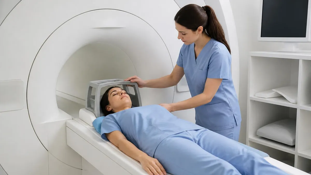
MRI vs. CT: Advantages and Disadvantages at a Glance
- +MRI produces superior soft-tissue contrast without ionizing radiation, making it safer for repeated imaging
- +MRI can acquire images in any plane (axial, coronal, sagittal, oblique) without moving the patient
- +Functional MRI techniques (DWI, perfusion, spectroscopy) provide physiological information beyond anatomy
- +CT is dramatically faster than MRI, completing full-body surveys in seconds
- +CT is compatible with virtually all metallic implants and does not require extensive screening
- +CT is more widely available and less expensive per scan than MRI in most US markets
- +CT excels at bone detail, lung evaluation, and calcification detection where MRI struggles
- +Both modalities support three-dimensional reconstruction and post-processing for surgical planning
- −MRI is contraindicated for patients with certain ferromagnetic implants, pacemakers, and cochlear implants
- −MRI scans are significantly longer, increasing motion artifact risk and patient discomfort
- −MRI is substantially more expensive than CT both for facilities to operate and for patients out-of-pocket
- −CT delivers ionizing radiation that accumulates with repeated studies, raising long-term cancer risk concerns
- −CT soft-tissue contrast is inferior to MRI for the brain, spinal cord, joints, and abdominal organs
- −CT iodinated contrast carries allergy and nephrotoxicity risks requiring pre-screening and patient selection
- −MRI is highly susceptible to metallic artifact from orthopedic hardware that can render images non-diagnostic
- −CT is limited in detecting hyperacute ischemic stroke where MRI diffusion sequences excel
Patient Preparation Checklist for CT and MRI Imaging
- ✓Screen every MRI patient for ferromagnetic implants, pacemakers, neurostimulators, and foreign bodies using standardized MRI safety forms before entering the controlled area.
- ✓Review renal function (eGFR) for any patient scheduled to receive gadolinium-based contrast — withhold in patients with eGFR below 30 unless absolutely necessary.
- ✓Instruct patients undergoing abdominal or pelvic MRI to fast for four to six hours before the appointment to reduce bowel peristalsis and improve image quality.
- ✓Remove all metallic objects (jewelry, hairpins, hearing aids, body piercings) from the patient before entering the MRI suite.
- ✓Screen for claustrophobia and arrange for anxiolytic pre-medication or open-bore scanner alternatives when appropriate.
- ✓Confirm contrast allergy history for both CT and MRI patients; pre-medicate known reactors according to ACR guidelines using corticosteroids and antihistamines.
- ✓Verify that patients receiving iodinated CT contrast have not taken metformin within 48 hours if eGFR is below 60, to prevent lactic acidosis risk.
- ✓Educate patients to hold their breath during CT acquisition to minimize respiratory motion artifact.
- ✓Provide hearing protection (earplugs or MRI-compatible headphones) to all MRI patients before entering the scanner bore.
- ✓Document the indication, relevant clinical history, and prior imaging on the requisition so the radiologist can tailor the protocol and provide a complete interpretation.
The 'Right Tool' Principle in Diagnostic Imaging
No single imaging modality is universally superior. The best scan is the one chosen for the right clinical question, performed with the correct protocol, and interpreted by a radiologist with appropriate subspecialty expertise. MRI technologists who understand both CT and MRI physics and applications are better prepared to participate in protocol optimization discussions, communicate with ordering providers, and counsel patients — skills that elevate technologist practice from technical to clinical partnership.
MRI safety is a subject that every technologist, student, and anyone working in or near the MRI suite must take seriously, because the consequences of safety failures can be catastrophic and irreversible. The MRI environment presents four primary hazard categories: projectile risk from ferromagnetic objects drawn into the magnetic field, implant interaction with the static field or radiofrequency energy, acoustic noise injury, and thermal injury from radiofrequency-induced tissue heating. Unlike CT, where the primary safety concern is radiation dose management, MRI safety is a complex, multi-factorial discipline requiring both knowledge and constant procedural vigilance.
The static magnetic field of an MRI scanner is always on — there is no equivalent of switching off an X-ray tube between exposures. This means that any ferromagnetic object brought near the scanner bore can become a dangerous projectile, accelerating toward the magnet at tremendous force.
Documented projectile incidents have involved oxygen cylinders, wheelchairs, stretchers, floor buffers, and even scissors — objects that seemed safely distant but were within the field's gradient force range. This is why ACR guidance divides the MRI environment into four zones, with Zone IV (the scanner room itself) accessible only to patients who have been fully screened and personnel who have completed MRI safety training.
Implant compatibility has become an increasingly nuanced topic as medical device technology advances. Older cardiac pacemakers were universally contraindicated in MRI environments, but a growing number of modern MRI-conditional pacemakers now exist that can be safely scanned under specific conditions — defined field strength, specific absorption rate (SAR) limits, and programming modes set by a cardiologist before and after the scan.
The MR Safe, MR Conditional, and MR Unsafe classification system adopted by the American Society for Testing and Materials (ASTM) provides a standardized framework, and technologists must consult implant manufacturer labeling and resources such as MRIsafety.com before proceeding with any scan involving an implanted device.
Radiofrequency energy deposition — quantified as the specific absorption rate (SAR) in watts per kilogram — can cause tissue heating if SAR limits are exceeded. The FDA limits whole-body average SAR to 4 W/kg for patients over 15 minutes; local SAR limits also apply. In practice, SAR is automatically monitored and limited by the scanner software, but technologists must understand that factors such as patient size, body habitus, coil selection, and pulse sequence parameters all affect SAR.
Certain implanted devices, including elongated leads and metallic objects forming loops, can concentrate RF energy and create focal heating far exceeding the whole-body average — a phenomenon that explains why even some non-ferromagnetic devices are classified MR Unsafe.
Acoustic noise generated by rapidly switching gradient coils is another legitimate safety concern. Peak sound pressure levels in MRI scanners routinely reach 100–120 dB depending on the sequence, approaching the threshold at which prolonged exposure without hearing protection causes permanent damage. All patients and accompanying personnel must wear hearing protection inside the bore, and MRI-compatible music or communication systems can help reduce patient anxiety while providing an additional sound attenuation layer. Parents accompanying pediatric patients during sedated exams must also be fitted with hearing protection before entering the room.
Cryogen safety is a specialized but important area of MRI facility management. Superconducting MRI magnets operate at approximately 4 Kelvin, maintained by liquid helium. A quench — a sudden loss of superconductivity — releases large volumes of helium gas that can displace oxygen in the scanner room, creating an asphyxiation hazard if the magnet room's quench pipe system fails to vent the gas safely outdoors. Technologists working in MRI facilities should know the location of the emergency quench button (used in extreme safety emergencies only), the location of oxygen monitors, and evacuation procedures for a quench event.
For registry examination purposes, MRI safety questions appear consistently across all major MRI credentialing examinations and are weighted heavily because they reflect real-world patient and staff protection responsibilities. The ARRT MRI examination blueprint devotes a significant portion of content to safety, and the ARMRIT examination dedicates an entire domain to it. Candidates should be familiar with the ACR Manual on MR Safety, the ASTM implant classification standards, and the four-zone model as frameworks that organize the broad body of safety knowledge into a testable and clinically applicable structure.
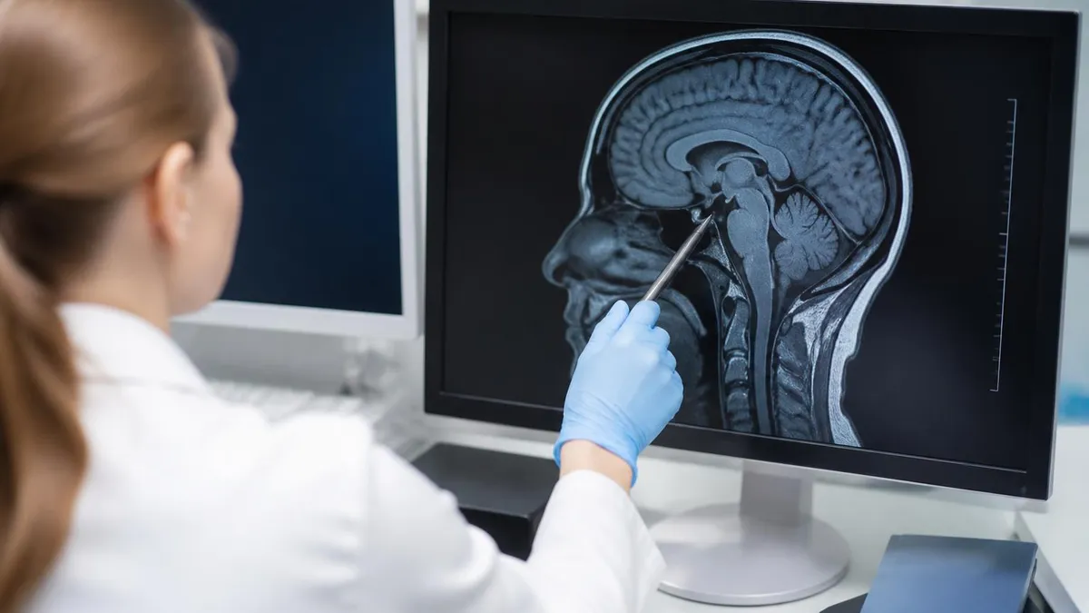
Never assume an implant is MRI-safe based on age, appearance, or patient report alone. Always verify implant MRI compatibility using the manufacturer's documentation or a validated reference source such as MRIsafety.com before proceeding. An incorrect assumption can result in device migration, heating, malfunction, or patient death — incidents that are entirely preventable with thorough pre-screening protocols.
Preparing for the MRI registry examination requires a structured, systematic approach that covers physics, instrumentation, patient care, safety, and image production — the five major content domains assessed by the ARRT and ARMRIT examinations. Many candidates underestimate the breadth of knowledge required, focusing narrowly on pulse sequences while neglecting safety, patient care, or quality assurance content that collectively comprises a substantial portion of exam questions. A balanced study plan that allocates time proportionally to each domain's examination weight is essential for consistent success.
Physics forms the conceptual foundation of MRI practice, and a solid understanding of magnetic resonance fundamentals pays dividends across multiple exam domains. Key topics include the Larmor frequency and its relationship to field strength, the behavior of longitudinal (T1) and transverse (T2) magnetization following an RF pulse, the role of TR and TE in determining image contrast, the k-space trajectory and how it relates to image reconstruction, and the sources and mitigation strategies for common MRI artifacts such as chemical shift, aliasing (wrap-around), Gibbs ringing, and susceptibility artifact.
Understanding these principles at a mechanistic level — not just memorizing which artifact looks like what — allows candidates to reason through novel clinical scenarios on the examination.
Image quality optimization is another heavily tested area that bridges physics and clinical practice. Spatial resolution, signal-to-noise ratio (SNR), and contrast-to-noise ratio (CNR) are interdependent parameters that technologists manipulate through choices of field of view (FOV), matrix size, slice thickness, number of excitations (NEX/NSA), and coil selection. Increasing spatial resolution by reducing FOV or increasing matrix size reduces SNR; recovering that SNR by increasing NEX extends scan time. These tradeoffs are not abstract — they reflect real decisions that technologists make every time they adjust a protocol, and exam questions test whether candidates understand the directional relationships between parameters.
Quality control and quality assurance in MRI require regular phantom testing to verify system performance metrics including signal uniformity, spatial linearity, geometric accuracy, slice thickness accuracy, and ghosting ratio. The ACR MRI Accreditation Program specifies phantom testing protocols and performance thresholds that accredited facilities must meet. Technologists responsible for QC testing should understand what each phantom test measures and what corrective actions are indicated when measurements fall outside acceptable limits. This content appears on registry examinations and also reflects practical competencies expected of entry-level MRI technologists in clinical settings.
Anatomy and pathology knowledge rounds out the competencies tested on MRI registry examinations. Candidates must be able to identify major anatomical structures on MRI in multiple planes, recognize common pathological findings, and understand how different conditions alter MRI signal characteristics. Brain tumors, white matter disease, acute stroke, spinal cord compression, meniscal tears, rotator cuff pathology, and liver lesion characterization are all representative topics. The key is to connect anatomy, pathophysiology, and MRI signal behavior into an integrated understanding rather than memorizing isolated image descriptions.
Practice examinations are among the most effective study tools available for registry preparation. Repeated exposure to board-style questions builds both content knowledge and test-taking stamina, helps identify knowledge gaps before the actual examination, and familiarizes candidates with the phrasing conventions and question structures used by the ARRT and ARMRIT. The most effective practice is active — reviewing every question regardless of whether you answered it correctly, understanding why the correct answer is correct and why distractors are incorrect, and revisiting missed topics in reference materials before moving to the next question set.
Time management during the actual examination is a skill that practice tests help develop. The ARRT MRI examination allows 3.5 hours for 200 questions (175 scored, 25 unscored pilot questions), giving an average of approximately 63 seconds per question. Most questions can be answered in 30 to 45 seconds with solid preparation, leaving time to flag and revisit questions where you were uncertain.
Avoiding the trap of spending excessive time on one difficult question — which costs time that could be used to answer several easier questions — is a test-taking strategy that practice under timed conditions helps internalize before examination day.
Practical preparation for your MRI registry examination should combine multiple study modalities rather than relying on any single resource. Textbooks such as Westbrook's Handbook of MRI Technique, Brown and Semelka's MRI: Basic Principles and Applications, and the ARRT content specifications document provide the authoritative content base. Online question banks and practice tests — particularly those organized by domain to mirror the actual exam blueprint — allow targeted reinforcement of weak areas. Joining study groups with peers preparing for the same examination creates accountability and surfaces questions and perspectives you might not encounter studying alone.
Creating concise summary sheets for high-yield topics is a time-tested study technique that forces active recall and synthesis rather than passive reading. For MRI physics, a one-page summary might include the relationship between field strength and Larmor frequency, the effect of TR and TE on T1 and T2 contrast, common artifact causes and solutions, and SAR limits. For safety, a summary might list the four ACR zones and their access rules, the ASTM implant classification definitions, and contraindications to gadolinium contrast. Reviewing these summaries in the final days before the examination refreshes high-density information efficiently.
Understanding the examination day logistics reduces anxiety and allows you to perform at your best. ARRT examinations are administered at Pearson VUE testing centers nationwide. You will need your ATT (Authorization to Test) number and a valid government-issued photo ID. No study materials, electronic devices, or personal items are permitted in the testing area. Arrive at least 15 minutes early to complete check-in procedures. The testing center provides scratch paper or a whiteboard for calculations; use it to jot down formulas or relationships you want to reference during the examination without relying on memory under pressure.
Nutrition and sleep in the days before the examination deserve as much attention as any study session. Sleep deprivation impairs working memory, attention, and the ability to retrieve recently learned information — exactly the cognitive functions the examination demands. Aim for seven to nine hours of sleep in the three nights before the test.
On examination day, eat a balanced meal beforehand to maintain stable blood glucose and avoid the cognitive fog that accompanies hunger or a heavy, digestion-demanding meal. Caffeine in moderate amounts can maintain alertness, but avoid amounts that cause jitteriness or that you are not already accustomed to consuming regularly.
In the weeks leading up to your examination, simulated full-length practice tests under timed conditions are the single highest-value study activity. Sitting for 200 questions in a single session builds the mental endurance needed to maintain concentration through the later portions of the real examination when fatigue typically increases error rates. After each practice test, conduct a systematic error analysis: categorize missed questions by domain, identify whether errors were due to knowledge gaps or misreading the question, and address each root cause specifically before the next practice session.
For MRI technologists who have been in clinical practice for several years before seeking credentialing, the challenge is often filling in theoretical gaps that clinical experience does not address. Years of scanning can build strong practical skills and anatomical intuition, but registry examinations test the underlying physics and safety rationale that experienced technologists may have never formally studied or may have learned only empirically. A pre-exam course or structured review program designed for experienced technologists can efficiently address these gaps in a compressed timeframe without redundantly reviewing material the candidate already knows well.
The MRI profession continues to evolve rapidly, with 7 Tesla clinical scanners expanding into broader clinical use, artificial intelligence-assisted reconstruction algorithms reducing scan times and improving image quality, and emerging applications in areas such as MR-guided radiation therapy and MRI-guided focused ultrasound creating new career opportunities for credentialed technologists.
Earning and maintaining your registry credential through continuing education not only demonstrates professional commitment but also positions you to participate in these advanced applications as the field advances. The knowledge you build preparing for the registry examination is not a one-time credential — it is the foundation of a lifelong clinical practice.
MRI Questions and Answers

MRI of Cervical Spine: Complete Guide to Imaging, Anatomy, Pathology, and Patient Preparation

Knee MRI Images: A Complete Guide to Reading, Understanding, and Interpreting Knee Scans
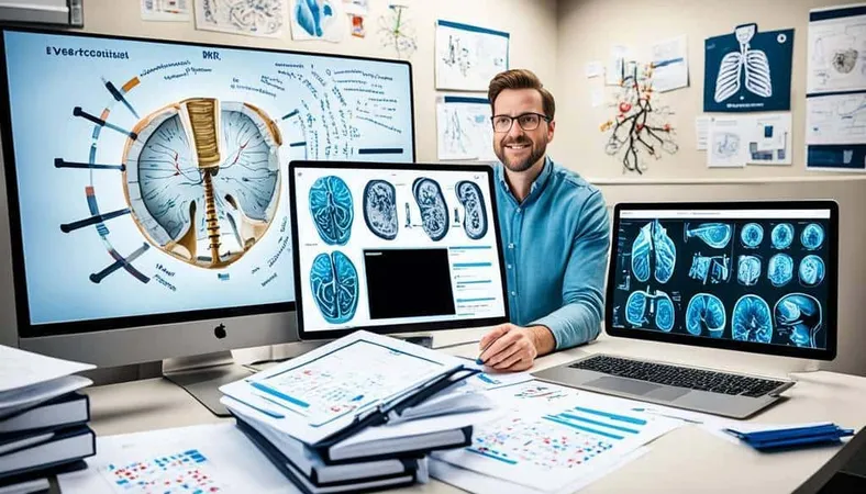
Ace Your Exam with MRI Practice Test 2026 Prep

MRI Tech School 2026 — Programs, Requirements, and What to Expect
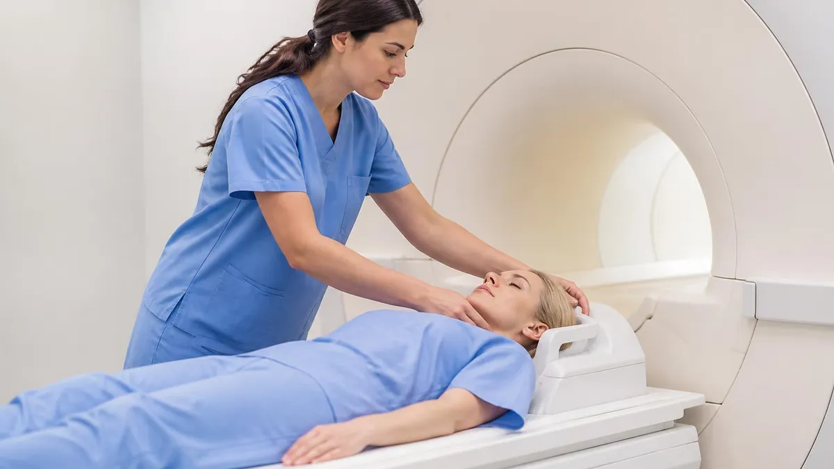
Man Dies in MRI Machine: Understanding MRI Safety Incidents, Risks, and Prevention
About the Author

Medical Laboratory Scientist & Clinical Certification Expert
Johns Hopkins UniversityDr. Sandra Kim holds a PhD in Clinical Laboratory Science from Johns Hopkins University and is certified as a Medical Technologist (MT) and Medical Laboratory Scientist (MLS) through ASCP. With 16 years of clinical laboratory experience spanning hematology, microbiology, and molecular diagnostics, she prepares candidates for ASCP board exams, MLT, MLS, and specialist certification tests.
Join the Discussion
Connect with other students preparing for this exam. Share tips, ask questions, and get advice from people who have been there.
View discussion (8 replies)