
Man Dies in MRI Machine: Understanding MRI Safety Incidents, Risks, and Prevention
Learn why a man dies in an MRI machine, what causes MRI fatalities, 🎯 and how facilities prevent deadly accidents. Essential safety knowledge for patients and...
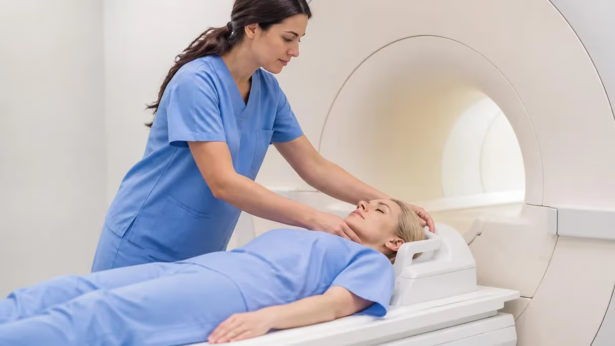
The phrase "man dies in MRI machine" captures one of the most alarming headlines in medical imaging history, yet it reflects a real and preventable tragedy. MRI scanners generate magnetic fields thousands of times stronger than Earth's own, and when ferromagnetic objects enter the scan room, the consequences can be catastrophic. In 2001, a six-year-old boy named Michael Colombini died at a New York hospital after an oxygen tank was pulled into the bore of an MRI machine and struck his head — a tragedy that fundamentally changed how facilities approach mri machine safety protocols across the United States.
MRI fatalities are exceedingly rare, but they are not zero. The FDA's Manufacturer and User Facility Device Experience (MAUDE) database logs hundreds of MRI-related adverse events every year, ranging from minor burns to life-threatening projectile incidents. Understanding why these accidents occur is not just academic curiosity — it is foundational knowledge for every MRI technologist, radiologist, nurse, and patient who sets foot inside a zone III or zone IV environment. The physics of magnetic attraction do not forgive careless exceptions.
Most MRI-related deaths and serious injuries fall into three broad categories: projectile accidents caused by ferromagnetic objects entering the magnetic field, burns resulting from conductive loops or implanted devices heating during RF pulse delivery, and adverse reactions in patients with contraindicated implants such as pacemakers or cochlear implants. Each category has its own risk profile, its own warning signs, and its own set of evidence-based prevention strategies that every imaging professional must master.
The 2001 Colombini case prompted the American College of Radiology to publish the first comprehensive MRI safety guidelines, which have since been updated multiple times. The Joint Commission, the ACR, and the International Society for Magnetic Resonance in Medicine all now require facilities to implement four-zone safety frameworks, conduct rigorous pre-screening, and maintain continuous staff education. Yet incidents still occur, most often when protocols are bypassed under time pressure or when a non-MRI staff member enters the scan room without proper screening.
For MRI technologists preparing for the ARRT registry examination, understanding MRI safety is not optional — it is one of the most heavily tested domains on the certification exam. Questions about ferromagnetic projectile physics, specific absorption rate (SAR) limits, implant classifications, and emergency quench procedures appear consistently across multiple exam forms. This article walks through the real-world science behind MRI fatalities, the regulatory landscape that governs safety practice, and the concrete steps that prevent tragedy.
This guide is also designed for patients and families who have heard frightening headlines and want accurate, science-based information. Fear of MRI is common, but it is largely unfounded when appropriate screening protocols are followed. Millions of MRI scans are performed safely in the United States every year. The goal here is not to frighten, but to educate — because informed patients ask better questions, and better questions save lives inside the magnet room.
MRI: upcoming dates and official changes
- Aug 17, 2026: ARRT launched a separate entry-level Imaging Assistant, Magnetic Resonance (I.A.) credential. A reduced $75 fee applied to applications made Aug. 17–Oct. 14, 2026, for testing Oct. 15–Nov. 30, 2026. Later applicants pay $225 and can test on or after Jan. 1, 2027.[3]
- The ARRT MRI exam for postprimary-pathway candidates has 220 questions: 200 scored and 20 unscored pilot items.[1]
- Postprimary-pathway candidates get 210 minutes for the ARRT MRI exam, inside a 230-minute (3 hours, 50 minutes) appointment.[1]
- To pass the ARRT MRI exam you need a total scaled score of 75 on ARRT's 1–99 scale. This is not a percentage.[2]
- The ARRT MRI exam through the postprimary pathway costs $225, or $450 for candidates whose supporting credential is from NMTCB or ARDMS.[3]
All verified MRI facts and sources
Numbers in brackets link to the official sources listed under Sources & last verified. Dates that have passed are removed automatically.
MRI Safety by the Numbers
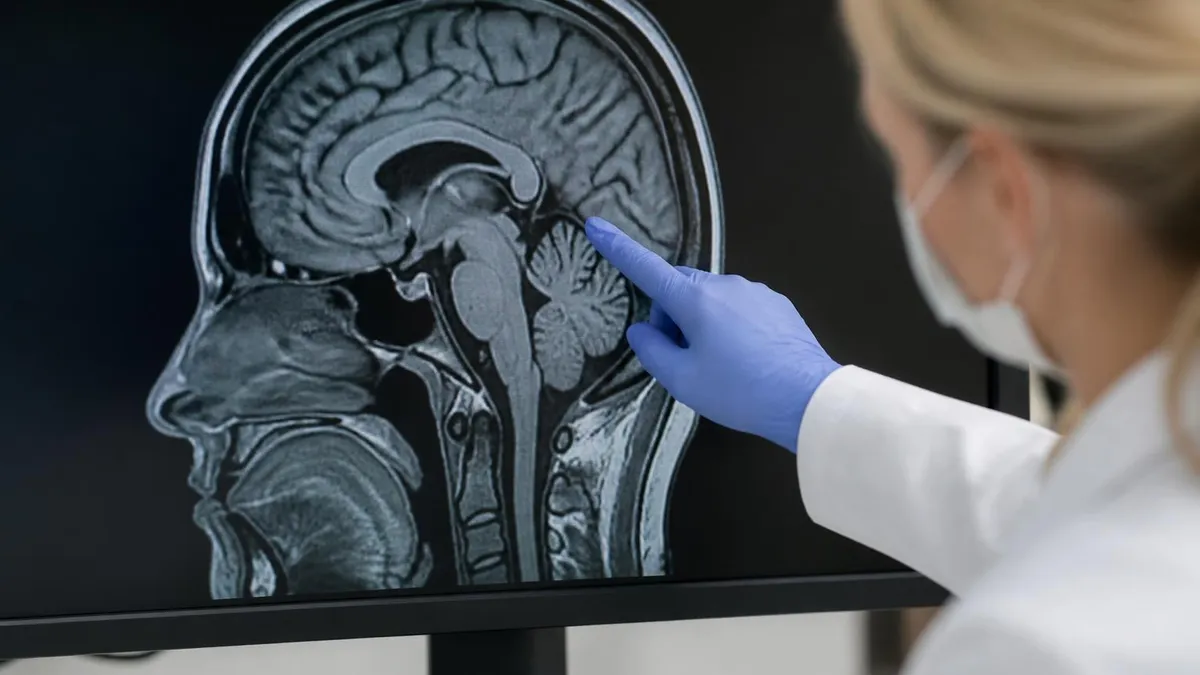
How MRI Fatalities and Serious Injuries Occur
Ferromagnetic Object Enters Zone IV
Projectile Accelerates Toward the Bore
RF Energy Causes Thermal Injury
Implant Interacts with Magnetic or RF Fields
Emergency Response Is Delayed or Absent
The American College of Radiology's four-zone framework is the cornerstone of modern MRI safety architecture, and understanding it is essential for anyone studying for the MRI registry exam or working in a clinical imaging department. Zone I is the general public area — the hospital lobby, waiting rooms, hallways — where no magnetic field restrictions apply. Zone II is the interface area between the uncontrolled public space and the strictly controlled MRI environment, typically where patient screening and registration occur. This is where the first critical safety checkpoint must happen.
Zone III is a restricted area accessible only to MRI personnel and pre-screened individuals. The fringe magnetic field in Zone III can already exert measurable force on ferromagnetic objects, which means unauthorized entry at this level poses real risk. Facilities are required to keep Zone III locked at all times when not actively supervised by qualified MRI staff. This often includes the MRI control room, where the technologist operates the scanner console. Even in Zone III, loose ferromagnetic items should never be present.
Zone IV is the MRI scan room itself — the highest-restriction space in any imaging facility. The magnetic field in Zone IV is always on in superconducting systems, even when no patient is being scanned. This is a point that surprises many people: turning off the MRI machine for a 1.5T or 3T superconducting scanner does not turn off the magnet. The only way to eliminate the field is an emergency quench, which releases cryogenic helium gas and requires costly recommissioning. Zone IV must be physically inaccessible without explicit clearance from MRI personnel.
The ACR recommends that every MRI facility designate a Medical MRI Safety Officer (MMSO) and an MRI Safety Officer (MRSO). The MMSO is typically a radiologist or physician with advanced training in MRI bioeffects, while the MRSO is usually the senior MRI technologist responsible for day-to-day protocol enforcement. These roles are not ceremonial — they carry real authority to halt any scan that poses a safety risk, even if clinical pressure to proceed is intense. Documented authority without genuine institutional support is a common precursor to adverse events.
Patient and non-MRI staff screening must occur before anyone enters Zone III. The ACR recommends a two-tier screening process: a written questionnaire covering implants, surgical history, shrapnel exposure, occupational metal exposure, tattoos, and pregnancy status, followed by verbal confirmation with a qualified MRI team member. Patients who cannot communicate reliably — due to sedation, language barriers, or cognitive impairment — require additional verification steps, including review of prior imaging studies and, when available, implant identification cards or manufacturer documentation.
Implant classification under the ASTM International standard (F2503) divides devices into three categories: MR Safe (poses no known hazards in any MRI environment), MR Conditional (safe only under specific conditions of field strength, SAR, and gradient slew rate), and MR Unsafe (known to pose hazards and must never enter the MRI environment). The online implant reference database maintained by MRIsafety.com and the FDA's MRI safety labeling guidance are the authoritative sources technologists consult before scanning a patient with a device. Using an outdated or incomplete reference can have fatal consequences.
Training frequency matters enormously. Joint Commission data consistently shows that MRI incidents spike when facilities rely on annual safety training alone. Leading programs now conduct quarterly safety drills, including simulated projectile scenarios and mock emergency quench exercises, so that the correct response becomes automatic under stress. For technologist students, this training begins in MRI tech school and continues through clinical rotations, providing the hands-on experience that written study alone cannot replicate.
MRI Practice Test Questions
Prepare for the MRI - Magnetic Resonance Imaging exam with our free practice test modules. Each quiz covers key topics to help you pass on your first try.
MRI Knowledge
MRI Exam Questions covering Knowledge. Master MRI Test concepts for certification prep.
MRI Physics
Free MRI Practice Test featuring Physics. Improve your MRI Exam score with mock test prep.
MRI Anatomy and Pathology
MRI Test Prep for MRI Anatomy and Pathology. Practice MRI Quiz questions and boost your score.
MRI Anatomy and Positioning
MRI Questions and Answers on MRI Anatomy and Positioning. Free MRI practice for exam readiness.
MRI Contrast Agents
Free MRI Quiz on MRI Contrast Agents. MRI Exam prep questions with detailed explanations.
MRI Patient Care and Positioning
MRI Practice Questions for MRI Patient Care and Positioning. Build confidence for your MRI certification exam.
MRI Safety Risk Categories: Projectile, Burns, and Implants
Projectile incidents are the most dramatic MRI hazards and account for the majority of fatal MRI accidents on record. The static magnetic field follows an inverse-square relationship with distance, meaning attractive force increases dramatically as a ferromagnetic object approaches the bore. Common projectile culprits include oxygen tanks (the most frequently cited fatal object), IV poles, wheelchairs, floor buffers, mop buckets, surgical tools, and even hairpins. A 10-pound oxygen cylinder can accelerate to over 40 mph within a fraction of a second, delivering enormous kinetic energy to anyone in its path.
Prevention requires absolute enforcement of Zone IV access restrictions and comprehensive pre-entry screening of every person, cart, and piece of equipment. Facilities should use ferromagnetic detection systems (FMDS) at Zone III entry points — handheld wands and walk-through portal detectors that flag metallic objects before they reach the scan room. All portable equipment used inside Zone IV must be labeled MR Safe and verified before each use. Regular equipment audits, staff training on recognizing non-MRI equipment that has migrated into controlled zones, and strict lockout procedures when the scanner is unattended are non-negotiable safety layers.
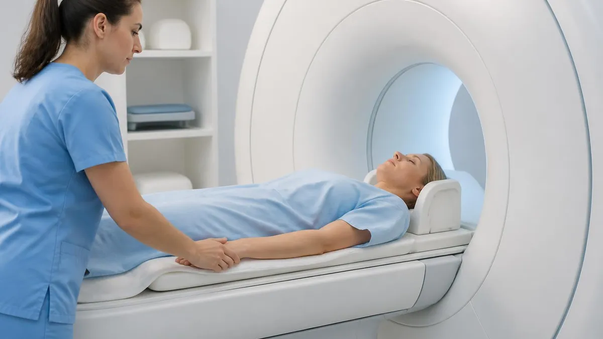
MRI Safety Protocols: Benefits of Strict Enforcement vs. Challenges of Implementation
- +Prevents projectile fatalities by keeping ferromagnetic objects out of Zone IV at all times
- +Reduces RF burn incidence through standardized coil placement and SAR monitoring protocols
- +Protects implant patients through evidence-based ASTM classification and pre-scan device verification
- +Builds staff confidence and reduces scan-day anxiety through regular safety drills and simulations
- +Supports accreditation and reduces institutional liability exposure from preventable adverse events
- +Ensures pediatric and sedated patients — who cannot self-report discomfort — receive maximum protection
- −Rigorous screening adds time to patient throughput, creating scheduling pressure in busy facilities
- −Ferromagnetic detection systems represent significant capital expenditure not all facilities can afford
- −Staff turnover requires continuous retraining investment that some departments chronically underfund
- −MR-Conditional implant scanning requires complex multidisciplinary coordination that delays access
- −Emergency quench destroys expensive liquid helium supply and requires costly magnet recommissioning
- −Non-MRI personnel — nurses, anesthesiologists, security staff — frequently lack adequate zone awareness training
Pros and Cons at a Glance
| Pros | Cons |
|---|---|
| Prevents projectile fatalities by keeping ferromagnetic objects out of Zone IV at all times | Rigorous screening adds time to patient throughput, creating scheduling pressure in busy facilities |
| Reduces RF burn incidence through standardized coil placement and SAR monitoring protocols | Ferromagnetic detection systems represent significant capital expenditure not all facilities can afford |
| Protects implant patients through evidence-based ASTM classification and pre-scan device verification | Staff turnover requires continuous retraining investment that some departments chronically underfund |
| Builds staff confidence and reduces scan-day anxiety through regular safety drills and simulations | MR-Conditional implant scanning requires complex multidisciplinary coordination that delays access |
| Supports accreditation and reduces institutional liability exposure from preventable adverse events | Emergency quench destroys expensive liquid helium supply and requires costly magnet recommissioning |
| Ensures pediatric and sedated patients — who cannot self-report discomfort — receive maximum protection | Non-MRI personnel — nurses, anesthesiologists, security staff — frequently lack adequate zone awareness training |
MRI Pre-Scan Safety Checklist for Technologists
- ✓Confirm all Zone III and Zone IV doors are locked before beginning patient escort from Zone II
- ✓Administer the standardized written implant and metal screening questionnaire to the patient or legal guardian
- ✓Verbally review each screening question with the patient, clarifying ambiguous responses before proceeding
- ✓Verify implant classification (MR Safe, MR Conditional, or MR Unsafe) using the current MRIsafety.com database or FDA labeling
- ✓Remove all ferromagnetic items from the patient including jewelry, piercings, hearing aids, dentures, and clothing with metal fasteners
- ✓Screen all accompanying personnel — family, nurses, anesthesiologists — before permitting Zone III or Zone IV entry
- ✓Inspect all equipment entering Zone IV with a handheld ferromagnetic detection wand and confirm MR Safe labeling
- ✓Position coils and cables to avoid conductive loops; insert foam padding between cables and patient skin
- ✓Set scanner SAR limits appropriate for patient weight and any MR-Conditional implant restrictions
- ✓Confirm emergency quench button location, helium vent path is unobstructed, and emergency contact numbers are posted
The Magnet Is Always On — Even When the Scanner Isn't Scanning
Superconducting MRI magnets at 1.5T and 3T maintain their magnetic field continuously — 24 hours a day, 7 days a week — regardless of whether a scan is in progress. The only way to eliminate the field is an emergency quench, which releases cryogenic helium and costs $30,000–$100,000 to recommission. This means Zone IV is never safe for unscreened entry, even during scheduled downtime, maintenance windows, or facility closures.
MRI safety knowledge is not only a clinical imperative — it is a core competency domain tested on the ARRT MRI registry examination, which is the primary credentialing pathway for MRI technologists in the United States. The ARRT exam content specifications allocate a significant portion of questions to patient care, patient assessment, and contrast media, all of which incorporate safety screening and adverse event recognition.
Students preparing for the registry must be able to distinguish between MR Safe, MR Conditional, and MR Unsafe classifications, calculate relative SAR contributions from different pulse sequence parameters, and articulate the physiologic mechanisms behind known MRI bioeffects.
Beyond the exam, continuing education in MRI safety is mandatory for ARRT credential maintenance. Registered technologists must complete 24 continuing education credits every two years, and safety-related coursework is strongly encouraged by the ACR and ISMRM. Emerging safety challenges — including ultra-high-field 7T systems entering clinical use, new generations of MR-Conditional implants with complex conditional parameters, and the increasing use of MRI in interventional suites where metallic instruments are routinely present — demand that even experienced technologists update their knowledge regularly.
The ARRT registry examination for MRI draws on content categories that include instrumentation, image production, procedures, and patient care. Patient care questions specifically address contrast agent administration, screening procedures for implants and claustrophobia, monitoring requirements for sedated patients, and response to adverse events including contrast reactions and gradient-induced peripheral nerve stimulation. Candidates who approach these topics through the lens of real incident cases — such as the fatal events documented in peer-reviewed literature — tend to retain the material more effectively than those who study definitions alone.
Practice tests are one of the most effective preparation tools for the MRI registry, particularly when questions are drawn from clinical scenarios that mirror real exam formatting. The ARRT uses a competency-based model that emphasizes application of knowledge rather than simple recall, which means candidates must be able to reason through unfamiliar patient scenarios using foundational principles. A candidate who truly understands why a gradient echo sequence generates higher peripheral nerve stimulation risk than a spin echo sequence — not just that it does — will consistently outperform one who memorized the answer without the underlying physics.
For technologist students in clinical rotations, observing and participating in pre-scan safety screening is perhaps the single most valuable educational experience available. Watching an experienced technologist navigate the complexity of screening a patient with a cochlear implant, multiple orthopedic prostheses, and a history of occupational metal exposure demonstrates the multi-variable reasoning that no textbook can fully convey. Students should actively seek out these complex cases and request mentored guidance through the decision-making process rather than deferring entirely to senior staff.
MRI physics knowledge underpins safety understanding in ways that are not always immediately obvious. A technologist who understands the relationship between field strength and precession frequency, the role of T1 and T2 relaxation in tissue contrast, and the mechanism of gradient switching understands not just how to acquire images but why certain parameters improve safety risk. Field strength affects not only image quality but also the translational and rotational forces on ferromagnetic implants, the magnitude of RF-induced heating, and the threshold for peripheral nerve stimulation — all direct safety variables that technologists must manage in real time during complex scans.
The ARRT also tests knowledge of contrast agent safety, including the classification of gadolinium-based contrast agents (GBCAs) by risk of nephrogenic systemic fibrosis (NSF), appropriate screening thresholds for renal function before administration, and recognition of acute hypersensitivity reactions. While contrast-related adverse events are distinct from magnetic field safety incidents, they represent a meaningful share of MRI-related morbidity and are appropriately emphasized in registry preparation. Candidates should be comfortable with both the ACR Manual on Contrast Media and the current ESUR guidelines for GBCA use in patients with renal impairment.
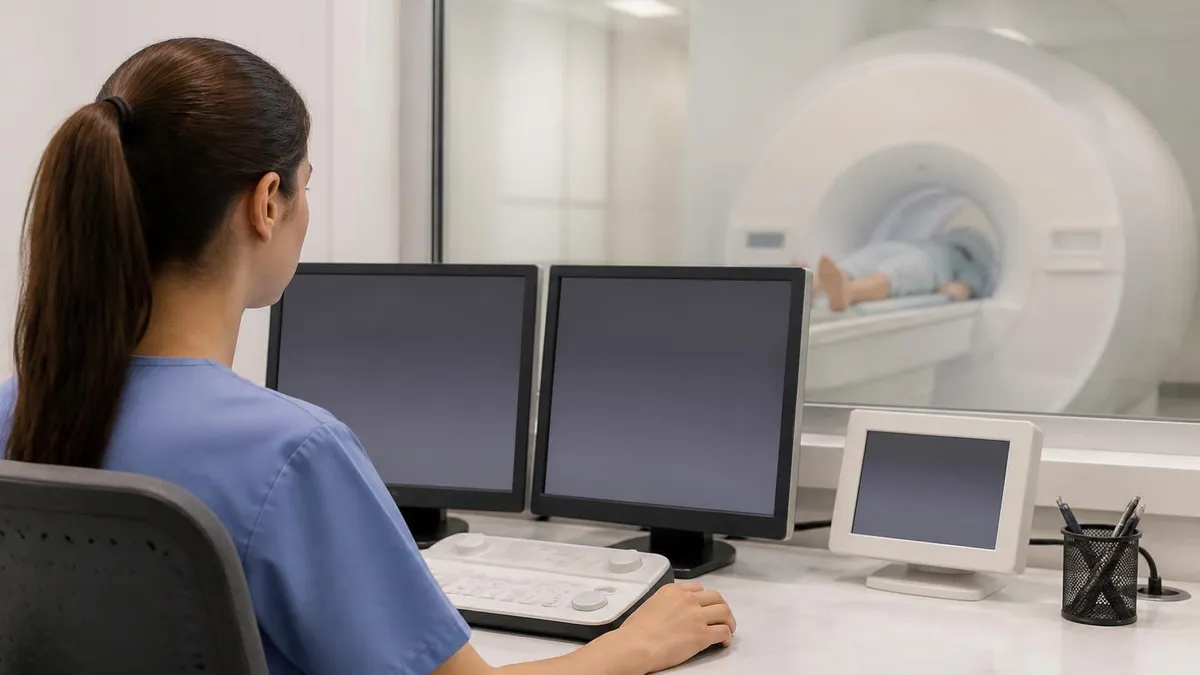
The Joint Commission's analysis of MRI sentinel events consistently identifies non-MRI personnel — emergency nurses, anesthesiologists, security officers, and housekeeping staff — as the individuals most likely to introduce ferromagnetic objects into Zone IV. Every facility must implement mandatory Zone III and Zone IV entry training for all hospital staff, not only radiology employees. A single lapse in access control by an unfamiliar staff member can turn routine equipment into a lethal projectile in under one second.
Emergency response inside an MRI facility requires a fundamentally different skill set than standard hospital emergency management, because the magnetic environment imposes hard constraints on what equipment and personnel can safely enter the scan room.
When a patient loses consciousness, experiences a cardiac arrest, or is trapped by a projectile event inside Zone IV, the immediate priority is to remove the patient from the bore and out of Zone IV — not to begin resuscitation inside the magnet room. Standard crash carts, defibrillators, and laryngoscopes contain ferromagnetic components and must never be brought into Zone IV during an active magnetic field.
Facilities must maintain MR-Conditional or MR Safe emergency equipment staged at the Zone III/Zone IV boundary. This includes MR-Conditional cardiac monitoring, MR-Conditional laryngoscopes with plastic blades, and MR Safe oxygen delivery systems. The emergency action plan must be posted conspicuously inside the control room, and all MRI staff must be able to recite the sequence of steps without consulting the document — because in a real emergency, there is no time to read a protocol from the wall.
An emergency quench — the intentional ramping down of the superconducting magnet by activating the quench button — is the option of last resort when a person is pinned to the magnet by a ferromagnetic object and cannot be freed by other means.
Quenching releases a large volume of cryogenic helium gas that vents through the roof port; if the vent is blocked or fails, the helium displaces oxygen in the scan room and creates an asphyxiation hazard on top of the existing emergency. Before quenching, staff must confirm the vent path is clear, evacuate all personnel from Zone IV, and call the magnet vendor's emergency line immediately after the quench is initiated.
Post-incident response is equally critical. Every MRI-related adverse event — including near misses in which a ferromagnetic object entered Zone III or Zone IV without causing injury — must be documented, investigated, and reported through the facility's patient safety reporting system. The ACR recommends root cause analysis for all Zone IV projectile events and near misses, with findings shared at departmental safety meetings. Facilities that normalize near-miss reporting and treat it as a learning opportunity rather than a punitive exercise consistently demonstrate lower rates of serious adverse events over time.
Cryogen incidents are a related emergency category that MRI technologists must understand. Liquid helium at 4 Kelvin maintains superconductivity in clinical MRI magnets, and a helium leak — whether from a planned quench, an unplanned quench caused by magnet fault, or a cryostat failure — creates an immediate oxygen depletion hazard. Facilities must install oxygen monitoring sensors in the scan room that alarm when oxygen concentration drops below 19.5%, and all staff must know to evacuate immediately without pausing to investigate. Oxygen-deficient atmospheres are odorless and can cause loss of consciousness within seconds.
For patients, the safest approach to MRI is active participation in the screening process. Patients should always disclose every surgical implant, regardless of how old it is or how confident they are that it is safe. Metallurgy and device design have changed significantly over decades, and assumptions made about devices implanted in the 1980s or 1990s based on current knowledge can be dangerously wrong.
Patients who work in metallic environments — welders, machinists, shipyard workers — should request orbital radiographs to screen for ocular metallic foreign bodies before any head or orbital MRI, per ACR guidance. For more detailed clinical preparation, review the complete guidance on mri machine safety in the context of cervical spine imaging.
Family members who wish to accompany patients into Zone III or Zone IV — for example, parents accompanying a pediatric patient — must undergo the same full screening process as the patient. No exceptions based on familial relationship, urgency, or staff familiarity are acceptable. A parent who entered the scan room dozens of times safely in the past may have received a new implant since the last visit, and assuming continuity of safety status is a known precursor to adverse events. The inconvenience of screening is always preferable to the irreversible consequences of a projectile event.
Preparing for the ARRT MRI registry examination requires a strategic approach that balances broad content coverage with deep understanding of the highest-yield domains. MRI safety — including projectile physics, implant classification, SAR management, and emergency response — consistently ranks as one of the most tested and most clinically consequential knowledge areas on the exam. Students who invest time in truly understanding the mechanisms behind safety incidents, rather than memorizing isolated facts, are far better prepared for both the registry and for the unpredictable scenarios they will encounter in clinical practice.
Effective registry preparation typically involves a combination of structured content review, targeted practice testing, and clinical case discussion with experienced technologists. Content review should be organized by the ARRT's published content specifications, which are publicly available and provide a transparent map of exam domains and their relative weighting. Students who build their study schedule around these specifications avoid the common mistake of over-studying low-yield topics at the expense of high-frequency safety and physics content.
Practice testing is not merely about memorizing correct answers — it is about calibrating reasoning under time pressure. The MRI registry allows approximately one minute per question, which means candidates must be able to access foundational knowledge rapidly and apply it to novel scenarios without hesitation. Timed practice under simulated exam conditions, reviewed immediately after completion with explanation-focused analysis of both correct and incorrect answers, produces far greater score improvement than untimed review alone.
Study groups offer an underutilized advantage for registry preparation. Explaining a concept to a peer — particularly a complex topic like the physics of gradient echo versus spin echo sequences, or the multi-step decision process for scanning a patient with an MR-Conditional pacemaker — forces the explainer to identify gaps in their own understanding that silent review does not surface. Students who participate in structured peer teaching sessions consistently demonstrate stronger retention and higher registry scores than those who study exclusively alone.
The week before the exam should focus on consolidation rather than new learning. Reviewing high-yield safety concepts, working through short timed practice sets to maintain cognitive pacing, ensuring adequate sleep, and arriving at the testing center with all required identification and scheduling confirmation documents are the priorities. Cramming new material in the final 48 hours reliably increases test anxiety without meaningful knowledge gains for most candidates.
After passing the registry, the learning does not stop. MRI technology continues to advance rapidly: 7T clinical systems are FDA-cleared and entering academic medical centers; AI-driven image reconstruction is reducing scan times and changing SAR profiles; new MR-Conditional implant categories are approved by the FDA on a monthly basis.
The technologists who remain safest throughout their careers are those who treat safety education as a permanent professional practice, not a one-time credentialing hurdle. Subscribing to the ISMRM safety committee updates, participating in ACR accreditation visits, and maintaining active engagement with facility safety officers are habits that distinguish excellent MRI technologists from merely competent ones.
Whether you are a patient trying to understand the risks before your first MRI, a student preparing for the registry, or an experienced technologist looking to reinforce foundational safety knowledge, the core message of this guide is consistent: MRI is overwhelmingly safe when protocols are followed, and preventable tragedies occur almost exclusively when protocols are bypassed.
Every step of the screening process, every locked door, every piece of MR Safe equipment exists because someone identified a failure mode and designed a solution. Honoring those solutions — every scan, every patient, every time — is the professional and ethical obligation of every MRI team member.
Sample MRI - Magnetic Resonance Imaging Practice Questions
Try these questions from our free MRI - Magnetic Resonance Imaging practice tests. The correct answer and an explanation follow each question.
Which classification of GBCAs carries the LOWEST risk for NSF based on ACR guidance?
- A. Group I (linear ionic)
- B. Group II (linear non-ionic)
- C. Group III (macrocyclic)
- D. All GBCAs carry equal NSF risk
Answer: C. Group III (macrocyclic)
Macrocyclic GBCAs (Group III) have a more stable cage-like structure that resists transmetallation and are associated with the lowest NSF risk.
Which MRI contrast agent class is associated with the highest risk of nephrogenic systemic fibrosis (NSF)?
- A. Macrocyclic ionic agents
- B. Linear non-ionic agents
- C. Blood pool agents
- D. Manganese-based agents
Answer: B. Linear non-ionic agents
Linear non-ionic (and some linear ionic) gadolinium agents release free Gd³⁺ more readily and carry the highest NSF risk in patients with severe renal impairment.
Which patient condition requires consultation with the ordering physician regarding MRI safety due to potential risks during the first trimester?
- A. Known diabetes mellitus
- B. Known pregnancy, especially first trimester
- C. Mild hypertension controlled with oral medication
- D. Prior abdominal surgery more than 1 year ago
Answer: B. Known pregnancy, especially first trimester
MRI in the first trimester is performed with caution; although no confirmed harm is established, ACR guidelines recommend physician consultation and clinical justification for first-trimester MRI.
On the ARRT MRI registry exam, which content area covers patient care, contrast agents, and MRI safety screening?
- A. Image Production
- B. Patient Care and Education
- C. Procedures
- D. Physics and Instrumentation
Answer: B. Patient Care and Education
Patient Care and Education encompasses screening, safety, contrast administration, and patient communication topics.
Take the full MRI - Magnetic Resonance Imaging practice test
MRI Questions and Answers
Sources & last verified
- assets-us-01.kc-usercontent.com — number of questions, time limit (official source). “MRI Scored Items 200 Pilot Items 20 Total Items 220 Test Time 210 min” Verified Oct 15, 2026.
- The American Registry of Radiologic Technologists — passing score (official source). “Our exam scoring scale ranges from 1 to 99, and you’ll need a total score of 75 to pass the exam.” Verified Oct 15, 2026.
- The American Registry of Radiologic Technologists — exam fee, other (official source). “Postprimary (M, CT, MR, BD, CI, VI, VS, BS, R.R.A.): $225 Postprimary (CT, MR, BD, or VS—using NMTCB as supporting category): $450 Postprimary (MR, VS,…” Verified Oct 15, 2026.
How we verify: editorial & fact-checking policy. Report an error: contact us.

MRI of Cervical Spine: Complete Guide to Imaging, Anatomy, Pathology, and Patient Preparation

Knee MRI Images: A Complete Guide to Reading, Understanding, and Interpreting Knee Scans
Ace Your Exam with MRI Practice Test 2026 Prep

MRI Tech School 2026 — Programs, Requirements, and What to Expect
MRI Procedure: What to Expect Before, During and After Your Scan
About the Author

Medical Laboratory Scientist & Clinical Certification Expert
Johns Hopkins UniversityDr. Sandra Kim holds a PhD in Clinical Laboratory Science from Johns Hopkins University and is certified as a Medical Technologist (MT) and Medical Laboratory Scientist (MLS) through ASCP. With 16 years of clinical laboratory experience spanning hematology, microbiology, and molecular diagnostics, she prepares candidates for ASCP board exams, MLT, MLS, and specialist certification tests.