
How Long Does a Thoracic Spine MRI Take? MRI Scan Duration by Body Part, CPT Codes and Open MRI Tips
🎯 How long does a thoracic spine MRI take? Usually 30-45 minutes. See scan times by body part, pelvic MRI CPT codes, and open MRI tips.

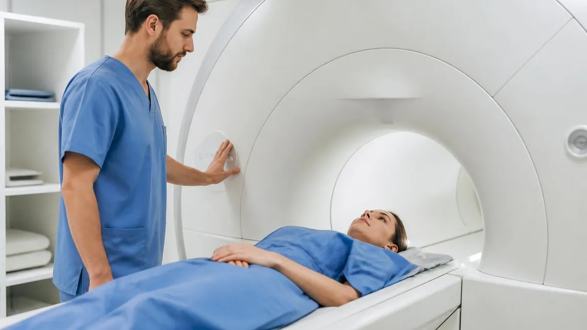
If you have been scheduled for a back scan, the first question is practical: how long does a thoracic spine mri take? For most patients the answer is 30 to 45 minutes of table time without contrast, and 45 to 60 minutes when contrast is added. Add check-in, screening, and positioning, and plan on one to one and a half hours at the facility. Billing staff will also reference a CPT code, such as the pelvic mri cpt code for pelvic studies.
Scan length is not random. It depends on the body part, the magnet strength, the number of imaging sequences your physician ordered, and whether a gadolinium-based contrast agent is needed. A thoracic spine covers twelve vertebrae and a long stretch of spinal cord, so technologists typically acquire sagittal and axial images in several weightings. Each sequence runs two to six minutes, and a protocol usually contains five to eight of them. That simple arithmetic explains most of the variation patients experience.
This guide breaks down typical times across the body, from a quick knee or brain screening to a lengthy cardiac study. You will find the logic behind each estimate, so you can judge whether your appointment seems normal. We also explain common CPT codes, including the pelvic coding family and the combined study often searched as mri pelvis w wo gyn cpt, because the code your facility bills often signals how many sequences are planned.
Understanding timing helps in practical ways. Patients who expect twenty minutes and receive ninety can grow anxious, tense, or claustrophobic, and movement is the single biggest cause of repeated sequences. Knowing the plan lets you arrange rides, childcare, and work breaks, and it helps you decide whether a sedative is worth discussing beforehand. Preparation also matters, and our mri scan duration by body part companion resource covers clothing, metal screening, and fasting rules in detail.
Students and aspiring technologists benefit too. Registry exams such as the ARRT MRI certification test ask about sequence selection, scan parameters, and how protocol choices change acquisition time. Understanding why a T2-weighted fast spin echo sequence takes minutes while a gradient echo localizer takes seconds builds the physics intuition you need on test day. Throughout this article, we connect real-world timing to the principles behind it, using concrete numbers rather than vague ranges wherever possible.
Finally, location matters. Many patients search for the closest open mri to my location because they feel anxious in a narrow bore or exceed the weight limit of a standard scanner. Open systems are real options, but they often use lower field strength and need longer acquisition times to reach comparable image quality. We cover that tradeoff later, along with advice for calling ahead and confirming that the site can perform the exam your doctor ordered.
Use the sections below as a roadmap. Start with the numbers at a glance, follow the step-by-step timeline of a thoracic exam, then compare body parts, contrast decisions, coding, and tips for staying still. Every estimate here reflects typical outpatient practice in the United States, and your own scan may differ. Your imaging center and ordering clinician always have the final word on your exact protocol, preparation instructions, and expected appointment length.
MRI Scan Duration by the Numbers
How a Thoracic Spine MRI Unfolds, Step by Step
Check-In and Safety Screening
Changing and Positioning
Localizer Images
Core Sequences
Optional Contrast Phase
Wrap-Up and Results
Typical scan times vary widely across the body. A brain MRI usually takes 30 to 45 minutes, although a limited stroke or screening protocol can finish in 15 minutes. Cervical and lumbar spine studies mirror the thoracic spine at 30 to 45 minutes. Joint exams such as the knee, ankle, or shoulder run 20 to 40 minutes because the anatomy is small and fewer slices are required. Larger regions take longer, since the scanner must cover more tissue.
Abdominal and pelvic exams generally run 30 to 60 minutes. Many require breath-hold sequences, where you hold your breath for 15 to 25 seconds while the scanner acquires data. Each breath-hold sequence is short, but the technologist pauses between them, so the total grows. Pelvic studies for gynecologic indications, prostate evaluation, or rectal cancer staging often include diffusion-weighted imaging and multiplanar T2 sequences, pushing the exam toward the upper end of that range.
Cardiac MRI is among the longest routine studies, commonly 60 to 90 minutes. The scanner synchronizes with your heartbeat through ECG leads, and the protocol evaluates function, scar, and sometimes stress perfusion. Breast MRI typically lasts 30 to 60 minutes and requires lying prone with contrast. MR angiography and magnetic resonance enterography also sit in the 45 to 75 minute range. Whole-body protocols can exceed two hours and are not routine for most patients.
Why does the thoracic spine land at 30 to 45 minutes? The protocol generally includes sagittal T1, sagittal T2, sagittal STIR or fat-suppressed T2, and axial T2 or T1 images through areas of concern. Sagittal sequences often take three to five minutes each. Axial stacks through suspected disc herniation or cord compression add another eight to twelve minutes. With contrast, a pre-contrast and post-contrast T1 pair adds roughly fifteen minutes, which is why the combined study takes close to an hour.
Magnet strength influences timing as well. Most hospitals use 1.5 tesla or 3 tesla scanners. A 3T system produces a stronger signal, which can shorten acquisition or improve resolution at the same length. Open magnets and older low-field machines, typically 0.3 to 1.2 tesla, collect less signal, so technologists average more excitations and the exam can run 45 to 90 minutes. Image quality can still be diagnostic, but the trade is more time lying still.
Patient factors add or subtract minutes. Motion from pain, coughing, or breathing forces repeat sequences, and a repeated four-minute sequence quickly adds up. Patients with scoliosis may need extra planning to cover a curved spine. Large body habitus can reduce signal and require protocol adjustments. Pediatric exams often need sedation or anesthesia, which extends the visit significantly. Conversely, a cooperative patient who stays still can complete a protocol at the faster end of the range.
Remember that table time is only part of the appointment. Arrive 15 to 30 minutes early for registration, a safety questionnaire, and changing into a gown. If contrast is planned, an IV is placed first, which takes five to ten minutes. After the scan, a radiologist usually interprets the images within one to two business days, and results go to your ordering physician. Budget at least two hours for the entire visit to avoid unnecessary stress.
Pelvic MRI CPT Code and Scan Time: Three Coding Scenarios
The pelvic mri cpt code 72195 describes magnetic resonance imaging of the pelvis without contrast material. It is commonly ordered for uterine fibroids, adenomyosis, adnexal masses seen on ultrasound, pelvic pain, and congenital anomalies. Because no contrast is involved, no IV is needed and the exam moves quickly. Expect roughly 30 to 40 minutes of table time, using axial, sagittal, and coronal T2 sequences plus T1 imaging to characterize fat, blood, and fluid.
Coverage for 72195 generally requires documented symptoms and often a prior ultrasound. Prior authorization is common with commercial insurers, so confirm approval before scheduling. The code covers the pelvis itself, not the abdomen, so a combined abdomen and pelvis study bills separately. If you are comparing it with the thoracic spine code 72146, remember that each anatomical region has its own code, which is why two scans on one day appear as separate line items.
Open MRI vs Closed MRI: Is the Closest Open MRI Worth It?
- +Less confining design reduces claustrophobia and panic for many patients.
- +Accommodates larger patients who exceed standard bore size or weight limits.
- +Often possible without sedation, avoiding the need for a driver afterward.
- +Side access lets a parent or caregiver stay close, which helps children.
- +Independent open centers frequently charge less than hospital departments.
- +Easier positioning for patients with painful or limited mobility.
- −Low-field magnets usually need longer scans, often 45 to 90 minutes.
- −Lower signal can reduce detail for small lesions and subtle cord changes.
- −Cardiac, breast, and advanced pelvic protocols may not be available.
- −Longer exams increase the chance of motion and repeated sequences.
- −Fewer open units exist, so travel distance can be significant.
- −Some insurers or physicians prefer high-field studies for complex cases.
Checklist Before Visiting the Closest Open MRI to My Location
- ✓Confirm the facility has an open or wide-bore scanner, not just a standard unit.
- ✓Ask for the magnet strength in tesla before you book.
- ✓Verify the unit can perform the exact exam your doctor ordered.
- ✓Check the weight limit and table width if body size is a concern.
- ✓Confirm the facility is in network and the CPT code is authorized.
- ✓Ask how long the exam takes on that specific scanner.
- ✓Bring your order, ID, insurance card, and medication list.
- ✓Request prior images be sent or bring them on disc.
- ✓Arrange a driver if you plan to take an anti-anxiety medication.
- ✓Wear metal-free clothing and arrive 20 minutes early.
Table time is not appointment time
A thoracic spine MRI takes 30 to 45 minutes on the table without contrast and up to 60 with it, but your total visit can reach two hours. Registration, screening, IV placement, and changing all happen outside the magnet. Plan the whole morning or afternoon, not just the scan.
Searching for the closest open mri to my location usually starts with discomfort. About 5 to 10 percent of patients experience enough claustrophobia to interrupt or abandon a standard closed-bore scan. Open MRI systems use a C-shaped or dual-pillar design with space on the sides, which feels less confining and can accommodate larger patients. They are a legitimate option, but they differ from conventional scanners in several ways that affect duration and image quality.
The main difference is field strength. Traditional open magnets operate between 0.2 and 1.2 tesla, while closed hospital scanners are 1.5 or 3 tesla. Lower field strength means a weaker signal, so the system must average more data, and sequences stretch out. A thoracic spine exam that takes 35 minutes on a 1.5T machine may take 50 to 70 minutes on a low-field open unit. Some modern high-field open systems narrow this gap considerably.
Wide-bore scanners are a middle path. These closed systems have a 70-centimeter opening, compared with 60 centimeters on older designs, and they maintain 1.5T or 3T strength. Many patients tolerate a wide-bore scanner without sedation, especially for spine or pelvis studies where the head can stay near the opening. If you need the quickest scan with the best detail, ask a facility whether a wide-bore option exists before committing to a low-field open unit.
To find a site, use a search tool such as the American College of Radiology accredited facility finder, your insurer's provider directory, or a map search. Call before booking. Ask about magnet strength, whether the scanner is truly open or wide-bore, the weight limit, and whether the exam your doctor ordered can be done on that unit. Not every study is suitable for low field; cardiac, breast, and advanced pelvic protocols often require high-field equipment.
Cost and insurance deserve attention. Independent imaging centers frequently charge far less than hospital outpatient departments for the same exam, sometimes by hundreds of dollars. Verify that the center is in network and that your authorization lists the specific CPT code. A facility that performs the pelvic mri cpt code 72195 on a low-field open unit may still be a poor fit if your physician needs high-resolution gynecologic detail, so confirm clinical suitability with the ordering provider.
Other options reduce anxiety without changing equipment. Ask for a mirror or prism glasses so you can see out of the bore, music or headphones, a fan, and an eye mask. Some centers allow a family member in the room for support. For severe claustrophobia, discuss a one-time anti-anxiety prescription with your physician. You must arrange transportation afterward, because sedatives impair driving. Practicing slow breathing at home for a week beforehand also helps many patients.
Do not choose an open scanner solely for comfort if your condition demands detail. Small lesions, subtle cord signal changes, and small tears of cartilage can be missed at low field. Your doctor can tell you whether a wide-bore 1.5T system is an acceptable compromise. If you do select an open unit, expect a longer visit, remain extremely still, and consider asking the radiology team whether a comparison with prior studies will be needed.
CPT codes and payer rules change, so confirm the exact code with your scheduler and insurer before the exam. Always disclose pacemakers, implants, shrapnel, and pregnancy during screening, since some devices make MRI unsafe or require special conditions.
Contrast is the most predictable reason one scan takes longer than another. Gadolinium-based agents are injected through an IV, and the technologist must obtain precontrast images, inject, and then wait for the agent to circulate before post-contrast sequences. For the thoracic spine, contrast is typically reserved for suspected tumor, infection, inflammatory disease like multiple sclerosis, or post-surgical scarring. Routine back pain without red flags rarely requires it, which keeps most spine studies at 30 to 45 minutes.
Each sequence has a purpose and a time cost. T1-weighted images show anatomy and fat, usually taking two to four minutes. T2-weighted fast spin echo images highlight fluid, disc hydration, and cord edema in three to five minutes. STIR suppresses fat to expose edema or marrow lesions. Diffusion-weighted imaging detects restricted motion in infarcts and some tumors. Adding even one sequence for an unexpected finding can extend the exam by five minutes.
Technologists also use scout and localizer images at the beginning, which take about 30 to 60 seconds. They plan slice positions so that every vertebral level from T1 to T12 is covered. For the thoracic spine, counting levels accurately is critical; many protocols include a cervical or lumbar localizer to verify numbering. These planning steps rarely appear on a printed time estimate, yet they account for three to five minutes of the appointment.
Acceleration techniques can shorten a scan. Parallel imaging, compressed sensing, and deep learning reconstruction allow scanners to acquire fewer data points and still reconstruct clear images. Facilities with newer software may complete a lumbar or thoracic spine study in 20 to 25 minutes. These tools are valuable for patients who struggle with pain or restlessness. However, they do not replace diagnostic quality, so radiologists balance speed against signal-to-noise ratio carefully for each protocol.
Coding and protocols are connected. A radiology order for a thoracic spine without contrast is billed as CPT 72146, with contrast as 72147, and without followed by with as 72157. Pelvic equivalents are 72195, 72196, and 72197. Cervical spine is 72141, 72142, and 72156, while lumbar spine is 72148, 72149, and 72158. Brain studies use 70551, 70552, and 70553. The code often predicts the minutes you will spend inside the scanner.
Sometimes one visit includes multiple regions. A patient with suspected spinal metastases may undergo cervical, thoracic, and lumbar imaging in one session, called a total spine study. Each region carries its own code and its own protocol, so a total spine exam can last 60 to 90 minutes or more. Ask your scheduler whether your order lists one region or several, because the difference can double your time on the table and change your out-of-pocket cost.
Equipment limitations can matter too. Metal implants, such as spinal fusion hardware, create artifacts that distort images near the metal. Technologists then use metal-artifact reduction sequences, which are slower. Pacemakers and defibrillators require MRI-conditional verification and cardiology coordination before the exam. Always disclose surgeries, implants, shrapnel, and tattoos during screening. Honest answers protect safety and also let the team plan realistic timing so that your appointment does not run unexpectedly long.
The best way to shorten a scan is to stay still. Before you enter the bore, use the restroom, get comfortable, and ask for extra padding under your knees or a bolster. Pain is the leading reason people move, so if you have severe back pain, ask your doctor whether a short-term pain reliever is appropriate beforehand. Relaxed muscles produce steadier images, and steady images mean fewer repeated sequences and fewer minutes lying on the table.
Dress for success. Wear loose, metal-free clothing such as sweatpants and a cotton shirt with no zippers, or change into the gown provided. Leave jewelry, watches, hair clips, and credit cards in a locker, because magnets can erase cards and metal can cause burns or image artifacts. Many centers provide lockers and gowns, but confirm in advance. Avoid cosmetics containing metallic particles, and remove medication patches if instructed by staff.
Manage the noise. MRI scanners produce loud knocking and buzzing sounds, often exceeding 100 decibels, so you will receive earplugs or headphones. The sounds change with each sequence, and the technologist will tell you how long each one lasts. Counting sequences helps many people because they can mentally track progress. If you feel panicky, you will have a squeeze ball or call button, and the team can stop immediately and speak with you through an intercom.
Prepare your paperwork. Bring your insurance card, photo ID, the order from your doctor, and a list of medications and allergies. If you have prior imaging from another facility, bring the disc or request transfer, since radiologists compare studies to detect change. Know your implant history, including dates of surgeries. Confirm the correct CPT code with the scheduler, whether it is 72146 for the thoracic spine or 72197 for a combined pelvic study, to avoid authorization denials.
Follow eating and drinking instructions precisely. Most spine exams require no fasting. Abdominal and pelvic exams often require four to six hours without food, and some pelvic protocols want a moderately full bladder while others want it empty. Follow the facility instructions rather than general advice, because the wrong bladder state can blur images. If you take diabetes medication, ask how fasting affects dosing, and bring a snack for afterward to prevent low blood sugar.
Plan the day around the exam. Schedule it when you are least likely to be rushed, arrange transportation if you are taking a sedative, and tell your employer you may need extra time. Eat normally afterward unless told otherwise. If contrast was given, drink plenty of water to help your kidneys clear it. Report any rash, swelling at the IV site, headache, or nausea to the facility, though serious reactions are uncommon.
After the scan, ask when results will be available and who will deliver them. Radiologists typically send a report to your ordering physician within one to two business days, and many portals release it sooner. Do not interpret findings alone, since many abnormalities are age-related and harmless. If you are studying for a certification exam, review sequence timing and protocol logic using practice questions, which reinforces how acquisition parameters shape real scan duration.
MRI Questions and Answers

MRI Preparation Guide: How to Prep for Your Scan Step by Step

Travel MRI Tech 2026: Salary, Requirements & ARRT MRI Exam

MRI Internal Auditory Canal: Complete Guide to IAC Imaging, Anatomy, Indications, and Interpretation
Healthy Brain MRI: What a Normal Scan Looks Like

MRI of Shoulder: A Complete Guide to Imaging, Anatomy, and Diagnosis
How this page was made: written with AI tools and published by Nathan Beja, founder of Practice Test Geeks, who is responsible for its content. Exam facts are checked against the official sources listed below. Not reviewed by a subject-matter expert. Editorial policy
Sources & references
Exam facts on this page (fees, format, dates, passing scores) are checked against these official sources; the date is our last check. Editorial policy
- arrt.org — Passing score, score release, fees, exam information · verified Oct 2026
- assets-us-01.kc-usercontent.com — Renewal, score validity, test delivery, eligibility, exam format, test dates, time limit · verified Oct 2026