
Can You Have an MRI With Dental Wire? Safety, Braces, Implants, Pelvic MRI CPT Codes and Open Bore Options
🎯 Can you have an MRI with dental wire? Learn which braces, fillings and implants are safe, plus pelvic MRI CPT codes and open bore MRI options.
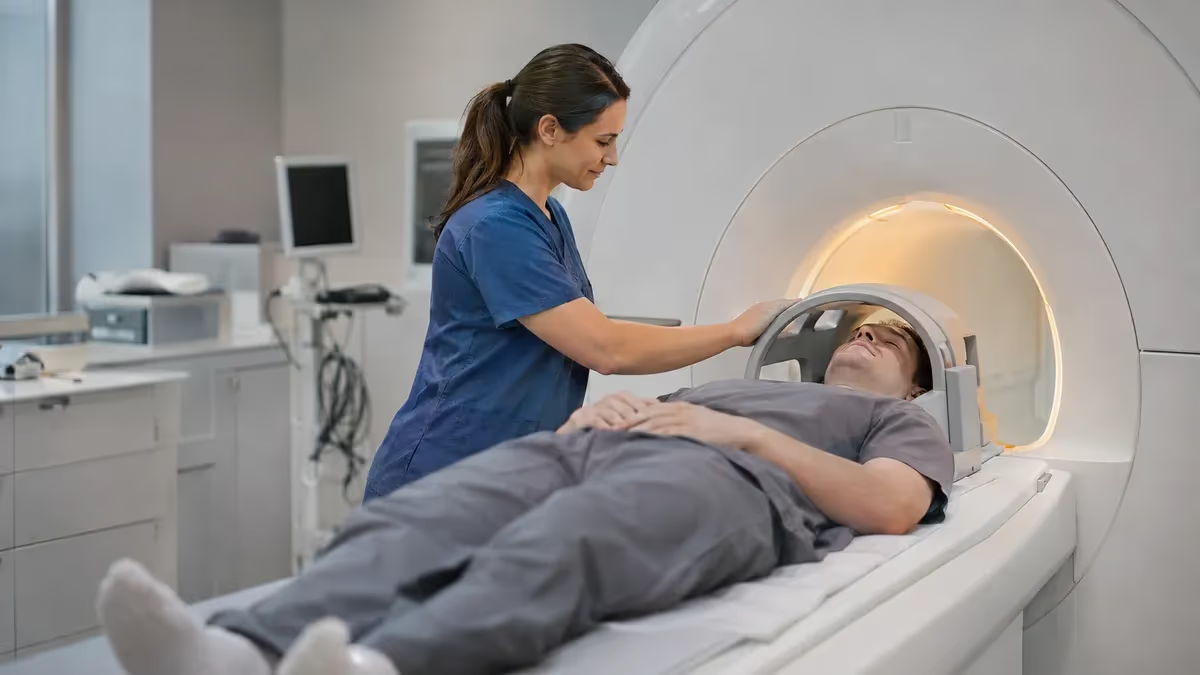
Can you have an MRI with dental wire? In most cases, yes. Orthodontic archwires, bonded retainers, fillings, crowns and titanium implants are scanned every day at hospitals and outpatient imaging centers across the United States. The real question is rarely whether the magnet will harm you. It is whether the metal will blur the pictures your radiologist needs. That distinction, safety versus image quality, drives nearly every decision a technologist makes when screening a patient with dental hardware.
MRI uses a strong static magnetic field, typically 1.5 tesla or 3 tesla, along with radiofrequency pulses and rapidly switching gradient coils. Objects made of ferromagnetic metals such as iron, some steels and cobalt can be pulled, twisted or heated inside that field. Most dental materials are not ferromagnetic. Stainless steel archwires are usually austenitic, nickel-titanium wires are only weakly magnetic, and gold, porcelain, composite resin and titanium respond so little that they exert no meaningful force on the body.
Where dental work does matter is image quality. Metal distorts the local magnetic field, creating signal voids and bright pile-up regions called susceptibility artifacts. Braces can wash out portions of the face, jaw, sinuses and even the lower brain on head and neck scans. A knee, spine, abdominal or pelvic scan, by contrast, sits far from the mouth, so your braces will not degrade those images. Understanding this difference helps you plan ahead and avoid needless worry before your appointment.
Because screening rules are built around the body part being imaged, this guide also covers related questions patients raise when booking. We explain how a pelvic MRI CPT code is assigned, what the MRI pelvis w wo gyn CPT listing on your paperwork actually means, and how to find open bore MRI locations if tight spaces make you anxious. If you are searching for the closest open MRI to my location, we also explain what to ask schedulers first.
For a broader comparison of scanner technology, see our overview of mri safety with dental work and how MRI contrasts with CT, which uses X-rays instead of magnets and carries a different set of metal considerations. CT is unaffected by magnetism, although dental metal can still streak the images. Knowing how both modalities treat metal helps you advocate for the right test when your physician offers a choice between the two.
Throughout the article we rely on widely accepted guidance, including the American College of Radiology manual on MR safety and device manufacturer labeling. Remember that your imaging facility always makes the final call. Bring your orthodontist's contact details, any implant cards and a list of past surgeries. A five-minute conversation before the appointment can prevent a rescheduled scan, an unnecessary wire removal, or a distorted study that has to be repeated at additional cost and inconvenience.
MRI and Dental Work by the Numbers
Dental Items and Their MRI Status
Stainless steel and nickel-titanium wires are generally MR conditional or safe in practice. They rarely move or heat dangerously, but they can create large artifacts on brain, face, jaw and neck scans. Many patients keep braces on for body scans.
Amalgam, gold, porcelain and composite restorations are not meaningfully magnetic. They are considered safe for MRI and almost never need removal. Large metal crowns may produce small local artifacts near the mouth only.
Implants are typically titanium or titanium alloy, which is non-ferromagnetic. They are safe at 1.5T and 3T and rarely cause serious distortion, though the abutment or healing cap material should be confirmed with your oral surgeon.
Removable retainers and dentures should come out before scanning. Fixed bonded lingual wires behave like braces. Dentures with magnetic keepers or attachments need review, because those magnets can be pulled or demagnetized.
Some overdentures, facial prostheses and orthodontic appliances use small rare-earth magnets. These are the dental items most likely to cause problems, including pain, displacement and severe image loss. Always disclose them during screening.
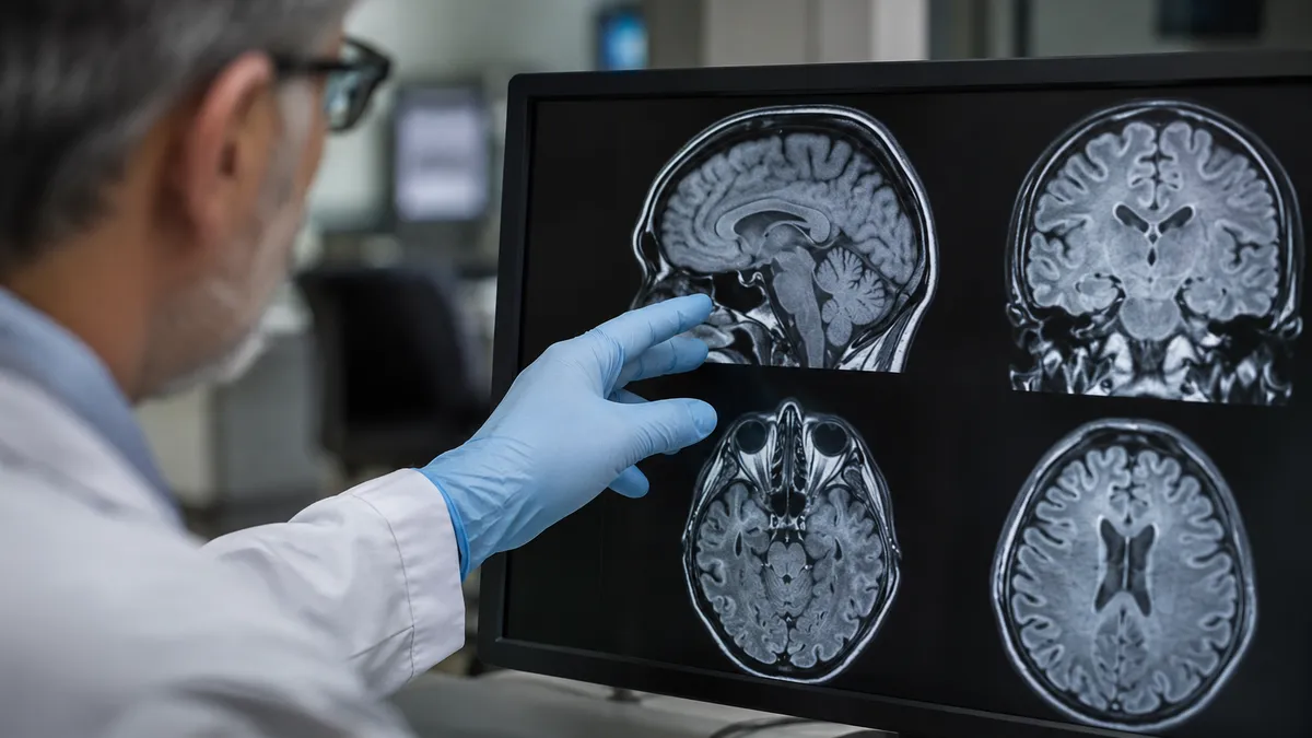
Susceptibility artifact is the main reason technologists ask about dental work. When metal sits inside the magnetic field, it changes the local field strength, and the scanner mislocates signal from nearby tissue. The result looks like a dark hole with a bright rim. Stainless steel braces typically distort an area extending several centimeters from the brackets, which is why a brain, orbit, sinus or temporomandibular joint study can become unreadable even though the wires themselves pose no real danger.
Distance is your best friend. A lumbar spine, shoulder, knee, ankle, abdominal or pelvic scan places the dental hardware outside the imaging volume, so artifact is not a concern. A cervical spine scan sits in a gray area, because the upper neck is close enough that the lowest slices may show some distortion. Radiologists often review the images immediately and decide whether anything diagnostic has been compromised before the patient leaves the table.
Field strength also plays a role. Higher fields such as 3 tesla amplify susceptibility effects, so the same set of braces may look worse at 3T than at 1.5T or on a lower-field open scanner. If you need a head or neck study and have fixed orthodontic appliances, ask whether a 1.5T system is available. Many centers keep both and will route patients with metal to the machine that gives the cleanest result for that particular body region.
Technologists can also adjust the protocol. Fast spin echo sequences resist metal artifact better than gradient echo sequences. Metal artifact reduction techniques, such as view angle tilting or slice encoding for metal correction, are available on many modern scanners. Increasing receiver bandwidth and using thinner slices further reduces distortion. These adjustments add a few minutes but can rescue an otherwise limited exam without requiring the patient to visit an orthodontist first.
Removal is occasionally discussed, but it is rarely the first choice. Orthodontists can take off archwires in a short visit, leaving the brackets in place, which cuts artifact substantially but not completely. Brackets and bands still contain metal. Because removal and replacement may cost money and interrupt treatment, most imaging teams recommend it only when a brain, face or neck study is essential and the preliminary images prove inadequate with the metal in place.
Heating is the other physical concern. Long metal loops can, in theory, concentrate radiofrequency energy and warm the surrounding tissue. Dental wires are short and not arranged as closed conductive loops of concerning size, so clinically significant heating is exceedingly rare. Patients should still report any burning, tingling or warmth during the scan immediately, using the squeeze ball or intercom, so the technologist can stop the sequence and reassess before any discomfort escalates.
Open Bore MRI Locations and Scanner Types for Patients With Dental Wire
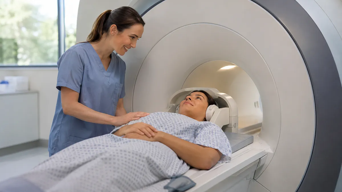
Traditional closed bore scanners use a tunnel roughly 60 cm wide and deliver the strongest magnetic fields, usually 1.5T or 3T. They produce the sharpest images and the shortest scan times for most body parts. For a patient with dental wire who needs a pelvic, abdominal, spine or extremity scan, a closed bore system is usually perfectly appropriate, because the mouth sits well outside the imaging region.
The tradeoff is comfort. The tunnel is long, the table is narrow and the machine is loud, with knocking noises reaching 100 decibels or more. Patients with claustrophobia may need earplugs, headphones, a prism mirror or mild anti-anxiety medication prescribed beforehand. Many people complete closed bore scans comfortably once they know the expected duration and can communicate with the technologist through the intercom.
Open Bore MRI vs Closed Bore MRI: Which Is Better With Dental Wire?
- +Open scanners reduce claustrophobia and anxiety for many patients
- +Lower field strength produces less metal artifact near braces
- +A family member can often stay in the room during the scan
- +Larger patients fit more comfortably on the wider table
- +Children and people with panic symptoms often avoid sedation
- +Open designs allow easier positioning for painful joints or injuries
- −Open magnets usually have lower field strength and lower image detail
- −Scan times can be longer, increasing the chance of motion blur
- −Fewer open bore MRI locations exist, so travel may be required
- −Some advanced exams are not available on low-field systems
- −Small lesions may be harder to see than on a 1.5T or 3T scanner
- −Insurance or referral rules may limit which facility you can use
Closest Open MRI to My Location: Pre-Scan Checklist for Dental Patients
- ✓Tell the scheduler about braces, wires, implants and retainers when booking.
- ✓Ask whether the scanner is closed bore, wide bore or true open bore.
- ✓Confirm the scanner's field strength, such as 0.3T, 1.5T or 3T.
- ✓Find out whether your body part is far enough from your dental metal.
- ✓Bring your orthodontist's name, phone number and any implant cards.
- ✓Remove removable retainers, dentures and night guards before arriving.
- ✓Complete the safety screening form honestly, including past surgeries.
- ✓Ask whether magnetic dental attachments are present in any appliances.
- ✓Request earplugs or headphones and ask about a prism mirror or music.
- ✓Report heat, pain or tugging in your mouth immediately during the scan.
Safety and image quality are two different questions
Almost all dental wire is safe inside an MRI scanner, but it can blur head and neck images. If your scan is of the pelvis, abdomen, spine or limbs, your braces are effectively invisible to the study. Always disclose every dental device and let the imaging team decide.
Many patients searching for dental safety information are also preparing for a pelvic study and want to understand the paperwork. In the United States, a pelvic MRI CPT code identifies the exam for billing and insurance authorization. Code 72195 describes MRI of the pelvis without contrast material. Code 72196 describes the study performed with contrast, and code 72197 describes imaging performed without contrast, followed by contrast and further sequences. The code depends on how the exam is performed, not on which condition is suspected.
The listing MRI pelvis w wo gyn CPT usually appears when an order is written for a gynecologic indication, such as an ovarian mass, fibroids, endometriosis, adenomyosis or suspected uterine abnormality. The phrase w/wo means with and without contrast, which maps to 72197. The gyn label tells the radiologist which protocol to use, including high-resolution T2 images of the uterus and ovaries, but it does not create a separate CPT code on its own.
Prior authorization is where mistakes happen. Insurers often require the exact CPT code at scheduling, and a mismatch between the ordered code and the performed study can trigger a denial or a billing correction. If your physician orders 72195 and the radiologist decides contrast is needed, the facility may need to obtain new approval before proceeding. Ask the scheduling office to verify the code, the facility and the contrast plan before your appointment date arrives.
Your dental wire has no effect on the CPT code, and it rarely affects pelvic image quality. The pelvis lies far below the mouth, so braces, crowns and implants cannot create artifact in that region. The screening form still asks about every implanted or attached device, because the same checklist applies to every MRI. Answering completely protects you if a different body part is added to the order later, such as a lumbar spine study.
Costs vary widely by facility and insurance plan. Hospital outpatient departments typically bill more than freestanding imaging centers for the same CPT code, and patients with high deductibles may notice a large difference. Asking for the facility's cash price or a good-faith estimate is reasonable. Contrast adds a separate supply charge and sometimes a separate administration line, and these items can raise the total even when the base code looks identical.
Contrast deserves a brief safety note. Gadolinium-based agents are used for the with-contrast portion of 72196 and 72197. They are generally well tolerated, but your care team will check kidney function when risk factors exist, and they will ask about pregnancy and prior reactions. Dental work does not interact with gadolinium. If you are or may be pregnant, tell the technologist early, because gynecologic MRI is often performed without contrast in that situation.
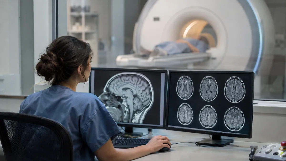
Rare-earth magnets in dentures, retainers or facial prostheses can be pulled, can heat, and can ruin image quality. Do not assume your dental device is safe. Tell the technologist before entering the scanner room, and bring written documentation whenever possible.
Choosing the right scanner begins with the body part and the clinical question. For a pelvic, abdominal or musculoskeletal exam, high-field closed or wide bore systems usually offer the best detail and speed, and dental wire is not a limitation. For brain, orbit, skull base or neck studies, the presence of braces may push the decision toward a lower-field scanner or a protocol with metal artifact reduction. Your ordering physician and the radiology team weigh these factors together.
Comfort should be weighed honestly. A poor-quality scan caused by motion is often worse than a slightly lower-field image captured while the patient is calm and still. If you have claustrophobia, say so when booking, because facilities can arrange a wide bore scanner, an open scanner, a mirror system or a visit to preview the room. Some patients benefit from a short practice session lying in a mock scanner before the real exam.
Finding the right facility takes a few phone calls. When looking up open bore MRI locations, start with your insurance network directory, your physician's referral list and the websites of large imaging chains. Search phrases such as closest open MRI to my location can help, but map listings are often outdated or label wide bore units as open. Always call to confirm the scanner type, field strength and the specific exam you need.
Ask pointed questions during that call. What is the bore diameter? What is the maximum table weight? Has the center scanned patients with fixed orthodontic braces before? Do they have metal artifact reduction software? How soon can they schedule the exam, and will the radiologist review images before you leave? Facilities that answer confidently and specifically are usually better prepared for patients with dental wire than those that give vague reassurances.
Travel distance matters more than people expect. A true open scanner 40 miles away may deliver a worse experience than a wide bore scanner five miles away, once parking, waiting and anxiety are factored in. Many patients find that a calm, well-prepared appointment on a wide bore 1.5T system provides the best balance of image quality and comfort. Lower-field open systems are best reserved for cases where closed designs truly are not tolerated.
Finally, remember that insurance and referral rules shape your options. Some plans require imaging at specific in-network sites, and prior authorization is tied to the facility as well as the CPT code. If an open scanner is medically necessary because of severe claustrophobia, ask your physician to document that need in the order. A clear note can support an exception request and shorten the time between ordering and actually completing the scan.
Start preparing at least a week ahead. Call the imaging center, describe every dental device in plain language, and ask whether anything requires documentation. If you have braces, mention the type, whether they are metal or ceramic, and whether a lingual wire is bonded behind your teeth. Staff would rather hear too much than too little, and early notice gives them time to choose the right scanner and protocol for your exam.
Gather paperwork before the appointment. Request a brief note or implant card from your orthodontist or oral surgeon that lists the device materials, manufacturer and dates. Carry a government-issued ID, your insurance card, the physician's order showing the CPT code, and a list of medications and allergies. Having these items ready speeds up check-in and screening, which reduces the waiting time that often fuels anxiety before the scan begins.
On the day of the scan, remove everything that can come out. Take out removable retainers, dentures, partial plates, night guards and any jewelry or piercings, including tongue and lip studs. Leave hairpins, hearing aids and wallets in a locker. Wear comfortable clothing without metal zippers or fasteners, or change into a gown. Most facilities provide a locker and a key, so you can keep valuables nearby but outside the scanner room.
During the exam, stay as still as possible. Motion degrades images much more than most dental artifact does. Breathe normally, follow any breath-hold instructions, and use the squeeze ball if you feel discomfort, heat or a pulling sensation in your mouth. Technologists can pause the sequence immediately. Many people find that focusing on slow breathing, counting the rhythm of the scanner noise, or listening to music through headphones makes a thirty to sixty minute exam pass quickly.
After the scan, ask when results will be available and who will review them with you. The radiologist sends a report to the ordering physician, usually within one to three business days, though urgent exams are read sooner. If the report mentions dental artifact limiting a portion of the study, ask whether a repeat scan, a different protocol or an alternative test such as CT is appropriate. Do not assume the exam failed without discussing it.
Finally, keep a personal record. Save the CPT code, the facility name, the scanner type and the date of your exam in your phone or health portal. If you need follow-up imaging, you can request the same scanner and protocol, which makes comparison easier for your doctor. Update your records whenever your dental work changes, including new braces, implants or retainers, so future screening forms are fast, accurate and complete.
MRI Questions and Answers

CT vs MRI Imaging: How They Differ, When Each Is Used, and What to Expect

MRI Prep: Complete Patient Guide to Preparing for Your MRI Scan

MRI Preparation Guide: How to Prep for Your Scan Step by Step

Travel MRI Tech 2026: Salary, Requirements & ARRT MRI Exam
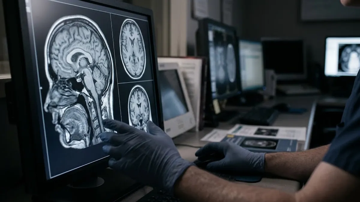
Dangers of MRI Machines: Safety Risks, Side Effects, Pelvic MRI CPT Codes, and Open Bore Options
How this page was made: written with AI tools and published by Nathan Beja, founder of Practice Test Geeks, who is responsible for its content. Exam facts are checked against the official sources listed below. Not reviewed by a subject-matter expert. Editorial policy
Sources & references
Exam facts on this page (fees, format, dates, passing scores) are checked against these official sources; the date is our last check. Editorial policy
- assets-us-01.kc-usercontent.com — Renewal, score validity, eligibility, exam format, retake policy, test dates, time limit · verified Nov 2026
- arrt.org — Fees, passing score, score release, exam information · verified Nov 2026
- home.pearsonvue.com — Test delivery · verified Nov 2026