
Can a Spine MRI Detect Lung Cancer? MRI Capabilities and Limitations Explained
Can a spine MRI detect lung cancer? Learn what spine MRI shows, 🟢 its lung imaging limitations, incidental findings, and when CT is the better choice.
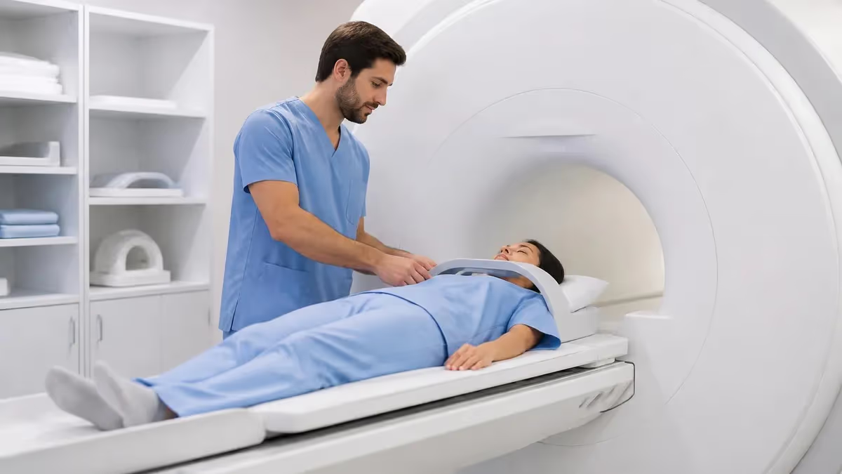
The question of whether can a spine MRI detect lung cancer comes up surprisingly often in radiology reading rooms and pulmonology consults. Patients scheduled for thoracic or lumbar spine MRI for back pain occasionally have lung lesions discovered incidentally on the same study. While spine MRI is not designed to evaluate lung tissue, the field of view often captures portions of the lungs, mediastinum, and chest wall. Understanding what spine MRI can and cannot show about the lungs is essential for both clinicians and patients navigating ambiguous imaging findings.
MRI excels at characterizing soft tissue contrast, making it the gold standard for evaluating spinal cord pathology, intervertebral discs, marrow edema, and paraspinal soft tissues. However, the physics of MRI imposes real limitations when imaging air-filled structures like the lungs. The lack of mobile protons in aerated lung parenchyma, combined with susceptibility artifacts at air-tissue interfaces and motion from breathing and cardiac pulsation, makes lung tissue notoriously difficult to image with conventional MRI sequences used for spine protocols.
Despite these limitations, spine MRI sometimes reveals lung pathology through indirect signs. Pleural-based masses, large peripheral lung tumors, mediastinal lymphadenopathy, and bone metastases from primary lung cancers can all become visible on properly windowed spine MRI images. Radiologists trained to systematically review the entire field of view often catch these incidental findings before symptoms develop. This makes the spine MRI a sometimes-unintentional gateway to early lung cancer diagnosis.
That said, no responsible radiologist would recommend a spine MRI as a screening tool for lung cancer. Low-dose CT chest screening remains the evidence-based modality for high-risk smokers, and dedicated chest imaging with CT or PET-CT provides far superior sensitivity for pulmonary nodules under 1 cm. The role of spine MRI in lung cancer detection is opportunistic rather than primary. Knowing the difference protects patients from false reassurance and unnecessary workups alike.
For technologists and radiologists preparing for boards, understanding what falls inside and outside the diagnostic envelope of each MRI protocol is foundational knowledge. The MRI with and without contrast distinction matters here too, since contrast enhancement patterns can change how visible an incidental lung mass becomes on a spine study. Recognizing the boundary between intended diagnostic targets and incidental findings shapes how radiologists report and recommend follow-up.
This article walks through the physics, anatomy, and clinical decision-making behind the question. We will look at exactly what portions of the lungs appear on cervical, thoracic, and lumbar spine MRI, what types of lung cancer can be visible, why MRI is poor at detecting small nodules, and when a finding on spine MRI should trigger a dedicated chest workup. Whether you are a patient with an incidental finding, a tech setting up a study, or a student preparing for the registry, this guide clarifies the real capabilities and limitations.
Throughout, we will return to the underlying principle that MRI is a problem-solving tool with strengths and weaknesses defined by hardware, sequences, and patient anatomy. No imaging modality is universally superior. The art of clinical radiology lies in matching the right test to the right question, and understanding when a spine MRI happens to answer a question it was never asked to address.
MRI Lung Detection by the Numbers
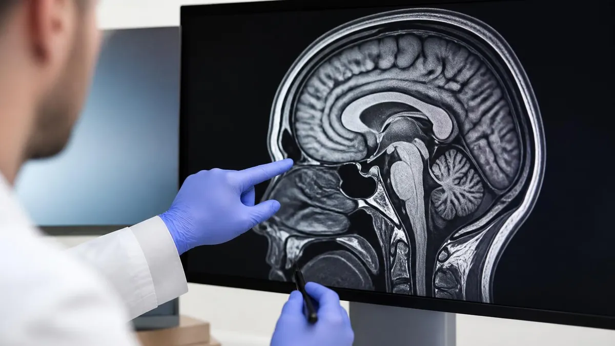
What Spine MRI Actually Shows
Spine MRI is exceptional at visualizing bone marrow signal changes, including edema, fatty replacement, infiltration by metastatic disease, and primary marrow disorders. Subtle marrow changes invisible on CT or radiography appear clearly on T1 and STIR sequences.
Direct visualization of the spinal cord, cauda equina, and exiting nerve roots is unmatched by any other modality. Myelomalacia, cord edema, syringomyelia, and nerve root impingement appear with excellent detail on dedicated cord sequences.
Disc hydration, height, herniation, and annular tears are clearly characterized on T2-weighted imaging. Modic endplate changes and discitis can be identified before structural collapse becomes visible on radiographs or CT.
Muscles, ligaments, epidural fat, and paraspinal soft tissue masses are well evaluated. This is where incidental findings of the lung apex, pleura, or chest wall sometimes appear on the periphery of the field of view.
Depending on coil placement and protocol, portions of the lungs, mediastinum, kidneys, liver, and adrenal glands may be partially imaged. These are not the target of the study but warrant systematic review.
Lung visibility on spine MRI depends heavily on which region of the spine is being imaged. Cervical spine MRI captures the lung apices, where Pancoast tumors classically develop in the superior sulcus. These tumors can invade the brachial plexus and vertebral bodies, producing shoulder and arm pain that often masquerades as cervical radiculopathy. A cervical spine MRI ordered for arm pain has a real chance of catching a Pancoast tumor before it is recognized as primary lung cancer, making careful review of the apical lung essential.
Thoracic spine MRI offers the most generous view of the lungs, with roughly 30 to 40 percent of the lung parenchyma falling within the field of view depending on coil size and patient body habitus. Peripheral and pleural-based lesions in the posterior segments are most likely to be detected because they sit closest to the vertebral column. Central lung tumors near the hila, however, often fall outside the imaging volume or are degraded by motion and susceptibility artifact from adjacent air-filled lung.
Lumbar spine MRI provides limited lung coverage, typically only capturing the lung bases through the costophrenic angles. Lower lobe peripheral lesions and pleural effusions can sometimes be seen here, but the diagnostic yield for lung cancer detection from lumbar studies is minimal. More commonly, lumbar MRI catches abdominal or pelvic incidental findings rather than thoracic pathology, including renal masses, adrenal lesions, and abdominal aortic aneurysms.
The fundamental physics challenge is that aerated lung parenchyma contains very few mobile protons compared to soft tissue. Hydrogen nuclei in air-filled alveoli generate minimal MRI signal, so normal lung appears nearly black on most spine sequences. This low baseline signal makes small soft tissue masses theoretically easier to detect against the dark background, but susceptibility artifacts at the millions of air-tissue interfaces in lung tissue create distortion and signal loss that obscure small lesions.
Motion is another significant barrier. Spine MRI sequences are typically not gated to respiration or cardiac cycles because the spine itself moves minimally during imaging. The lungs, however, move with every breath and the heart pulsates continuously, blurring lung structures across multiple slice positions. Without dedicated respiratory triggering or breath-hold sequences used in dedicated chest mri cpt code, small nodules can be smeared into invisibility by physiological motion artifacts.
The common MRI findings radiologists encounter on routine spine studies include incidental nodules, pleural thickening, and lymphadenopathy that fall on the edge between definite pathology and indeterminate findings requiring follow-up. Radiologists are trained to comment on these incidental observations and recommend dedicated chest CT when warranted, even when the spine itself appears normal. This safety net catches a meaningful number of early lung cancers each year.
Patient body habitus also influences lung visualization. Thin patients with less subcutaneous fat have more compact thoracic anatomy, often resulting in better lung coverage on spine sequences. Larger patients may have more lung tissue technically within the field of view but with more signal-to-noise compromises that reduce diagnostic confidence. Coil selection, with phased-array surface coils being standard for spine work, also dictates how much lateral lung tissue receives adequate signal coverage.
MRI Practice Test Questions
Prepare for the MRI - Magnetic Resonance Imaging exam with our free practice test modules. Each quiz covers key topics to help you pass on your first try.
MRI Knowledge
MRI Exam Questions covering Knowledge. Master MRI Test concepts for certification prep.
MRI Physics
Free MRI Practice Test featuring Physics. Improve your MRI Exam score with mock test prep.
MRI Anatomy and Pathology
MRI Test Prep for MRI Anatomy and Pathology. Practice MRI Quiz questions and boost your score.
MRI Anatomy and Positioning
MRI Questions and Answers on MRI Anatomy and Positioning. Free MRI practice for exam readiness.
MRI Contrast Agents
Free MRI Quiz on MRI Contrast Agents. MRI Exam prep questions with detailed explanations.
MRI Patient Care and Positioning
MRI Practice Questions for MRI Patient Care and Positioning. Build confidence for your MRI certification exam.
MRI vs CT for Lung Cancer Evaluation
CT chest is dramatically more sensitive than MRI for detecting small pulmonary nodules. A modern multidetector CT can resolve nodules as small as 2 to 3 millimeters in diameter, while MRI typically struggles below 8 to 10 millimeters even on dedicated lung sequences. For low-dose lung cancer screening in high-risk smokers, CT remains the only validated modality, with strong evidence from the National Lung Screening Trial supporting its use.
MRI does have a few areas where sensitivity approaches or exceeds CT. Evaluation of chest wall invasion by peripheral tumors, brachial plexus involvement in Pancoast tumors, and characterization of mediastinal cystic lesions are all situations where MRI may add information beyond CT. Bone marrow involvement from metastatic disease is also detected earlier on MRI than on CT in most cases.
Using Spine MRI for Lung Evaluation: Pros and Cons
- +No ionizing radiation exposure during imaging
- +Excellent detection of vertebral bone metastases from lung primaries
- +Superior characterization of Pancoast tumor extension into brachial plexus
- +Can identify pleural-based masses larger than 1 cm in field of view
- +Detects spinal cord compression from epidural tumor extension
- +Useful problem-solving tool for ambiguous CT findings near the spine
- +Captures incidental large lung masses without additional patient burden
- −Poor sensitivity for small pulmonary nodules under 8 millimeters
- −Limited lung field of view, especially on cervical and lumbar studies
- −Susceptibility artifacts at air-tissue interfaces degrade image quality
- −Respiratory and cardiac motion blur small lung structures
- −Not validated for lung cancer screening in any clinical guidelines
- −Central hilar tumors often fall outside spine MRI coverage
- −Reading focus is on spine, increasing risk of missed lung findings
Incidental Lung Finding Workup Checklist
- ✓Confirm the finding is real on multiple sequences and planes, not artifact
- ✓Measure the lesion in three dimensions using calipers on the workstation
- ✓Compare to any prior chest imaging available in the patient record
- ✓Document lesion location by lobe, segment, and proximity to pleura
- ✓Note any associated lymphadenopathy in the mediastinum or hila
- ✓Check for vertebral marrow abnormalities suggesting metastatic disease
- ✓Recommend dedicated chest CT for any solid nodule larger than 8 millimeters
- ✓Apply Fleischner Society criteria for incidental nodule follow-up
- ✓Communicate urgent findings directly to the ordering clinician same day
- ✓Document the recommendation and follow-up plan clearly in the report
Spine MRI is a Witness, Not a Detective
Spine MRI is optimized for spinal pathology and only opportunistically captures lung tissue. When a lung finding appears, it should trigger dedicated chest imaging rather than serving as the final diagnostic word. Up to 30 percent of lung cancers visible on spine MRI have associated bone metastases at presentation, making the spine study sometimes the first hint of disseminated disease.
Common pitfalls in interpreting spine MRI for lung findings start with the simple problem of tunnel vision. Radiologists focused on a back pain workup may dictate a thorough spine report while glossing over the lung apex or peripheral lung tissue at the edges of the image. Systematic search patterns that explicitly include peripheral structures help mitigate this, but time pressures and high study volumes mean that subtle lung abnormalities can be missed. Building lung review into every spine MRI dictation template is one effective countermeasure.
Pulsation artifacts from the heart and great vessels can mimic mass lesions on certain sequences, particularly in the posterior mediastinum and paraspinal region. These ghosting artifacts repeat at predictable intervals in the phase-encoding direction and can be distinguished from real masses by their geometric pattern. Recognizing motion ghosting is a core skill for any radiologist reading thoracic spine studies, because real lung lesions and motion artifacts can occupy similar locations on the image.
Susceptibility artifacts at the lung-soft tissue interface can also obscure small posterior lung lesions. Gradient echo sequences are particularly prone to this, while turbo spin echo sequences used in standard spine protocols are more forgiving. Knowing which sequences in your protocol are most diagnostic for lung tissue helps focus the review. T2-weighted fat-saturated sequences often provide the best contrast for distinguishing soft tissue masses from adjacent normal lung and pleura.
Another pitfall involves mistaking benign findings for malignancy. Atelectasis in dependent lung regions can appear as a soft tissue density on MRI, and pleural plaques from prior asbestos exposure can resemble pleural masses. Calcified granulomas appear dark on all sequences and can be mistaken for missing tissue or artifact. Familiarity with these benign mimics prevents unnecessary workups that consume resources and cause patient anxiety, while still capturing genuinely concerning findings.
Lymph node interpretation requires particular care on spine MRI. Mediastinal and hilar nodes that fall within the field of view can be measured, but the lack of dedicated chest sequences means that subtle pathologic enlargement may be missed. Most radiologists apply a threshold of 10 millimeters in short axis as a general guideline for abnormal node size, but this threshold has poor specificity and any concerning node should prompt dedicated chest evaluation rather than definitive characterization on the spine study.
Contrast enhancement patterns add another layer of complexity. When spine MRI is performed with gadolinium for suspected infection, tumor, or postoperative evaluation, any incidental lung lesion will also enhance to some degree. Avid arterial enhancement raises concern for hypervascular metastases or primary lung tumors, while peripheral enhancement with central necrosis suggests aggressive malignancy. However, definitive characterization always requires dedicated chest imaging because contrast timing and sequences are not optimized for the lungs.
Communication of incidental findings is itself a pitfall area. Even when a radiologist correctly identifies a suspicious lung lesion on spine MRI, the finding must reach the ordering clinician promptly and be clearly actionable. Burying a recommendation for chest CT in the middle of a long spine report risks the finding being missed by busy clinicians. Best practice is to flag actionable incidental findings at the top of the report and communicate directly for urgent concerns.
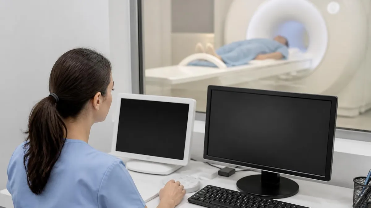
Any new soft tissue mass greater than 1 cm in the lungs on spine MRI, especially with associated lymphadenopathy or vertebral marrow changes, warrants direct communication with the ordering clinician and prompt dedicated chest CT. Do not wait for routine follow-up. Documented communication within 24 hours is the standard of care for actionable incidental findings.
Clinical decision pathways for incidental lung findings on spine MRI follow established frameworks that balance early detection with avoiding overdiagnosis. The Fleischner Society guidelines for incidental pulmonary nodules provide the foundation, though they assume CT-based detection. When MRI catches a lung lesion, the first step is almost always confirming the finding on dedicated chest CT before applying nodule management algorithms. This sequential approach prevents acting on artifacts and ensures accurate measurement.
For high-risk patients with smoking history, occupational exposure, or known primary malignancies, the threshold to recommend dedicated chest imaging is appropriately lower. A 5 millimeter nodule identified on spine MRI in a patient with breast cancer history warrants chest CT even though Fleischner guidelines would defer follow-up for a similar nodule in a low-risk patient. Risk stratification drives the urgency and intensity of follow-up recommendations more than the absolute imaging characteristics in many cases.
Multidisciplinary tumor boards play an important role when spine MRI findings suggest both spinal pathology and lung involvement. Patients with epidural disease and a lung mass on the same study may require coordinated workup involving pulmonology, oncology, radiation oncology, and spine surgery. Centralized review of complex incidental findings ensures that no contributor to the patient's presentation is overlooked and that staging is completed efficiently. Looking into your MRI scan near me options early can speed the workup when access to specialists is critical.
The biopsy decision pathway depends on accessibility and clinical urgency. Peripheral lung lesions are often amenable to CT-guided percutaneous biopsy, while central lesions may require bronchoscopic approaches or surgical biopsy. When vertebral metastases are also present, vertebral biopsy under fluoroscopic or CT guidance may provide tissue diagnosis with potentially lower procedural risk than thoracic biopsy. Coordinating biopsy approach across involved sites optimizes both diagnostic yield and patient safety.
Staging workup typically expands to include PET-CT after tissue diagnosis confirms lung cancer. PET-CT identifies additional sites of disease not visible on the original spine MRI or follow-up chest CT, including distant lymph node involvement, contralateral lung disease, and unsuspected bone or visceral metastases. Brain MRI is also typically added for staging because lung cancer commonly metastasizes to brain and CT is inadequate for early metastatic detection.
Treatment planning then integrates findings from all imaging modalities. Radiation oncology relies heavily on the original spine MRI to define spinal cord tolerance and target volumes when treating vertebral metastases. Medical oncology uses the full imaging picture to assign disease stage and select systemic therapy regimens. Surveillance imaging after treatment typically uses a combination of CT and MRI, with MRI prioritized when vertebral or cord involvement was part of the original presentation.
Patient communication about incidental findings deserves careful thought. Receiving a phone call about a possible lung mass found on a back pain study is jarring for patients. Clear explanations of why the finding was noticed, what additional imaging is needed, and the timeline for resolution help reduce anxiety and improve adherence to follow-up. The radiologist's role often extends beyond the report into supporting clinician-patient conversations about ambiguous findings and next steps.
Practical guidance for technologists begins with protocol optimization. When patients have known or suspected thoracic pathology and need spine MRI, adjusting the field of view to capture more lung tissue can pay dividends. Coordinating with the radiologist before the patient is on the table allows tailored sequences that better cover the chest, particularly for cervicothoracic studies in patients with shoulder or arm pain that might represent Pancoast tumors. Small protocol adjustments produce meaningfully better lung visualization without compromising the spine evaluation.
Patient positioning and breathing instructions also influence image quality. Standard spine MRI does not use breath-holding, but instructing patients to breathe quietly and shallowly reduces motion artifacts that smear lung structures. For patients who cannot lie still or who have severe pain, sedation protocols or alternative positioning may improve diagnostic quality. Documenting any motion or positioning compromises in the technologist notes helps the radiologist interpret limitations appropriately.
Coil selection deserves attention when lung evaluation is a secondary goal. The standard phased-array spine coil provides adequate coverage of paraspinal structures and pleural surfaces but may not optimally image lateral lung tissue. When clinical context suggests lung pathology should be specifically evaluated, switching to a body coil or adding a chest coil can improve coverage. These choices balance technical considerations against scan time and patient tolerance.
For students preparing for the ARRT MRI registry exam, the principles in this article appear in multiple question categories. Image production questions cover the physics of why lung tissue produces minimal signal. Procedure questions ask about appropriate protocols for various clinical scenarios. Patient care questions probe how to handle incidental findings and communicate concerns. Understanding the intersection of physics, anatomy, and clinical decision-making prepares candidates for the integrative questions that distinguish high scorers.
Continuing education for practicing technologists should include refreshers on incidental finding recognition. Many institutions now offer structured curricula that train technologists to flag concerning findings to radiologists in real time. This is not about technologists making diagnoses, but rather about ensuring that clinically important findings are not overlooked during the brief radiologist review of high-volume studies. The technologist-radiologist partnership improves patient outcomes when properly supported.
Quality assurance programs should specifically audit incidental finding reporting rates and follow-up compliance. Tracking how often spine MRI studies generate recommendations for chest CT, and what proportion of those recommendations are completed within recommended timeframes, identifies gaps in care coordination. Closing these gaps requires collaboration between radiology, primary care, and specialty services. Many missed cancers stem not from missed findings but from incomplete follow-up on recognized findings.
Finally, patients themselves benefit from understanding what their imaging can and cannot show. A patient told that their spine MRI was normal may incorrectly assume that lung cancer has been ruled out. Clear communication that spine MRI is not designed to evaluate the lungs, and that lung cancer screening requires different testing in eligible patients, prevents false reassurance. Empowered patients ask better questions and advocate more effectively for appropriate care when symptoms persist or new concerns arise.
MRI Questions and Answers
About the Author

Medical Laboratory Scientist & Clinical Certification Expert
Johns Hopkins UniversityDr. Sandra Kim holds a PhD in Clinical Laboratory Science from Johns Hopkins University and is certified as a Medical Technologist (MT) and Medical Laboratory Scientist (MLS) through ASCP. With 16 years of clinical laboratory experience spanning hematology, microbiology, and molecular diagnostics, she prepares candidates for ASCP board exams, MLT, MLS, and specialist certification tests.
Join the Discussion
Connect with other students preparing for this exam. Share tips, ask questions, and get advice from people who have been there.
View discussion (8 replies)