
How to Read a Brain MRI: Sequences, Anatomy, Open MRI Machine Options and CPT Codes
🧠 Learn how to read a brain MRI: T1, T2, FLAIR and DWI sequences, normal anatomy, red flags, plus open MRI machine options and CPT codes.
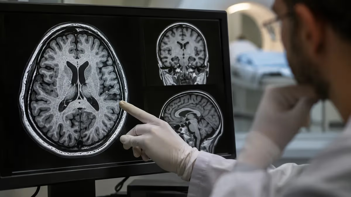
Learning how to read a brain MRI starts with understanding what you are actually looking at: a stack of thin grayscale slices, each built from radio signals emitted by hydrogen atoms in your tissue. Whether you are a student, a technologist preparing for registry exams, or a patient curious about your report, the logic is the same. This guide explains the sequences, the anatomy, and the patterns that matter, and it covers the open MRI machine options many people ask about.
A magnetic resonance image is not a photograph. Brightness reflects how quickly hydrogen protons in water and fat release energy after a radiofrequency pulse, and scanners vary that timing on purpose. Each timing recipe is called a sequence, and each sequence makes different tissues look bright or dark. Reading a scan therefore means asking two questions at once: which sequence am I looking at, and what should normal tissue look like on it?
Many readers arrive here comparing modalities first. If you want the bigger picture, our guide on how to read an mri alongside CT explains why MRI shows soft tissue so well while CT wins for fresh bleeding and bone. For the brain, MRI is usually the preferred test for tumors, multiple sclerosis, stroke, infection, and structural causes of seizures, because contrast between gray matter, white matter, and fluid is far richer than on CT.
Most people begin with the three planes. Axial slices cut horizontally, as if you were looking up from the patient's feet. Coronal slices cut front to back, like looking at the face. Sagittal slices cut left to right and show the midline structures, including the corpus callosum, brainstem, and cerebellum. A radiologist scrolls through all three planes because a lesion that hides in one view often stands out in another.
Orientation conventions matter, and they trip up beginners. On axial images the patient's right appears on the left side of the screen, the same convention used in CT. Marker letters or an annotation usually confirm this. A quick sanity check is the liver on abdominal studies, but on a brain scan you can use a known asymmetry, such as a surgical clip or the venous sinus pattern, to verify you are not reading the image backward.
Equally important is knowing the limits of self-interpretation. A radiologist spends four years of residency plus fellowship training learning to separate normal variants from disease, and even then uses clinical history, prior studies, and comparison across many sequences. Think of this article as a literacy guide. It teaches you the vocabulary so you can follow a report, study for an exam, or ask better questions at your appointment.
Along the way we will also answer practical questions that cluster around brain imaging: what an open MRI machine is, who benefits from one, how to find open bore MRI locations, and which billing codes apply, including pelvic MRI CPT code families that often appear in the same conversations. Use the table of contents to jump to whichever section fits your goal, then test yourself with the free practice questions linked throughout.
Brain MRI by the Numbers
How to Read a Brain MRI Step by Step
Confirm the Patient and Study
Identify the Sequence
Check Symmetry and Midline
Review Gray and White Matter
Add Diffusion and Contrast
The T1-weighted sequence is the anatomy map. Fat and subacute blood appear bright, white matter looks lighter than gray matter, and cerebrospinal fluid is dark. Because gadolinium contrast shortens T1 times, enhancing tissue lights up on T1 images after injection. If you see a clean, sharply detailed brain with black ventricles, you are very likely viewing T1, and any enhancing ring or nodule after contrast deserves careful attention.
The T2-weighted sequence flips the fluid appearance. Cerebrospinal fluid is brilliant white, gray matter is brighter than white matter, and most pathology, including edema, tumors, and inflammation, appears bright because it contains extra water. T2 is excellent for spotting disease but poor at telling where it ends, since normal fluid in the ventricles and sulci can hide lesions that sit right next to it.
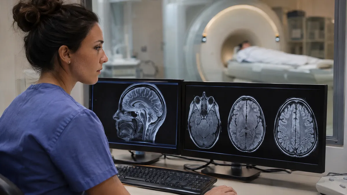
FLAIR solves that problem. Fluid-attenuated inversion recovery is essentially a T2 image with the cerebrospinal fluid signal suppressed to black. Lesions near the ventricles or cortex, which would blend into bright fluid on T2, now stand out clearly. FLAIR is the workhorse for multiple sclerosis plaques, small vessel ischemic change, subarachnoid abnormalities, and cortical lesions, and it is included in nearly every routine brain protocol.
Diffusion-weighted imaging, or DWI, measures how freely water molecules move. In acute stroke, cells swell and water movement becomes restricted within minutes, producing a bright signal on DWI. To avoid confusing this with T2 shine-through, radiologists compare it with the apparent diffusion coefficient map, where true restriction appears dark. Bright DWI plus dark ADC strongly suggests acute infarction, and it can persist for roughly one to two weeks.
Susceptibility-weighted imaging and gradient echo sequences detect blood products and mineral deposits. Old microhemorrhages, cavernous malformations, and calcifications show up as small black dots because they distort the local magnetic field. These sequences are valuable after trauma, in patients with suspected amyloid angiopathy, and when evaluating hypertensive bleeding patterns. A normal brain shows only a few dark vessels and some physiologic iron in deep gray structures.
Normal anatomy gives you the baseline. The lateral ventricles should be symmetric, the third ventricle is a narrow slit at the midline, and the fourth ventricle sits in front of the cerebellum. The corpus callosum arches as a bright band on sagittal T1. The brainstem, from midbrain to pons to medulla, descends toward the foramen magnum, and the cerebellar tonsils should not extend more than about five millimeters below it.
Technologists and students often memorize signal behavior in a table: fat is bright on T1 and moderately bright on T2, water is dark on T1 and bright on T2, air and cortical bone are dark everywhere, and flowing blood is usually dark because of flow voids. Once this pattern feels automatic, every scan becomes a pattern-matching exercise rather than a guessing game, and registry-style questions become far easier to answer.
Open MRI Machine Options: Which Scanner Fits You?
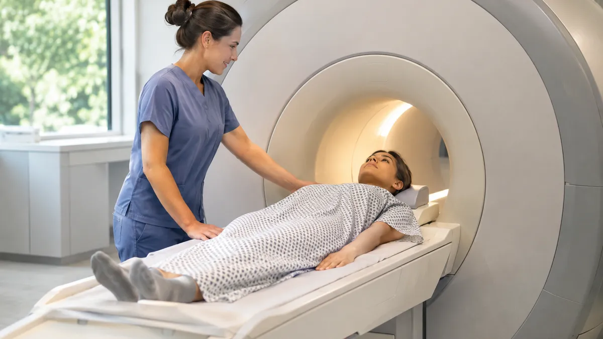
An open MRI machine uses a design that is open on the sides rather than a long enclosed tube. Traditional open scanners use a vertical magnet with a flat table between two pads, usually at a lower field strength of roughly 0.2 to 1.2 tesla. Patients who feel panic in tight spaces, who are larger-bodied, or who need a parent beside them often find this layout far more tolerable than a standard closed unit.
The trade-off is image quality and time. Lower field strength means a weaker signal, so exams often run longer and fine detail can be reduced, which matters most for small brain lesions, pituitary imaging, and advanced sequences. Many facilities steer complex neurologic questions toward high-field systems, while using open designs for joints, spines, and patients who otherwise could not complete the exam at all.
Is an Open MRI Machine Better Than a Closed Scanner?
- +Reduces claustrophobia and anxiety for many patients
- +Accommodates larger-bodied patients more comfortably
- +Allows a parent or caregiver to stay close to a child
- +Often avoids the need for sedation medication
- +Lets patients with limited mobility position more easily
- +Typically produces less intense scanner noise
- −Lower field strength reduces signal and fine detail
- −Scan times are often longer, increasing motion risk
- −Fewer advanced brain sequences may be available
- −Small lesions can be harder to detect confidently
- −Fewer facilities offer true open systems
- −Radiologists may recommend a repeat on a high-field unit
Brain MRI Review Checklist for Open MRI Machine Studies
- ✓Confirm the patient name, date, and study type.
- ✓Note whether gadolinium contrast was used.
- ✓Identify each sequence before judging brightness.
- ✓Check that the midline is straight and centered.
- ✓Compare ventricle size and symmetry side to side.
- ✓Scan FLAIR for white matter bright spots.
- ✓Review DWI and ADC together for restricted diffusion.
- ✓Look for dark blood products on SWI or gradient echo.
- ✓Inspect the posterior fossa and cerebellar tonsils.
- ✓Read the radiologist report before drawing conclusions.
Always Look at the CSF First
Cerebrospinal fluid is your built-in sequence label. Bright fluid means T2, black fluid on a bright-water image means FLAIR, and dark fluid with detailed anatomy means T1. Identify the fluid, and you instantly know what every other shade of gray should mean.
Acute ischemic stroke is the pattern most worth recognizing. On DWI, the affected territory glows bright within minutes, while the matching ADC map turns dark. FLAIR may still look normal during the first few hours, which is why a bright DWI with unremarkable FLAIR suggests a very recent event. Typical territories follow arteries, such as a wedge-shaped region in the middle cerebral artery distribution, and that wedge shape helps separate stroke from tumors.
Multiple sclerosis produces a different signature. Look for ovoid, bright FLAIR lesions oriented perpendicular to the lateral ventricles, often called Dawson fingers on sagittal images. Lesions also favor the corpus callosum, juxtacortical white matter, brainstem, and cerebellum. After gadolinium, active plaques may enhance, often in an open ring, while older lesions do not. The combination of location, shape, and enhancement pattern guides the diagnosis.
Small vessel ischemic change is extremely common with age and hypertension. Scattered bright spots appear in the periventricular and deep white matter on FLAIR, often symmetric and nonspecific. A few spots can be normal for a 65-year-old, while confluent disease raises concern for vascular cognitive impairment. Radiologists often grade severity using the Fazekas scale, which ranges from zero to three, and they interpret the score alongside patient age and symptoms.
Brain tumors usually announce themselves through mass effect. A mass pushes on adjacent structures, shifts the midline, and surrounds itself with bright vasogenic edema on T2 and FLAIR. After contrast, glioblastoma often shows an irregular rim of enhancement around a necrotic center, while meningiomas tend to enhance brightly and uniformly along the dura. Metastases frequently appear as multiple round enhancing nodules near the gray-white junction.
Hydrocephalus and volume loss look opposite but can confuse beginners. In hydrocephalus, the ventricles enlarge disproportionately, the temporal horns widen, and periventricular fluid may leak into surrounding white matter as a bright halo on FLAIR. In generalized atrophy, ventricles and sulci both widen proportionally. Normal pressure hydrocephalus shows enlarged ventricles with relatively tight high-convexity sulci, a pattern that points toward a treatable cause of gait trouble.
Hemorrhage changes appearance over time, which is a favorite exam topic. Hyperacute blood is isointense on T1 and bright on T2, acute blood turns dark on T2, early subacute blood becomes bright on T1 as methemoglobin forms, and chronic hemosiderin leaves a dark rim on SWI. Memorizing this progression helps you estimate the age of a bleed and explains why the same hematoma looks different from week to week.
Always remember normal variants and artifacts. A prominent perivascular space, a pineal cyst, or a developmental venous anomaly can look dramatic but is often benign. Motion blur, metal artifact from dental work, and CSF flow pulsation can mimic disease. Experienced radiologists weigh these possibilities daily, which is why a single bright spot on one slice should never be interpreted in isolation.
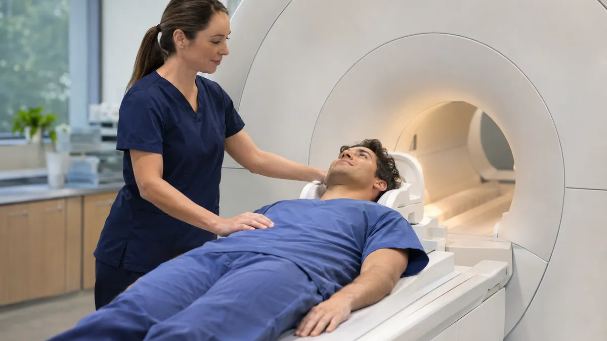
Reading a scan for study purposes is very different from diagnosing a medical condition. Only a board-certified radiologist working with your clinician can interpret your images in context. If a report mentions something unfamiliar, ask your ordering provider to walk you through it.
Billing codes confuse patients and students alike, so it helps to know the structure. Brain MRI uses three Current Procedural Terminology codes: 70551 for the study without contrast, 70552 for the study with contrast, and 70553 for the study without contrast followed by contrast. The third code is chosen when the clinician needs pre- and post-injection comparison, such as when evaluating a known mass or suspected demyelinating disease.
Pelvic imaging follows a parallel pattern. The pelvic MRI CPT code family is 72195 for the exam without contrast, 72196 for the exam with contrast, and 72197 for without followed by with. When people search for the MRI pelvis w wo gyn CPT, they are almost always looking for 72197, used for gynecologic evaluations such as fibroids, endometriosis, adnexal masses, and cancer staging where both phases are needed.
Why does this matter for a brain article? Because many outpatient centers, including those with open scanners, schedule head and pelvic studies on the same equipment, and quotes depend on the exact code. If an order is written for the wrong code, insurers may deny the claim or require a new authorization. Always confirm that the code on your order matches the exam your provider actually intends.
Prior authorization is the next hurdle. Many commercial plans and Medicare Advantage products require approval for advanced imaging, and a missing authorization can leave you responsible for the full bill. Ask the ordering office to submit clinical notes, symptoms, and prior imaging results. Approvals typically arrive within a few business days, although urgent studies for suspected stroke or tumor bypass the process entirely.
Locating a facility works best when you combine cost and equipment. Search for open bore MRI locations through your insurer's directory, then verify with a phone call. Independent imaging centers frequently charge less than hospital outpatient departments for the same code, sometimes by a factor of two or more, because hospitals add facility fees. Price transparency tools and cash-pay quotes can save hundreds of dollars on a single scan.
Equipment limits also deserve attention. Open systems have table weight limits, commonly between 350 and 550 pounds, and some cannot perform contrast-enhanced brain protocols with the same quality as 1.5 or 3 tesla units. Implants require a separate safety review, since pacemakers, certain aneurysm clips, and cochlear devices may be restricted. Our guide on MRI safety risks and open bore options covers screening in depth.
Finally, request a copy of your images on disc or through a patient portal, along with the report. Having both lets a specialist compare studies years later, which is invaluable for conditions like multiple sclerosis or slow-growing tumors. Radiologists reading a new scan against a prior one catch subtle change far more reliably than they can from a single snapshot, so keep your records organized and accessible.
Start your practice with a fixed routine rather than random scrolling. Choose one normal brain MRI, identify the sequence, trace the ventricles, find the corpus callosum, and name the major lobes. Repeat with a second normal case until the layout feels automatic. Only then add abnormal cases. Learners who build the normal baseline first recognize disease faster, because abnormality becomes a departure from a familiar pattern instead of a puzzle.
Use public teaching resources to build volume. Open radiology libraries, university teaching files, and case-based platforms provide thousands of de-identified studies with answers. Spend about twenty minutes per case: commit to your own read before checking the answer, then write down what you missed. This active recall approach produces much better retention than passively viewing finished interpretations, and it mirrors how radiology residents train.
Link every finding to its physics. If a lesion is bright on T2 and FLAIR but dark on T1, ask what that implies about water content. If DWI is bright, ask why diffusion restricts. Understanding the mechanism lets you answer unfamiliar questions on exams instead of memorizing isolated facts. Technologists preparing for the ARRT MRI exam especially benefit, since the test blends physics, safety, and anatomy into scenario-based questions.
Create a one-page cheat sheet for sequences. List T1, T2, FLAIR, DWI, ADC, SWI, and post-contrast T1, with columns for what looks bright, what looks dark, and what it is best at detecting. Review it for five minutes each morning. Spaced repetition over three or four weeks moves this information into long-term memory, and you will find yourself identifying sequences at a glance within a month.
Practice with timed questions to simulate exam pressure. Our free knowledge, physics, and registry question banks let you track weak areas, such as k-space, contrast agents, or artifact recognition. When you miss a question, read the explanation and rewrite the concept in your own words. Aim for at least eighty percent on a topic before moving on, and revisit weaker topics every few days.
If you are a patient preparing for your own scan, focus on logistics instead. Remove all metal, wear comfortable clothing, tell the technologist about implants, tattoos, and possible pregnancy, and ask about earplugs or headphones. Anxious patients can request an open or wide-bore scanner, practice slow breathing, or discuss mild sedation with their provider. Holding still is the single biggest factor in image quality. Our MRI prep patient guide walks through the full process.
Finally, stay humble about interpretation. Even experienced clinicians consult radiologists for brain studies, and second opinions are routine for complex findings. Use what you learn here to ask sharper questions, understand your report, and study efficiently. If a result worries you, contact your care team promptly. Knowledge of sequences and anatomy is empowering, but it complements professional interpretation and never replaces it.
MRI Questions and Answers

CT vs MRI Imaging: How They Differ, When Each Is Used, and What to Expect

MRI Prep: Complete Patient Guide to Preparing for Your MRI Scan

Travel MRI Tech 2026: Salary, Requirements & ARRT MRI Exam
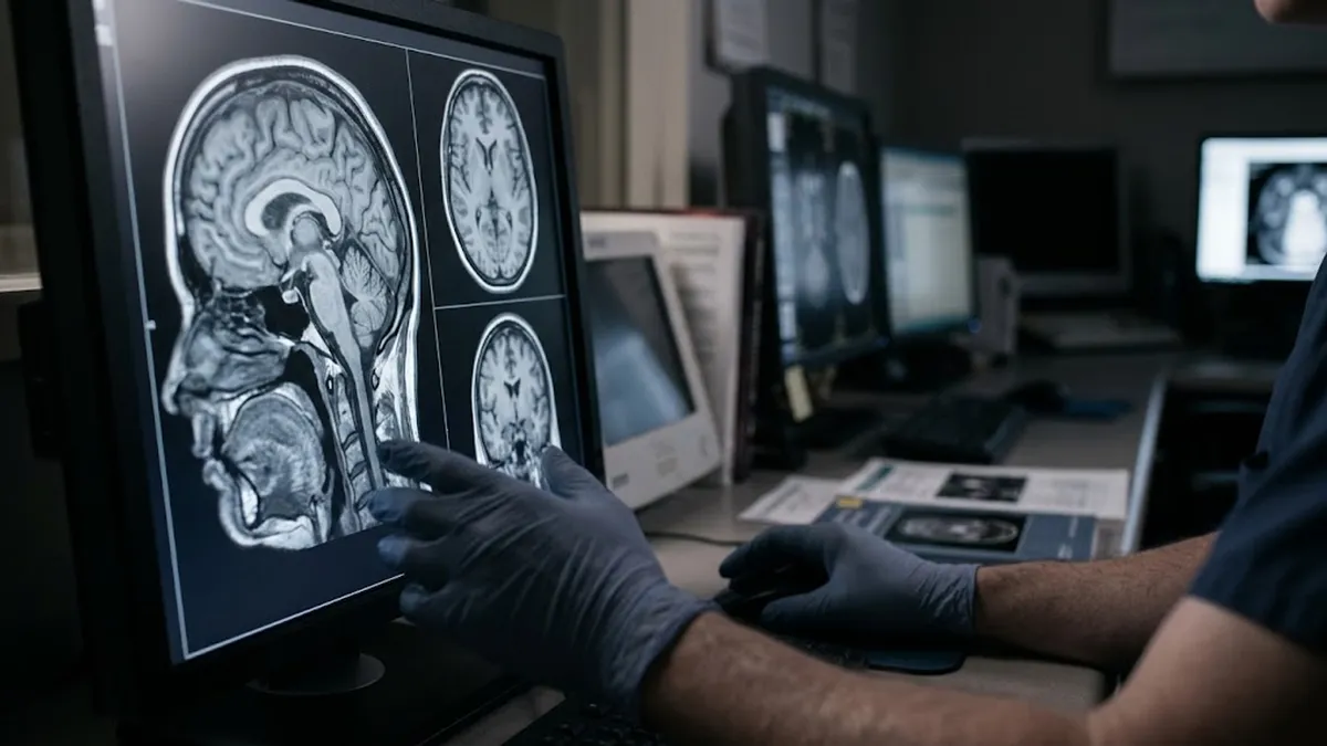
Dangers of MRI Machines: Safety Risks, Side Effects, Pelvic MRI CPT Codes, and Open Bore Options
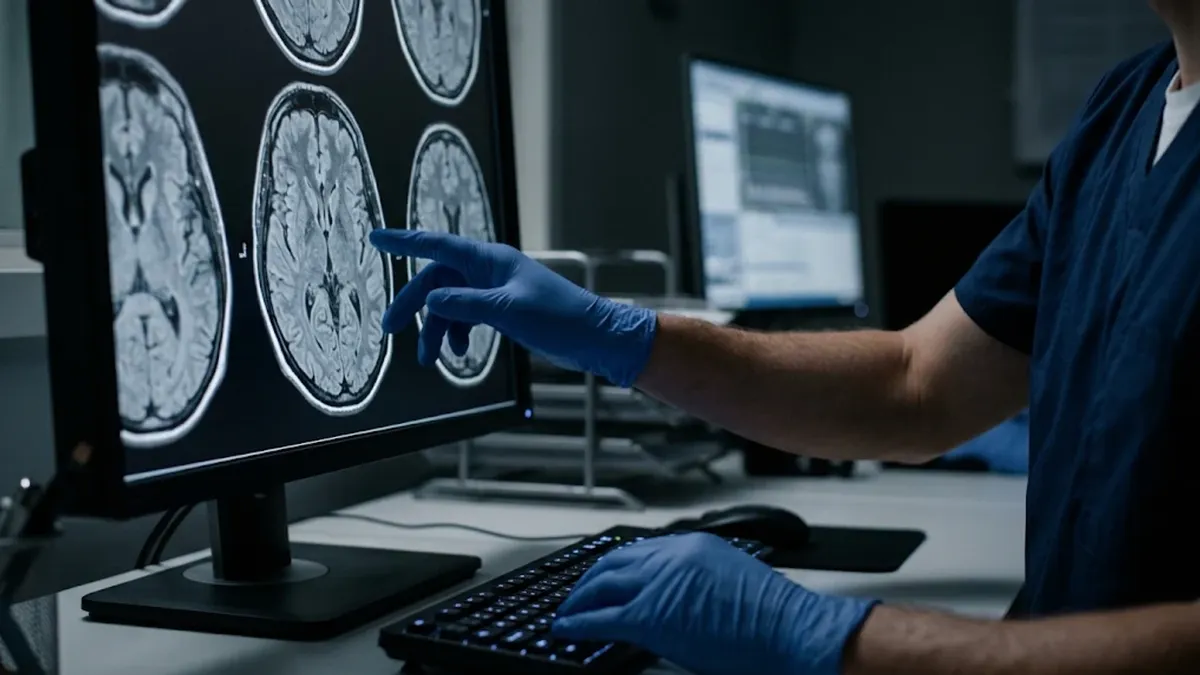
GE 1.5 MRI Machines: Models, Pelvic MRI CPT Codes, and Open Bore Options
How this page was made: written with AI tools and published by Nathan Beja, founder of Practice Test Geeks, who is responsible for its content. Exam facts are checked against the official sources listed below. Not reviewed by a subject-matter expert. Editorial policy
Sources & references
Exam facts on this page (fees, format, dates, passing scores) are checked against these official sources; the date is our last check. Editorial policy
- assets-us-01.kc-usercontent.com — Renewal, score validity, eligibility, exam format, retake policy, test dates, time limit · verified Nov 2026
- arrt.org — Fees, passing score, score release, exam information · verified Nov 2026
- home.pearsonvue.com — Test delivery · verified Nov 2026