
Can I Eat Before MRI With Contrast? Complete Patient Guide to Fasting Rules
Can I eat before MRI with contrast? ✅ Learn exact fasting rules, what to avoid, and how to prep for a contrast MRI safely.
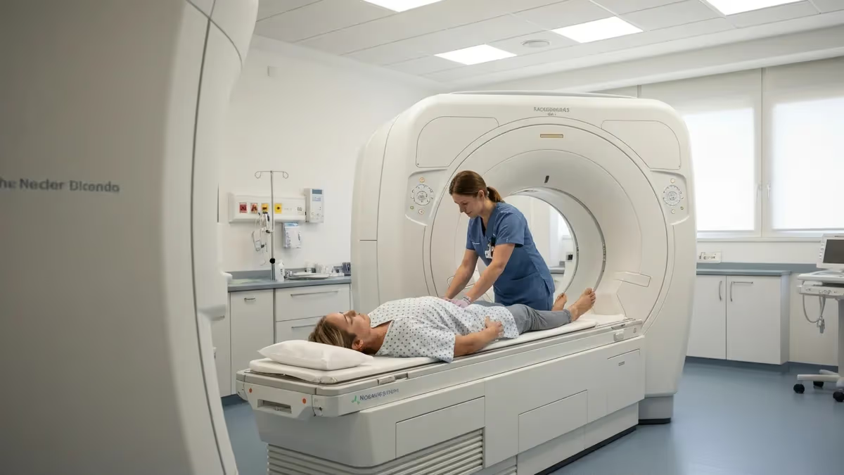
If you have a contrast MRI scheduled, one of the first questions you will ask your imaging center is: can I eat before MRI with contrast? The short answer is that it depends on the type of contrast agent being used, the body region being scanned, and your facility's specific protocol — but most patients undergoing gadolinium-based contrast MRI are allowed a light meal beforehand, while certain abdominal and pelvic studies require at least four hours of fasting.
Getting this right matters because eating the wrong foods or drinking the wrong beverages at the wrong time can degrade image quality and, in rare cases, increase the risk of nausea during the injection.
Gadolinium contrast agents are injected intravenously during the scan to improve the visibility of blood vessels, tumors, inflammation, and areas of abnormal tissue breakdown. Unlike iodinated contrast used in CT imaging, gadolinium agents carry a much lower risk of severe allergic reaction, which is one reason fasting requirements tend to be less strict for MRI than for CT.
That said, nausea is still reported in a small percentage of patients — estimates range from one to five percent — and arriving with a very full stomach can amplify that discomfort when you are lying still inside the scanner bore for thirty to ninety minutes.
The clinical guidelines that imaging centers follow most often come from the American College of Radiology (ACR), which recommends that patients having routine brain, spine, or musculoskeletal MRI with gadolinium contrast may eat and drink normally up to the time of the exam. For abdominal or pelvic MRI with contrast, the ACR and many academic medical centers recommend a four-hour fast from solid food, though clear liquids such as water, black coffee, and plain tea are usually permitted until two hours before the scan. Always confirm the exact instructions with your ordering provider and the facility itself.
Patients with chronic kidney disease, diabetes, or a history of contrast reactions need to pay special attention to pre-scan nutrition. Those with reduced kidney function (eGFR below 30) may require a blood test before the appointment to confirm it is safe to administer gadolinium, because impaired kidneys clear the agent more slowly. Diabetic patients taking metformin are sometimes instructed to hold the medication before and after the scan because, although the interaction with gadolinium is far less pronounced than with iodinated CT contrast, some facilities maintain a precautionary protocol. Discuss any chronic conditions with your doctor well in advance.
Hydration plays an equally important role as food restriction. Drinking adequate water — roughly 16 to 24 ounces — in the hours before your MRI with contrast helps dilute the gadolinium agent after injection and supports faster renal clearance, which is especially important for patients with mild kidney impairment. Dehydration can also make IV access more difficult, leading to multiple needle attempts and patient discomfort. Unless you are fasting for an abdominal or pelvic protocol that restricts fluids, you should actively encourage yourself to drink water on the morning of your appointment.
For eating before mri with contrast procedures involving diffusion-weighted sequences, preparation requirements can intersect in important ways, since bowel motion artifact in abdominal DWI studies is a well-documented image quality problem that fasting directly mitigates. Understanding how different MRI techniques interact with patient preparation helps radiologists and technologists choose the right protocol and helps patients arrive fully prepared for whatever sequence their physician has ordered.
This guide covers every dimension of eating and drinking before MRI with contrast: the science behind fasting rules, how requirements differ by body region and patient population, what foods and medications to avoid, and a practical step-by-step checklist to follow in the 24 hours leading up to your appointment. Whether you are a patient preparing for your first contrast scan or a radiology student studying patient care protocols, the information here will give you a complete, evidence-based picture of what to expect.
MRI With Contrast: Key Numbers
Fasting Timeline Before MRI With Contrast
24 Hours Before: Avoid High-Fat and Gas-Producing Foods
4 Hours Before: Stop Eating Solid Food
2 Hours Before: Limit Fluids
1 Hour Before: Take Regular Medications With Water
At Arrival: Report All Food and Fluid Intake
Understanding exactly what you can and cannot eat before MRI with contrast requires separating two distinct concerns: the risk of nausea from the contrast injection itself, and the risk of image degradation caused by bowel contents or gastric motion. These concerns apply differently depending on which part of the body is being imaged.
For brain and spine MRI with gadolinium, neither concern is significant enough to restrict food, and you can eat a normal breakfast or lunch before the appointment. For abdominal MRI targeting the liver, pancreas, kidneys, or adrenal glands, both concerns are relevant, and a structured fast is strongly recommended.
Foods that are explicitly discouraged before abdominal or pelvic contrast MRI include high-fat meals such as fried foods, dairy-rich dishes, red meat, and fast food consumed within four to six hours of the scan. These foods slow gastric emptying, keep the stomach fuller for longer, and increase the likelihood of nausea when the gadolinium agent causes a brief sensation of warmth spreading through the body. High-fiber foods — beans, lentils, raw cruciferous vegetables, and bran cereals — generate intestinal gas that creates susceptibility artifacts and motion in the bowel loops adjacent to your target organ.
Foods that are generally safe the morning of a brain, spine, or extremity contrast MRI include a light breakfast such as toast, a banana, oatmeal, or yogurt. Avoid anything greasy or very high in fat, even for non-abdominal studies, simply because nausea is more likely on a heavy stomach. Clear liquids — water, diluted fruit juice without pulp, plain tea, and black coffee — are almost universally permitted for all types of contrast MRI up until approximately two hours before the scan. Some facilities allow clear liquids right up to the appointment time for non-abdominal protocols.
Alcohol deserves special mention. You should avoid alcohol for at least 24 hours before any contrast MRI. Alcohol dehydrates the body, potentially making IV access harder, and can interact unpredictably with the vasodilatory effects of gadolinium. Patients who arrive with signs of intoxication may be refused the scan entirely for safety reasons. Energy drinks and highly caffeinated beverages are also discouraged in the hours before the scan, as they can elevate heart rate and blood pressure, which some technologists note in their pre-scan assessment.
Medications are a separate category from food. Most prescription and over-the-counter medications should be taken as normal with a small sip of water, even during a fasting window. The exception most commonly discussed is metformin (brand names Glucophage and others), the first-line oral medication for type 2 diabetes.
The concern with metformin is not a direct interaction with gadolinium but rather a theoretical risk of lactic acidosis if kidney function is temporarily impaired following contrast exposure. While this risk is much lower with gadolinium than with iodinated CT contrast, many facilities still ask patients to hold metformin on the day of the scan and for 48 hours afterward. Verify this instruction with your prescribing physician before making any changes.
Iron supplements and antacids containing bismuth are sometimes recommended to be held before abdominal MRI because they can cause susceptibility artifacts that mimic pathology on certain sequences. Oral contraceptives and blood thinners do not typically interfere with the MRI or the contrast agent and should be taken as prescribed. If you are unsure whether any specific supplement or herbal remedy could affect your scan, the safest approach is to list everything you take and ask both your referring physician and the MRI facility coordinator before the appointment day.
Pediatric patients follow modified guidelines because children are generally more sensitive to fasting-related discomfort and hypoglycemia. For infants and toddlers, contrast MRI often requires sedation, and anesthesia protocols require a minimum fast of two to four hours from breast milk and four to six hours from formula or solid food to reduce aspiration risk. For older children and adolescents, guidelines generally mirror adult recommendations, adjusted for body weight and the emotional stress of fasting before a medical procedure. Parents should confirm all instructions with the pediatric radiology team well in advance of the scheduled appointment.
How Gadolinium Contrast Agents Affect Your Body
Gadolinium-based contrast agents (GBCAs) work by shortening the T1 relaxation time of nearby hydrogen protons, causing tissues that accumulate the agent to appear brighter on T1-weighted sequences. This enhancement effect is particularly valuable for detecting brain tumors, areas of active inflammation, blood-brain barrier breakdown, and abnormal vascularity in organs throughout the body. The agent is injected intravenously, reaches peak tissue concentration within seconds, and begins clearing through the kidneys within minutes of injection.
The brightness enhancement that gadolinium produces depends on the local concentration of the agent in tissue, which in turn depends on blood flow, vascular permeability, and the timing of image acquisition relative to injection. Dynamic contrast-enhanced (DCE) MRI exploits these timing differences to characterize lesions: highly vascular tumors enhance rapidly and wash out quickly, while fibrotic tissue enhances slowly and retains contrast longer. This kinetic behavior provides information that unenhanced MRI cannot — which is precisely why gadolinium is ordered so frequently despite the additional cost and injection requirement.
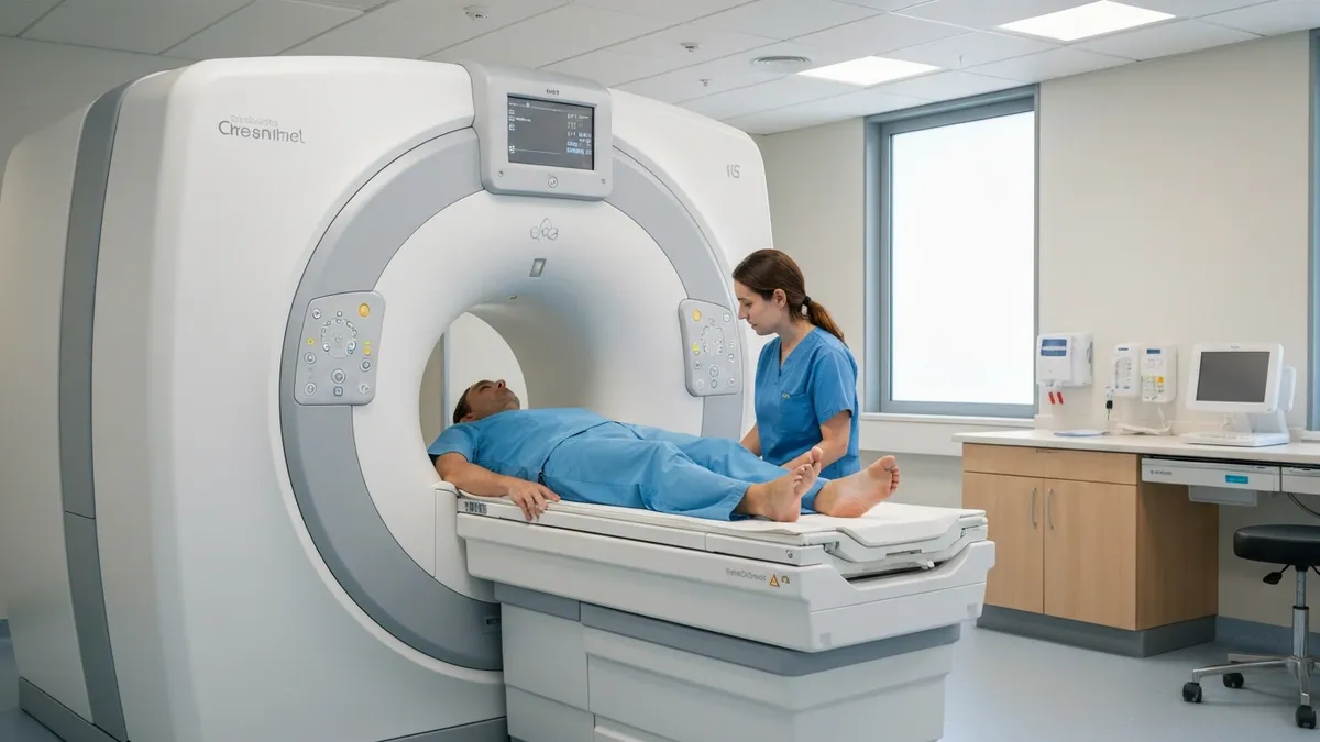
Fasting Before Contrast MRI: Benefits vs. Drawbacks
- +Reduces nausea and vomiting risk during gadolinium injection
- +Minimizes bowel motion artifact for abdominal and pelvic sequences
- +Improves image sharpness and diagnostic quality for liver and pancreatic MRI
- +Decreases gastric volume, lowering aspiration risk in sedated or anxious patients
- +Allows more accurate dynamic contrast timing in DCE-MRI protocols
- +Easier venous access when the patient is adequately hydrated but not overfull
- −Unnecessary for brain, spine, and extremity studies where fasting provides no image benefit
- −Can cause hypoglycemia in diabetic patients, requiring careful glucose management
- −Patient discomfort and stress, especially for anxious or pediatric patients
- −Risk of patients misunderstanding instructions and over-restricting fluids, causing dehydration
- −Fasting delays can create scheduling difficulties for early-morning or back-to-back appointments
- −No proven reduction in allergic reaction rate, so strict fasting may provide false reassurance about safety
Pre-Scan Preparation Checklist for MRI With Contrast
- ✓Confirm with your imaging center whether your specific scan requires fasting at least 48 hours before your appointment.
- ✓Avoid high-fat, fried, or gas-producing foods for 24 hours before abdominal or pelvic contrast MRI.
- ✓Stop eating solid food at least four hours before abdominal MRI with contrast.
- ✓Drink 16–24 ounces of water in the hours leading up to your scan to stay hydrated and support IV access.
- ✓Take all prescribed daily medications with a small sip of water unless told to hold specific drugs like metformin.
- ✓Avoid alcohol and energy drinks for at least 24 hours before the appointment.
- ✓Bring a complete list of all medications, supplements, and known allergies to your appointment.
- ✓Inform the scheduling team if you have a history of kidney disease, diabetes, or prior contrast reactions.
- ✓Wear comfortable, loose-fitting clothing with no metal fasteners to minimize undressing delays.
- ✓Arrive 15–20 minutes early to allow time for screening paperwork and IV line placement before the scan begins.
Most Brain and Spine Contrast MRI Patients Can Eat Normally
Unless your MRI is targeting the abdomen, pelvis, or a region where bowel motion causes image problems, you do not need to fast before gadolinium contrast administration. The ACR guidelines allow normal food and fluid intake for brain, spine, and extremity studies. When in doubt, call your imaging center the day before — a two-minute phone call can prevent unnecessary discomfort and ensure you arrive in the best possible condition for a high-quality scan.
Special patient populations require individualized preparation instructions that go beyond standard fasting guidelines. Pregnant patients represent one of the most carefully managed groups. Gadolinium contrast agents cross the placental barrier and can enter fetal circulation, where they may persist longer than in adult tissue due to immature renal function.
The ACR advises that gadolinium should be used during pregnancy only when the potential benefit clearly outweighs the risk, and when an unenhanced examination would be insufficient to answer the clinical question. When contrast MRI is performed during pregnancy, no additional dietary restrictions beyond routine prenatal guidance are typically imposed, but the decision to proceed requires an informed consent discussion between the patient and referring physician.
Breastfeeding patients often ask whether they need to pause nursing after receiving gadolinium. Current evidence indicates that less than 0.04 percent of the administered gadolinium dose is excreted into breast milk, and less than one percent of that small amount is absorbed by the nursing infant's gastrointestinal tract.
The ACR's most recent guidance acknowledges that the risks to the infant are extremely low and that it is safe to continue breastfeeding without interruption following gadolinium administration. However, some patients choose to pump and discard breast milk for 12 to 24 hours after the scan for personal peace of mind, and this choice should be respected and supported by the care team.
Elderly patients may have reduced kidney function that is not yet reflected in an elevated creatinine level, simply because they have less muscle mass and therefore produce less creatinine as a baseline. Age-related decline in GFR means that a standard creatinine reading that appears normal may mask a significantly lower actual filtration rate.
For patients over 65 undergoing contrast MRI, many facilities request a recent serum creatinine or eGFR result before the appointment. Dietary preparation for elderly patients should also account for polypharmacy, as many older adults take multiple medications that could be affected by fasting or that could interact with contrast-induced changes in fluid balance.
Patients with a history of prior contrast reactions face a more complex pre-procedure pathway. A documented mild prior reaction — such as hives or mild nausea — typically triggers a pre-medication regimen consisting of prednisone given 12 hours, 7 hours, and one hour before the scan, plus diphenhydramine one hour before. Eating with prednisone is actually recommended, since corticosteroids taken on an empty stomach can cause significant gastrointestinal irritation. This creates a direct conflict with fasting protocols for abdominal MRI, requiring the care team to coordinate meal timing around the pre-medication schedule carefully.
Pediatric sedation protocols are among the most strictly enforced fasting guidelines in all of radiology. Children under two years who require general anesthesia or deep sedation for MRI must typically fast from breast milk for four hours, from formula for six hours, and from solid food for eight hours prior to induction.
These rules exist to prevent pulmonary aspiration, which is a life-threatening complication. Anesthesia teams verify compliance with these rules before proceeding, and scans are routinely cancelled when fasting instructions have not been followed. Parents should plan the child's last meal carefully, especially if the appointment is scheduled for late morning or afternoon.
Patients with claustrophobia or severe anxiety sometimes receive oral anxiolytic medications such as lorazepam or diazepam before MRI. These sedatives interact with food in important ways — taking a benzodiazepine on an empty stomach results in faster absorption and stronger sedation, while a full meal delays and blunts the effect. If your facility prescribes oral sedation, ask specifically whether you should eat beforehand to modulate the timing and intensity of sedation, and arrange for a driver since you will not be able to operate a vehicle after taking these medications regardless of meal status.
Contrast MRI for cardiac imaging, including cardiac MRI stress testing with gadolinium, involves specific preparation that mirrors cardiac stress test guidelines. Patients are generally asked to fast for three to four hours before the study, avoid caffeine for 24 hours if pharmacological stress is being used, and ensure adequate hydration. The cardiac protocol often runs longer than standard MRI — sometimes 60 to 90 minutes — making pre-scan comfort and a settled stomach particularly important for image quality and patient tolerance throughout the entire examination.
Many readers pair this guide with the open mri near me to check their readiness before exam day.
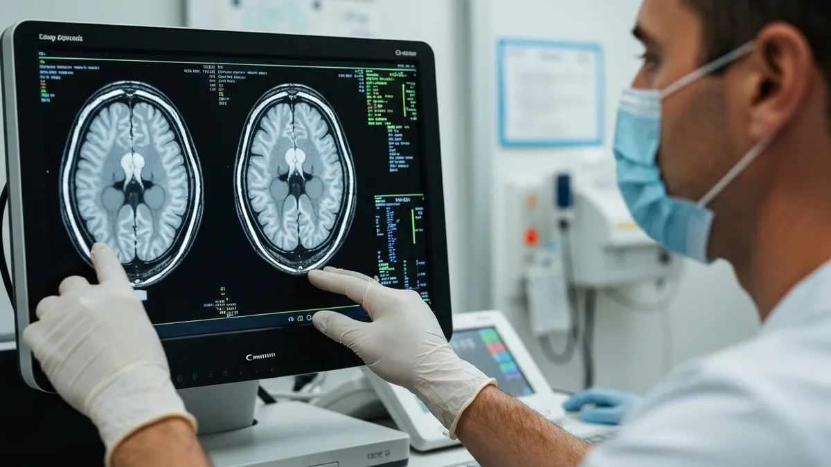
Fasting protocols for MRI with contrast are not universally standardized and vary significantly between institutions, individual radiologists, and even specific scan types within the same department. The guidelines summarized in this article reflect current ACR recommendations and common clinical practice, but your imaging center may have different rules based on their equipment, contrast agents used, or patient population. Always call the facility directly 24 to 48 hours before your appointment to confirm the exact preparation instructions you are expected to follow.
The day of your MRI with contrast should begin with a clear understanding of what you are allowed to eat and drink based on the specific protocol ordered for your scan. If you are having a brain or spine MRI with gadolinium, you can eat breakfast as you normally would — just choose something light and avoid greasy or very heavy foods out of an abundance of caution.
A bowl of oatmeal, toast with a light spread, a banana, or yogurt are all excellent choices that will keep you comfortable during the scan without creating any risk of nausea or image degradation. Drink water freely and stay well hydrated throughout the morning.
If you are having an abdominal or pelvic MRI with contrast, your preparation looks very different. You should have had your last solid meal at least four hours before your scheduled scan time. If your appointment is at 10:00 AM, that means finishing breakfast no later than 6:00 AM.
A light, easily digestible breakfast — plain toast, clear broth, a small portion of rice or crackers — is preferable to anything rich in fat or fiber. After 8:00 AM, transition to clear liquids only: water, black coffee, plain tea, or apple juice without pulp. Avoid orange juice, milk, smoothies, and anything carbonated or that contains fiber.
When you arrive at the imaging center, you will be asked to complete a safety screening questionnaire that covers metal implants, prior surgeries, allergies, and kidney function. Be prepared to answer questions about when you last ate and drank. The technologist will review your preparation, confirm your IV access site, and explain what to expect during the contrast injection.
Most patients feel a brief wave of warmth and a mild metallic taste that lasts ten to thirty seconds after the gadolinium bolus — these sensations are normal and expected. Serious reactions are rare but the team is trained to respond immediately if they occur.
During the scan itself, you will be positioned on the table and moved into the bore of the magnet. For most contrast studies, the injection is timed to a specific point during the scan sequence to capture peak tissue enhancement. You may be asked to hold your breath for several seconds at intervals — especially for abdominal studies — to freeze respiratory motion. Fasting helps here because a full stomach pushes against the diaphragm and makes breath-holding more uncomfortable, and stomach gurgling creates noise and motion in the image.
After the scan, there are no dietary restrictions unless your facility provides specific post-contrast instructions for patients with kidney issues. Drink plenty of water — at least 32 to 48 ounces over the next several hours — to help your kidneys flush the gadolinium from your system efficiently. If you feel nauseous after the exam, crackers, plain toast, or a small glass of ginger ale can help settle your stomach. Most patients feel completely normal within minutes of the injection and have no post-scan dietary issues at all.
Results from an MRI with contrast are typically interpreted by a radiologist within 24 to 72 hours, depending on the urgency of the clinical question and the facility's workload. A preliminary report may be available sooner for urgent or emergent studies. Your referring physician will receive the radiology report and contact you to discuss findings and next steps. In some cases, follow-up imaging with or without contrast may be recommended, and you can apply everything you have learned in this guide to prepare for that subsequent appointment as well.
If you want to deepen your understanding of how MRI contrast studies are interpreted and what the sequences actually show, reviewing related content on topics like diffusion-weighted imaging can provide important complementary knowledge. Understanding the full scope of MRI technique — including how preparation choices affect image quality — is valuable both for patients navigating their own care and for radiology professionals building clinical expertise. The more you know about how preparation interacts with image acquisition, the better equipped you are to ask the right questions and advocate for a high-quality diagnostic experience.
Practical preparation for MRI with contrast starts well before the day of the exam. The most common mistake patients make is not confirming fasting instructions with the facility, assuming that internet-sourced information applies to their specific scan type.
Call your imaging center two to three days before the appointment, identify yourself, give your scan time and order details, and ask specifically: do I need to fast, for how long, and can I have liquids? Write down what you are told, including the name of the staff member who gave you the instructions, so you have a clear reference if questions arise on the day of the scan.
Planning your meals on the day before abdominal contrast MRI is also worthwhile. Avoid beans, lentils, broccoli, cauliflower, cabbage, and raw onions at dinner the evening before, since these foods generate intestinal gas that takes many hours to clear. A dinner of plain rice, grilled chicken or fish, steamed non-cruciferous vegetables, and water is an ideal preparatory meal.
Avoid heavy restaurant meals, especially anything fried or cream-based, on the evening before your appointment. This light eating pattern reduces the amount of gas and solid content in your bowel at scan time, which directly improves the diagnostic quality of the images your radiologist will interpret.
On the morning of your abdominal MRI appointment, keep your pre-fast breakfast simple and finish it within the instructed time window. Set an alarm or phone reminder to stop eating at the right time — it is surprisingly easy to lose track when you are going about your morning routine. After the cutoff, switch to clear liquids and focus on staying hydrated. If you feel hungry while waiting, sipping water or plain broth can take the edge off discomfort without violating your fasting protocol.
Pack a small snack for after the scan. Most patients are hungry immediately following an MRI, and having crackers, a granola bar, or a piece of fruit in your bag means you can eat as soon as you leave the imaging suite rather than having to find food while you are already tired and hungry.
For diabetic patients, having a glucose-containing snack readily available is a patient safety measure — hypoglycemia after a fasting period can occur, and being prepared is essential. Inform the MRI technologist if you are diabetic so they can monitor you and have glucose available if needed during the procedure.
Wear comfortable clothing with no metal elements: elastic waistbands rather than belts with metal buckles, sports bras instead of underwire bras, slip-on shoes rather than boots with metal zippers. Metal clothing items must be removed before entering the scanner room, and having to undress entirely adds time and stress to the appointment. Many facilities provide hospital gowns, but wearing appropriate clothing from home allows you to remain dressed throughout the scan, which most patients find more comfortable during a 30-to-90-minute procedure.
If you are bringing a companion — a family member, friend, or caregiver — inform them in advance that they will also need to complete a metal screening questionnaire before entering the scanner room. Pacemakers, cochlear implants, certain aneurysm clips, and embedded shrapnel are absolute contraindications to entering the MRI room, regardless of whether the person is the patient or a support companion. The magnetic field is always on in an MRI suite, even when no patient is being scanned, and ferromagnetic objects can become dangerous projectiles without warning.
Finally, manage your expectations about how the scan will feel. Most patients report the experience as louder than expected — MRI scanners produce knocking and banging sounds that can reach 100 decibels — but not painful. Earplugs or headphones with music are routinely offered by MRI facilities. The contrast injection creates a brief flush sensation but does not cause significant discomfort.
If you experience chest tightness, difficulty breathing, severe nausea, or a feeling that something is wrong after the injection, alert the technologist immediately through the intercom or call button provided inside the bore. These symptoms are uncommon but require prompt assessment by the clinical team.
MRI Questions and Answers
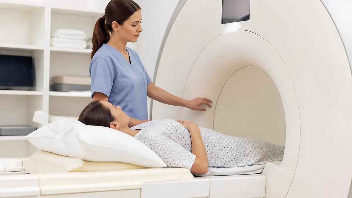
DWI MRI: Diffusion-Weighted Imaging Explained (2026 Guide)
MRI Lumbar Spine Without Contrast: Complete Guide to the Scan, What It Shows, and How to Prepare
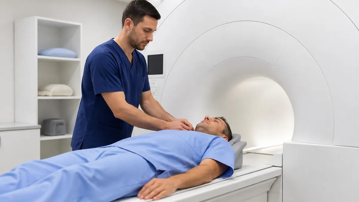
MRI History: From Nuclear Resonance to Modern Scanners

MRI of Cervical Spine: Complete Guide to Imaging, Anatomy, Pathology, and Patient Preparation
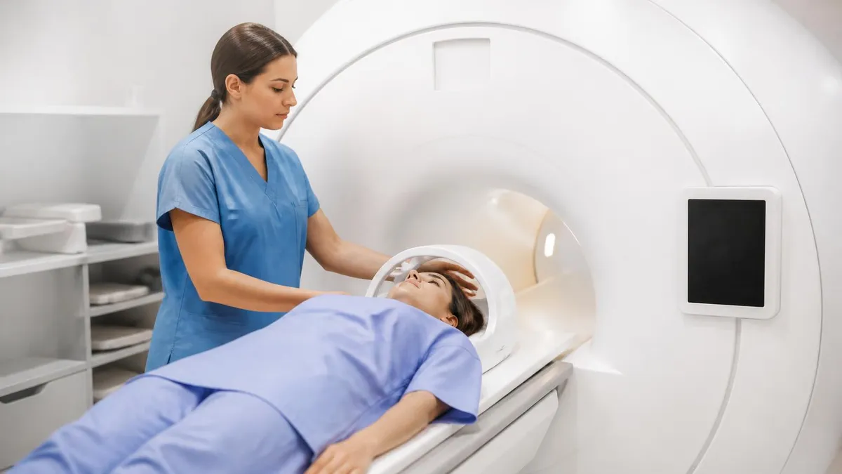
Where to Get an MRI: Facilities, Costs, and How to Choose
About the Author

Medical Laboratory Scientist & Clinical Certification Expert
Johns Hopkins UniversityDr. Sandra Kim holds a PhD in Clinical Laboratory Science from Johns Hopkins University and is certified as a Medical Technologist (MT) and Medical Laboratory Scientist (MLS) through ASCP. With 16 years of clinical laboratory experience spanning hematology, microbiology, and molecular diagnostics, she prepares candidates for ASCP board exams, MLT, MLS, and specialist certification tests.
Join the Discussion
Connect with other students preparing for this exam. Share tips, ask questions, and get advice from people who have been there.
View discussion (8 replies)