Diffusion Weighted MRI: How DWI Works, Sequences and Clinical Uses 2026 July
Diffusion weighted MRI explained for technologists. 📗 Learn DWI physics, sequences, b-values, ADC maps, stroke imaging, artifacts and reading findings.
Diffusion weighted MRI, often shortened to DWI, is a sequence that maps how water molecules move inside tissue. Most MRI sequences look at where protons sit and how fast they relax. DWI looks at something different. It looks at motion. Water that moves freely loses signal. Water that is trapped keeps its signal. That single contrast trick reshaped stroke imaging, tumor characterization and a long list of body and pelvic exams.
You will see DWI almost every shift if you scan brains. You will also see it in liver, prostate, breast and whole-body oncology protocols. The sequence is short, it does not need contrast, and it gives a yes-or-no kind of contrast that radiologists love. This guide breaks down the physics, the sequences, the b-values, the ADC map, the common artifacts and the day-to-day workflow. It is written for MRI technologists studying for boards and for new readers learning the basics. If you want a wider refresher, our magnetic resonance imaging overview pairs well with this read.
Diffusion is everywhere in the body. Inside a cell, water bumps into membranes. Between cells, water moves a bit more freely. In a ventricle, water is almost free. DWI uses paired motion-sensitizing gradients to label that motion. If a proton sits still between the two gradients, it keeps its phase. If it moves, the gradients do not cancel, and the signal drops. That drop is the contrast. The more diffusion, the darker the voxel. The less diffusion, the brighter the voxel. Restricted diffusion equals bright DWI.
The math behind DWI is the Stejskal-Tanner equation. A pair of equal gradients straddles a 180-degree refocusing pulse. The first gradient labels each proton with a phase shift based on its position. The second gradient reverses that shift. Stationary protons end the pair with no net phase change. Moving protons end with a leftover phase, and the signal scales with how much they moved. The strength of the labeling is called the b-value. Bigger b-values produce stronger diffusion weighting and weaker raw signal.
Single-shot echo planar imaging, or SS-EPI, is the standard readout. It grabs an entire slice in one shot, usually under 100 milliseconds. That speed beats motion, which is why DWI can be done in unsedated children, restless adults and even fetal exams. The trade-off is that EPI is sensitive to susceptibility, eddy currents and B0 inhomogeneity. You will see geometric distortion near the skull base, the orbits and any metal. New readout schemes such as readout-segmented EPI and multi-shot DWI reduce those issues at the cost of scan time.
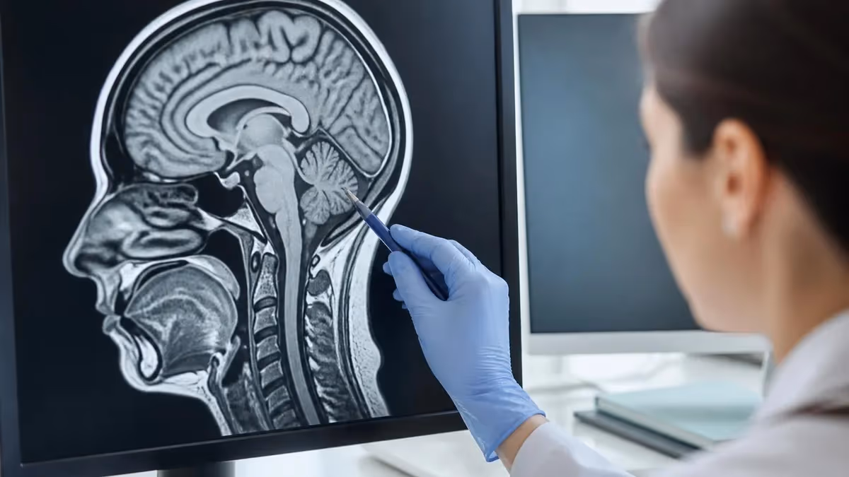
Some MRI techs are surprised to learn that DWI piggybacks on a standard spin echo. The 180-degree pulse refocuses T2 signal. Add the paired diffusion gradients and you have DWI. That T2 root is the reason for one of the most common reading errors. A bright spot on DWI may not be true restriction. It may just be very bright T2 bleeding through. The cure is the ADC map. Real restriction is bright on DWI and dark on ADC. T2 shine-through is bright on both. We dig into ADC, b-zero images and shine-through later in this article.
Restricted Diffusion vs T2 Shine-Through
True restricted diffusion is bright on DWI and dark on the ADC map. T2 shine-through is bright on DWI but also bright on the ADC map. Always read the pair. Never call restriction from DWI alone.
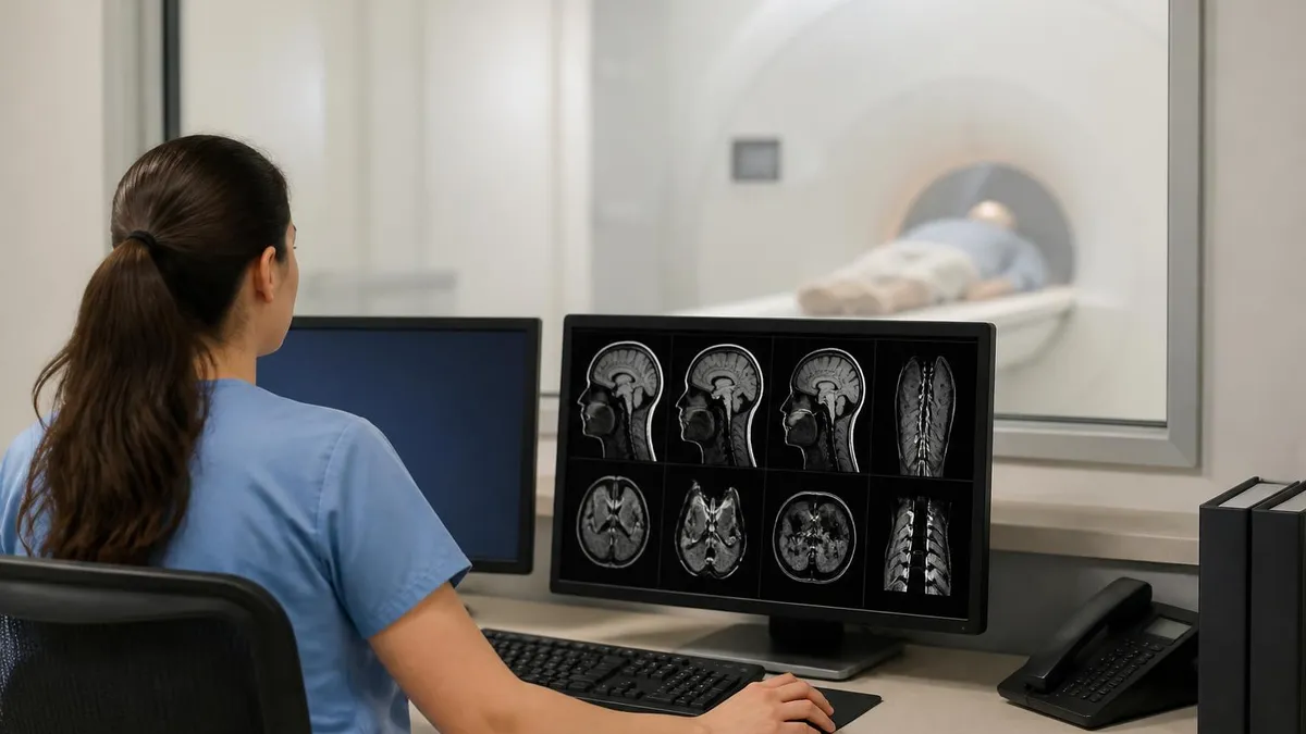
A standard DWI series almost always has three acquisitions stacked together. The b-zero image looks like a slightly noisy T2. The intermediate b-value image, often b500 in body work, gives mid-range contrast. The high b-value image, usually b1000 in brain and b800 to b1000 in body, gives the punchy diffusion contrast you see on read-outs. The scanner then calculates an ADC map by fitting an exponential decay through those acquisitions, voxel by voxel.
ADC stands for apparent diffusion coefficient. The word apparent matters. True molecular diffusion would need a perfect free-water reference. Tissue has cells, membranes, fibers and perfusion. The number we measure is the apparent value the whole microenvironment produces. ADC is reported in mm-squared per second, and it usually falls between 0.5 and 3.0 times ten to the minus three. Acute infarct typically reads around 0.3 to 0.5. Normal white matter sits near 0.7. CSF runs above 3.0. These ranges are useful, but you should always compare to the contralateral side rather than to a textbook number.
Diffusion tensor imaging, or DTI, builds on DWI by sampling at least six gradient directions instead of three. The tensor result lets you measure fractional anisotropy and reconstruct white-matter tracts. Diffusion kurtosis imaging samples even more directions and fits non-Gaussian curves. Both are research-leaning tools, but DTI is becoming routine for pre-surgical mapping and for some pediatric epilepsy workups. If your site has a neurosurgical service, expect to run DTI several times a week.
Common DWI B-Values and Their Use
T2-weighted reference image. Used to anchor ADC math and to spot T2 shine-through.
Mid-range body contrast. Common in prostate, liver and breast DWI protocols.
Standard high b-value for brain stroke, infection and most adult neuro DWI.
High contrast in white matter lesions. Used in multiple sclerosis research and pediatric epilepsy.
Body oncology, especially prostate PI-RADS reads. Pushes lesion conspicuity higher.
Stroke is the killer use case for DWI. Within minutes of an ischemic infarct, the failing cell pumps stop, sodium and water rush in, and cells swell. That swelling traps water. The trapped water shows up as a bright spot on DWI and a dark spot on ADC long before any CT change. A modern stroke protocol runs DWI first so the radiologist can call the infarct while the rest of the exam keeps acquiring. Time-to-diagnosis is now measured in single-digit minutes at high-volume centers.
Beyond stroke, brain DWI helps with abscess versus tumor. Pyogenic abscess shows striking central restriction because pus is packed with cells and proteins. A high-grade glioma may show some peripheral restriction but rarely the same dense central pattern. Epidermoid cysts also light up on DWI, which separates them from arachnoid cysts that follow CSF. In demyelination, acute multiple sclerosis plaques sometimes restrict at the leading edge, which can help date a lesion. For the full timing story, see our breakdown of how long does a brain MRI take across stroke versus elective protocols.
Body DWI is now a routine part of cancer staging. PI-RADS scoring for prostate cancer leans heavily on the DWI/ADC pair, with high b-values pushing tumor conspicuity. Liver lesions are often characterized using DWI plus dynamic contrast. Breast DWI is a leading non-contrast alternative for high-risk screening. Whole-body DWI with background suppression, sometimes called DWIBS, has matured into a strong tool for multiple myeloma and lymphoma surveillance. None of this would have been practical fifteen years ago. Faster gradients, parallel imaging and multi-band acceleration made it routine.
DWI Clinical Applications
DWI is artifact-heavy. The big four are susceptibility distortion, eddy currents, Nyquist ghosting and motion. Susceptibility distortion warps the image near air-tissue and bone-tissue interfaces. The orbits, sinuses, temporal lobes and posterior fossa all suffer. Eddy currents come from the strong, fast switching of diffusion gradients. They cause shearing and shifting between the directional images. Modern scanners run eddy-current compensation, and most workstations apply registration in post-processing.
Nyquist N over 2 ghosting is a periodic phantom of the brain shifted by half the field of view. It usually means the phase correction failed. A repeat shim or a calibration scan fixes it. Motion in DWI is a strange beast because the sequence is so sensitive to phase. A small head shift between gradient directions can cause bizarre signal loss in the ADC map. If your ADC has a streaky pattern that does not match anatomy, suspect motion before you call disease.
Chemical shift is less of a problem on EPI than on conventional readouts, but fat signal can still ghost across the image if fat saturation fails. Always check that the fat sat band is set correctly and that the patient is not too far off-isocenter. For body DWI, breathing is the enemy. Free-breathing acquisitions with respiratory triggering, navigator pulses or simple multiple averages can clean things up. We cover the full pre-scan setup checklist on our MRI scanner reference page.
Common DWI Artifacts
Warps the image near air, bone or metal. Worst at orbits, sinuses and posterior fossa.
Cause shifting and shearing between gradient directions. Modern scanners apply compensation in real time.
Half-FOV phantom of the brain. Fix with a fresh shim or recalibration.
Patient motion between directions creates streaky ADC patterns that do not match anatomy.
Failed fat saturation leaves bright fat ghosts across the image. Check fat sat band placement.
Around day 7 to 14 post-stroke, ADC returns toward normal even as the infarct remains.
Diffusion EPI is one of the most metal-sensitive sequences on the scanner. A small ferromagnetic clip can wipe out a whole hemisphere of signal. Always check the implant list against the conditional labeling before you load DWI. When in doubt, swap to a multi-shot or readout-segmented DWI sequence with reduced distortion.
Reading the ADC map is a skill. A good reader checks four things in order. First, is the abnormality real or is it an artifact at a known trouble spot. Second, is the DWI signal truly restricted, meaning bright on DWI and dark on ADC. Third, what is the absolute ADC value, and is it consistent with the suspected pathology. Fourth, does the pattern match an anatomic territory or a structural rule. Stroke follows vascular territories. Diffuse axonal injury crosses them.
Quantitative ADC is becoming standard. A region of interest measurement gives a number in mm-squared per second times ten to the minus three. Many sites use 0.6 as a rough threshold for acute infarct. In prostate, values below 0.9 raise suspicion. In liver, hemangiomas usually run above 2.0 while metastases sit between 0.8 and 1.5. These are guideposts, not hard rules. Always compare side-to-side and across sequences.
Pseudonormalization is the trap that catches new readers. Around 7 to 14 days post-stroke, the restricted region starts to look normal on ADC because the cell membranes break down and water moves more freely. The DWI may still look bright due to T2 shine-through, but the ADC is no longer dark. Without clinical context, that infarct can be missed. Always check the date of symptom onset before you call an infarct acute or subacute.
DWI Quality Control
- ✓Run a daily phantom DWI to track ADC stability across the bore.
- ✓Verify b-zero matches a standard T2 weighted image with no obvious distortion.
- ✓Check ADC values for normal white matter fall between 0.6 and 0.8 times ten to the minus three.
- ✓Look for ghosting, shearing or signal loss in air-tissue interface regions.
- ✓Confirm fat saturation is uniform across the field of view.
- ✓Review the multi-direction images for motion before calculating the ADC map.
- ✓Set the phase-encode direction to minimize distortion across the lesion of interest.
- ✓Document the b-value pair and the number of directions in the report.
Routine brain DWI runs three orthogonal directions with b zero and b 1000. Slice thickness is 4 to 5 millimeters. Voxel size is around 1.5 by 1.5 in-plane. The acquisition usually takes about a minute. For stroke, that minute can sit at the front of the scan so that the radiologist can call the infarct early. Body DWI uses b 50, b 500 and b 1000 in three directions, with breath-hold or respiratory triggering. Prostate DWI pushes to b 1400 or higher and may compute a synthetic b 2000 image for PI-RADS reading.
Pediatric DWI deserves a special note. Newborn brains have much higher water content, so ADC values are higher than in adults. A neonatal stroke may show restriction at higher ADC values than a textbook adult cutoff. Always use age-matched reference data when reading pediatric scans. The same caution applies to elderly brains, where small-vessel changes shift the baseline ADC across white matter.
Field strength matters. At 1.5 tesla, EPI distortion is milder and SNR is lower, so you may need more averages. At 3 tesla, distortion is worse but signal is much higher, which is good for high b-values. At 7 tesla, distortion becomes a real challenge, and most clinical 7 T sites use parallel transmit and multi-shot DWI. The sequence is the same conceptually across field strengths, but parameter tuning is different at every step.
DWI Reading Workflow
- ✓Open the b zero image and check for normal brain anatomy and obvious distortion.
- ✓Scroll the high b-value DWI to find any bright focus that stands out from surrounding tissue.
- ✓Cross-check each bright focus on the ADC map for true dark signal.
- ✓Compare the abnormality with conventional T1, T2 and FLAIR sequences for shape and edema.
- ✓Confirm the lesion sits within a known vascular territory if you suspect stroke.
- ✓Measure ADC values with a small region of interest and compare to the contralateral side.
- ✓Document b-values, number of directions and any motion or artifact concerns in the report.
- ✓Correlate with patient symptom onset time to decide if the infarct is hyperacute, acute or subacute.
- ✓Look for additional foci that may suggest embolic shower rather than a single occlusion.
- ✓Recommend follow-up imaging when ADC findings are ambiguous or symptoms are progressing.
DWI Strengths and Limits
- +Catches acute stroke within minutes of onset
- +No contrast needed, safe for renal patients
- +Short scan time, often under 90 seconds
- +Adds value to almost every brain and body protocol
- +Quantitative ADC gives an objective number for reporting
- +Works across 1.5 T, 3 T and 7 T scanners
- +Supported by every modern MRI vendor
- +Becoming standard in oncology staging
- −Heavy susceptibility distortion near air, bone and metal
- −T2 shine-through can mimic restriction without ADC check
- −Low spatial resolution compared to T1 or T2
- −EPI is sensitive to motion and eddy currents
- −Body DWI requires careful fat suppression and triggering
- −Lower spatial resolution than structural sequences
- −ADC absolute values vary by vendor and field strength
For MRI technologists, DWI shows up on every certification path. The ARRT MRI registry has direct questions about b-values, ADC interpretation and the difference between DWI and DTI. The ARMRIT exam tests sequence basics. The MRSO and MRSE safety exams ask about ferromagnetic risk and how DWI distortion can hide pathology. If you want a focused refresher, our MRI images page covers contrast windows and reading basics, while the how does MRI work guide explains the underlying physics in plain language.
If you supervise scanning, build a DWI bin into your daily QA. Phantom ADC values drift over time. Gradient performance degrades with age. Eddy-current compensation parameters need recalibration after major service. A simple ice-water phantom with a known ADC of 1.1 times ten to the minus three gives you a stable reference. Track it weekly. The first sign of a gradient problem often shows up as a quiet shift in measured ADC long before the service alarm fires.
Finally, talk to your radiologists. Ask them which b-value they actually use when they read. Ask them whether they want computed b-values or true acquired b-values. Ask whether they want trace DWI or each direction kept separate. These small protocol choices have a big impact on read confidence. A two-minute conversation at the start of a new protocol can save weeks of repeat scans down the line.
Many programs now teach DWI as one of the first sequences a new MRI technologist must master. The reason is simple. Stroke calls happen every shift in a busy hospital, and the DWI series is what the radiologist will open first. Getting the geometry, the b-values and the fat saturation right on the first try keeps the workflow moving. Learning to spot a motion artifact in seconds rather than minutes also saves a repeat scan, which matters when the patient is fragile and the timer is ticking.
Vendor differences add a small but real layer of complexity. Some scanners label the b-value series with the direction index. Others give you only the combined trace image. Some workstations let you generate computed b-values on the fly. Others lock you to acquired b-values. Spend a shift exploring your console with a senior technologist and you will save hours of confusion later. Document those quirks in your site protocol manual so new staff onboard faster.
Looking ahead, multi-band DWI is changing the speed-quality balance. With four-band acceleration on a modern 3 T, you can get a full brain in 30 seconds while keeping b 1000 SNR adequate. That speed is opening new use cases such as serial perfusion-diffusion mismatch maps in stroke and rapid pediatric exams without sedation. Synthetic high b-value imaging, where the scanner extrapolates to b 2000 or higher from lower acquired values, is another growing trend in prostate and breast imaging.
Sites that invest a few hours in DWI protocol tuning every quarter consistently report fewer repeat scans and faster radiologist turnaround. It is one of the highest-leverage QA habits in any MRI department, big or small.
MRI Questions and Answers
About the Author
Medical Laboratory Scientist & Clinical Certification Expert
Johns Hopkins UniversityDr. Sandra Kim holds a PhD in Clinical Laboratory Science from Johns Hopkins University and is certified as a Medical Technologist (MT) and Medical Laboratory Scientist (MLS) through ASCP. With 16 years of clinical laboratory experience spanning hematology, microbiology, and molecular diagnostics, she prepares candidates for ASCP board exams, MLT, MLS, and specialist certification tests.
Join the Discussion
Connect with other students preparing for this exam. Share tips, ask questions, and get advice from people who have been there.
View discussion (6 replies)