
Breast MRI Results: What They Mean, How to Read Them, and What Happens Next
Understand your breast MRI results — what findings mean, BI-RADS scores, timelines, and next steps. ✅ Complete 2026 September guide.
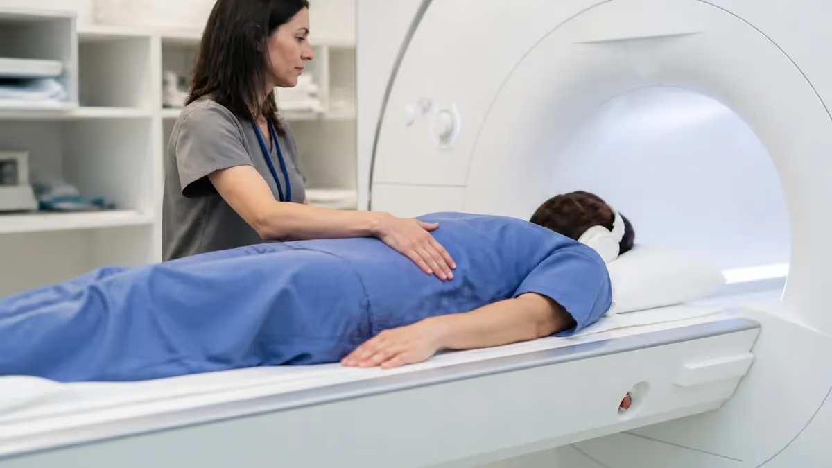
Breast MRI results can feel overwhelming to receive, especially when you are waiting anxiously after a scan ordered for screening, diagnosis, or surgical planning. Understanding what your report actually says — and what each finding means for your health — is one of the most empowering steps you can take. Breast MRI uses powerful magnetic fields and radio waves to produce highly detailed cross-sectional images of breast tissue, and the resulting report contains terminology that may be unfamiliar to most patients.
The radiologist who reads your scan will classify findings using the ACR BI-RADS (Breast Imaging Reporting and Data System) scale, a standardized scoring system that ranges from 0 to 6. Each category carries a specific recommendation — from routine annual screening to immediate biopsy. Knowing what these categories mean before your follow-up appointment allows you to ask informed questions and participate actively in your own care decisions.
Breast MRI is significantly more sensitive than mammography, detecting cancers in dense breast tissue that other modalities frequently miss. However, this sensitivity comes with a trade-off: MRI also has a higher rate of false-positive findings, meaning it sometimes flags areas of concern that turn out to be benign on biopsy. This is why understanding context is so important when reviewing your results.
Your report will describe the appearance of any enhancing lesions — areas where contrast dye accumulated — in terms of shape, margin, and kinetic behavior. A mass that shows rapid initial enhancement followed by a plateau or washout pattern is more concerning than one with slow, persistent enhancement. Radiologists weigh these features together rather than relying on any single characteristic to reach their assessment.
Background parenchymal enhancement (BPE) is another key concept you will encounter in breast MRI results. This refers to the normal enhancement of breast glandular tissue in response to contrast. BPE is categorized as minimal, mild, moderate, or marked, and it can affect how clearly abnormal lesions stand out. High BPE is more common in premenopausal women and during certain phases of the menstrual cycle, which is why timing the scan correctly matters.
For those studying medical imaging or preparing for board exams, learning to interpret breast mri results alongside diffusion-weighted imaging principles builds a more complete clinical picture. DWI adds functional information that complements the morphological and kinetic data captured on standard contrast-enhanced sequences, helping radiologists differentiate benign from malignant findings with greater confidence.
This guide walks you through every major component of a breast MRI result — from the standardized BI-RADS categories and enhancement patterns to what happens after an abnormal finding is identified. Whether you are a patient trying to understand your own scan, a student preparing for the MRI registry exam, or a technologist looking to deepen your clinical knowledge, you will find clear explanations and practical context throughout this article.
Breast MRI by the Numbers
What Happens After Your Breast MRI Scan
Scan Completed and Images Transmitted
Radiologist Interprets the Study
Report Finalized and Delivered
Referring Physician Reviews and Contacts Patient
Next Steps Determined Based on BI-RADS
Biopsy or Additional Imaging If Indicated
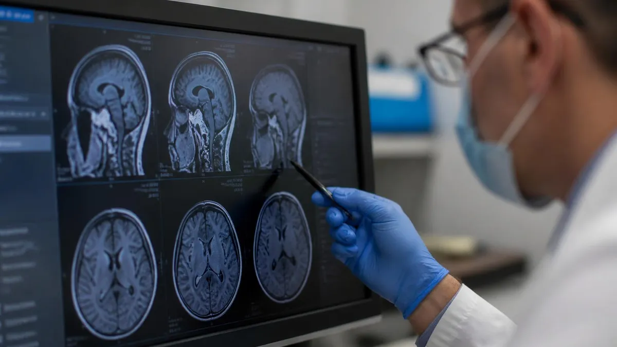
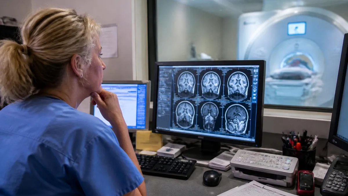
When a radiologist sits down to interpret a breast MRI study, the process is far more systematic than simply looking for a bright spot on a screen. The standard protocol begins with a review of the patient's clinical history — including personal and family history of breast cancer, BRCA mutation status, prior biopsies, and the reason the scan was ordered. This context profoundly shapes how findings are weighted and reported.
The radiologist first evaluates background parenchymal enhancement on the pre-contrast and early post-contrast sequences. High background enhancement can partially obscure lesions, reduce specificity, and occasionally lead to recall for short-interval follow-up when nothing pathological is actually present. This is one reason breast MRI is ideally scheduled on days 7 through 14 of the menstrual cycle, when hormonal influence on breast tissue is at its lowest.
Next, the radiologist assesses the fibroglandular tissue composition — whether the breasts are almost entirely fatty, scattered, heterogeneously dense, or extremely dense. Dense breast tissue has implications not just for MRI interpretation but for overall cancer risk and the limitations of mammographic screening, which is why many women with dense breasts are referred for supplemental MRI screening.
Any enhancing focus, mass, or non-mass enhancement is then characterized in detail. A focus is a tiny dot of enhancement too small to fully characterize. A mass has a defined three-dimensional shape and margin. Non-mass enhancement (NME) is enhancement that occupies a region without forming a distinct mass — and NME carries its own descriptors for distribution (linear, segmental, regional, diffuse) and internal pattern (homogeneous, heterogeneous, clumped, clustered ring).
Kinetic curve analysis is one of the most important tools in breast MRI interpretation. After contrast injection, the radiologist tracks how quickly a lesion enhances (the wash-in rate) and what happens to signal intensity over time. A type 1 (persistent) curve, where signal keeps rising, is typically benign. A type 2 (plateau) curve is indeterminate. A type 3 (washout) curve, where signal drops after initial peak, is the most suspicious pattern and significantly increases the probability of malignancy.
The morphological features of a mass — its shape (oval, round, or irregular), margin (smooth, irregular, or spiculated), and internal enhancement pattern (homogeneous, heterogeneous, rim enhancing, or dark internal septations) — are combined with kinetic data to generate an overall BI-RADS assessment. No single feature is determinative; it is the constellation of findings that drives the final impression and recommendation.
Radiologists also look for secondary signs that can support or refute a diagnosis. Skin thickening, nipple retraction, chest wall involvement, and axillary lymphadenopathy are all important ancillary findings that must be documented. When a suspicious mass is found, documenting these features helps surgeons and oncologists plan treatment, determine whether neoadjuvant chemotherapy is appropriate, and establish baseline measurements for monitoring treatment response.
Understanding Enhancement Patterns in Breast MRI
A mass on breast MRI is a space-occupying lesion with definable margins seen in three dimensions. Radiologists describe masses by shape (oval, round, or irregular), margin character (smooth, irregular, or spiculated), and internal enhancement pattern. Oval or round masses with smooth margins and homogeneous enhancement are strongly associated with benign fibroadenomas, lymph nodes, or cysts, typically earning a BI-RADS 2 or 3 assessment with no urgent intervention required.
Irregular masses with spiculated margins and rim enhancement are among the most concerning findings on breast MRI, correlating strongly with invasive carcinoma in published literature. The rim enhancement pattern — where the outer shell enhances more intensely than the center — reflects central necrosis or fibrosis, a hallmark of aggressive malignancy. When these morphological features combine with a washout kinetic curve, the positive predictive value for cancer exceeds 85%, making biopsy mandatory regardless of other clinical factors.
Breast MRI: Advantages and Limitations for Screening and Diagnosis
- +Highest sensitivity of any breast imaging modality — detects cancers missed by mammography and ultrasound in dense tissue
- +No ionizing radiation, making it safe for repeated use in high-risk patients who need annual screening
- +Accurately measures tumor size and extent, critical for surgical planning and determining eligibility for breast conservation
- +Detects multicentric and multifocal disease that would change surgical approach if found before the initial operation
- +Highly effective for evaluating implant integrity, detecting rupture, and assessing post-treatment response to chemotherapy
- +Can identify the primary tumor in patients who present with axillary metastasis but have no palpable breast mass
- −Higher false-positive rate than mammography leads to unnecessary biopsies and patient anxiety in a meaningful percentage of cases
- −Requires intravenous gadolinium contrast, which carries a small risk of allergic reaction and is avoided in patients with severe kidney disease
- −Long scan time (30–60 minutes in the magnet) combined with claustrophobia and noise can make the experience difficult for some patients
- −Expensive compared to mammography or ultrasound — many insurance plans require documented high-risk criteria before covering annual MRI screening
- −Cannot reliably detect microcalcifications, which are a key early sign of DCIS on mammography — MRI and mammography are complementary, not interchangeable
- −Results are highly operator- and reader-dependent — interpretation quality varies significantly between facilities and radiologist experience levels
What to Do After Receiving Your Breast MRI Results
- ✓Write down your BI-RADS score and the specific recommendation before your follow-up appointment so you can refer to it during the conversation.
- ✓Ask your ordering physician to explain every finding in plain language and confirm whether any areas require biopsy, additional imaging, or only routine follow-up.
- ✓Request copies of both the written radiology report and the actual images on a disc or through your patient portal for your personal medical records.
- ✓If biopsy is recommended, ask whether it will be performed under MRI guidance, ultrasound guidance, or stereotactic guidance — and why that method was chosen.
- ✓Schedule any recommended follow-up MRI within the specified timeframe (usually 6 months for BI-RADS 3) to avoid losing the timing window.
- ✓Confirm that the radiologist who reads your follow-up study has access to your prior breast MRI images for direct comparison of any previously identified findings.
- ✓Discuss your personal and family history with your provider to determine whether you qualify for annual high-risk MRI screening covered under your insurance plan.
- ✓Ask about genetic counseling if you have a family history of breast or ovarian cancer, as BRCA1/2 testing may influence your long-term screening protocol.
- ✓Avoid making major health decisions based on a single MRI result — seek a second opinion from a fellowship-trained breast radiologist if you have any doubts.
- ✓Keep a personal log of all breast imaging studies including dates, facilities, findings, and BI-RADS scores to maintain continuity of care across providers.
A BI-RADS 3 Finding Carries Less Than a 2% Risk of Malignancy
Many patients receiving a BI-RADS 3 result assume the worst, but this category is defined as "probably benign" with a malignancy risk of less than 2%. The standard recommendation is a 6-month short-interval follow-up MRI — not biopsy — because the goal is to demonstrate stability over time. A finding that remains unchanged or regresses at 6 months and 12 months is almost certainly benign and may ultimately be downgraded to BI-RADS 2.
Comparing breast MRI to other breast imaging modalities helps put results into proper clinical context. Mammography remains the gold standard for population-level screening in women of average risk because it is widely available, low-cost, and has decades of evidence supporting its ability to reduce breast cancer mortality. It is particularly effective at detecting microcalcifications — tiny calcium deposits that are often the earliest sign of ductal carcinoma in situ — which breast MRI frequently misses entirely.
Ultrasound is typically used as a complementary tool to mammography, particularly for evaluating palpable lumps, characterizing masses detected on mammogram or MRI, and guiding biopsies. Ultrasound has no radiation, is well tolerated, and can distinguish fluid-filled cysts from solid masses with high accuracy. However, it is operator-dependent and less reliable as a standalone screening tool because it cannot image the entire breast systematically in the same way that MRI or digital breast tomosynthesis can.
Digital breast tomosynthesis (DBT), often called 3D mammography, represents a major advance over conventional 2D mammography. By acquiring images at multiple angles and reconstructing thin cross-sectional slices, DBT reduces tissue overlap — the main reason conventional mammography produces false positives and misses cancers in dense tissue. Many facilities now offer DBT as their primary screening tool, and for women at average risk, DBT combined with conventional mammography outperforms either modality alone.
Contrast-enhanced mammography (CEM) is an emerging technique that uses iodinated contrast to highlight enhancing lesions in a way analogous to breast MRI, but at a fraction of the cost and with far greater availability. Early studies suggest CEM may approach breast MRI sensitivity for cancer detection while maintaining better specificity. For patients who cannot undergo MRI due to claustrophobia, pacemakers, or cost barriers, CEM is becoming an increasingly viable alternative in centers equipped with the technology.
Molecular breast imaging (MBI) uses a radiotracer to detect metabolically active lesions and can find cancers in dense breasts that mammography misses. Though it involves a small radiation dose, MBI has demonstrated sensitivity approaching that of MRI in multiple prospective trials. It is particularly useful in patients who cannot receive gadolinium contrast, as the radiotracer used in MBI is not affected by kidney function in the same way gadolinium is.
When interpreting your breast MRI results, it is important to understand that no imaging test is perfect. A negative breast MRI does not guarantee the absence of cancer — particularly lobular carcinoma, which can infiltrate breast tissue without forming a discrete enhancing mass. Conversely, a positive or suspicious finding does not confirm cancer without pathological confirmation from a biopsy. The radiologist's role is to assess probability; the pathologist's report provides the definitive answer.
For MRI students and radiology technologists, understanding how each modality's strengths and limitations interact helps build clinical judgment about when breast MRI is the right tool and how to contextualize its findings within a broader diagnostic workup. This integrated knowledge base is precisely what the MRI registry exam tests — not rote memorization of protocols, but the ability to apply principles to real patient scenarios with multiple variables in play.
Gadolinium-based contrast agents used in breast MRI have been shown to deposit small amounts of gadolinium in brain tissue and other organs with repeated use, even in patients with normal kidney function. While the clinical significance of this retention is still under investigation and no harm has been definitively established in patients with healthy kidneys, the FDA recommends that providers consider the necessity of contrast for each scan. Patients who have received many prior contrast MRI studies should discuss this with their ordering physician before scheduling additional contrast-enhanced examinations.
Preparing for your follow-up appointment after receiving breast MRI results is one of the most practical things you can do to reduce anxiety and ensure you leave the office with a clear plan. Before the appointment, review the written radiology report carefully and write down every term you do not understand. Bring this list to the appointment so your provider can walk through each item with you. Do not rely on your memory alone — breast MRI reports are dense with technical terminology, and it is easy to misremember details in a stressful clinical encounter.
If biopsy has been recommended, ask your provider specifically which facility will perform the procedure and whether that facility has MRI-compatible biopsy equipment. Not all imaging centers are equipped to perform MRI-guided core needle biopsy, which is required when a suspicious finding is only visible on MRI and cannot be targeted by ultrasound. Being sent to a facility that lacks this capability can result in delays and the need for a repeat diagnostic workup, so confirming logistics upfront saves significant time and frustration.
Understanding your personal risk profile is essential context for interpreting any breast MRI result. Women at greater than 20% lifetime risk of breast cancer — based on family history, genetic mutations, prior chest radiation, or validated risk models like Tyrer-Cuzick — are eligible for annual screening MRI as a supplement to annual mammography. If your MRI was ordered for high-risk screening and returns a BI-RADS 1 or 2, that is genuinely reassuring news within the context of your elevated baseline risk.
For women who undergo breast MRI as part of surgical planning after a recent cancer diagnosis, the results serve a different purpose than screening. The primary goal is to determine the extent of disease — whether the cancer is unifocal or multifocal, whether it involves both breasts, and whether there is any chest wall or skin involvement. These findings directly influence the choice between lumpectomy and mastectomy and help the surgical team achieve clear margins in a single operation when possible.
Post-treatment MRI plays an important role in monitoring response to neoadjuvant chemotherapy. Radiologists compare tumor size and enhancement characteristics between baseline and interim or post-treatment scans to determine whether the cancer is responding to systemic therapy. A pathologic complete response — where no viable cancer cells remain in the breast or lymph nodes after chemotherapy — is associated with significantly improved long-term outcomes, and MRI can predict this response with reasonable accuracy before surgery takes place.
For technologists preparing for the MRI registry exam, the clinical applications of breast MRI — including protocol selection, contrast timing, patient positioning, and artifact recognition — are tested heavily. Understanding the relationship between coil selection, spatial resolution, and imaging time is particularly important. Breast MRI requires dedicated bilateral breast coils, a prone patient position, and a carefully timed dynamic contrast protocol to capture the enhancement curves that radiologists rely on for accurate BI-RADS categorization.
Finally, remember that a single breast MRI result exists within a continuum of care. Prior imaging studies, pathology reports, clinical examination findings, and your personal risk profile all feed into every radiologist's interpretation of your scan. Bringing as much of this historical information as possible — including outside films from prior studies at different institutions — gives your radiologist the complete picture needed to provide the most accurate and actionable report. Gaps in the imaging record can genuinely change a BI-RADS category in either direction.
For students and technologists approaching the MRI registry examination, mastering breast MRI content requires a blend of technical physics knowledge, anatomy recall, and clinical reasoning. The registry exam tests your understanding of coil selection and placement, fat suppression techniques, dynamic contrast protocols, and the physics underlying T1 and T2 weighting in breast tissue. Each of these concepts connects directly to how findings appear on the images that radiologists ultimately interpret and report.
Fat suppression is particularly critical in breast MRI because the breast contains large amounts of adipose tissue that would otherwise overwhelm the signal from enhancing lesions. Techniques such as CHESS (chemical shift selective saturation), STIR, and Dixon water-fat separation are all used in breast MRI protocols, each with distinct advantages and failure modes. Understanding why a fat suppression technique fails — for example, near the chest wall or in the presence of metallic implants — helps technologists troubleshoot artifacts and produce diagnostic-quality images even in challenging cases.
The dynamic contrast-enhanced (DCE) sequence is the cornerstone of breast MRI interpretation. It typically consists of one pre-contrast series and at least three to five post-contrast series acquired at 60–90 second intervals after gadolinium injection. The timing must be precise: acquiring the early post-contrast series too late means missing the peak enhancement window that best distinguishes malignant from benign kinetics. Technologists who understand this timing relationship take an active role in producing data that enables accurate radiological interpretation.
Diffusion-weighted imaging (DWI) is increasingly incorporated into breast MRI protocols as a non-contrast technique for characterizing lesions. Malignant tumors restrict diffusion of water molecules, resulting in low apparent diffusion coefficient (ADC) values compared to benign tissue. DWI adds diagnostic value particularly when gadolinium contrast is contraindicated, and it can improve specificity for breast cancer diagnosis when used in combination with DCE sequences. This multiparametric approach to breast MRI is rapidly becoming the new standard of care at academic medical centers.
Artifact recognition is another high-yield registry topic. Motion artifacts from breathing or patient movement are the most common issue in breast MRI and can simulate or obscure pathology. Susceptibility artifacts from prior biopsy clips, surgical staples, or metallic debris cause local signal loss that may render adjacent tissue uninterpretable. Chemical shift artifact at fat-water interfaces can create false boundaries that mimic mass margins. Knowing the appearance, cause, and corrective strategy for each artifact type is essential both for producing quality images and for answering registry questions correctly.
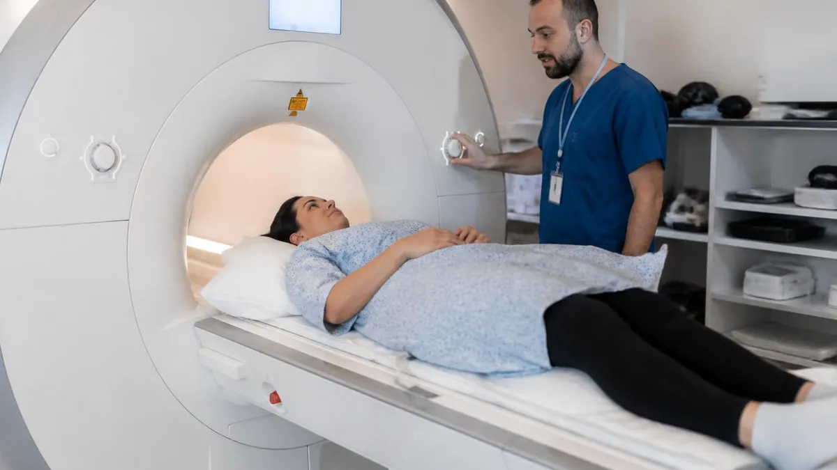
Patient communication is a non-technical skill that is increasingly recognized as important in the MRI technologist's role. Many women presenting for breast MRI are understandably anxious — some are being evaluated for a new cancer diagnosis, others are known high-risk patients who have been screening annually for years. Taking time before the scan begins to explain what the patient will experience, how long the study will take, and what happens after the images are acquired can meaningfully reduce patient anxiety, improve cooperation, and ultimately result in fewer motion artifacts and better image quality.
Building a comprehensive knowledge base for the MRI registry exam means integrating all of these threads — physics, protocols, anatomy, pathology, artifacts, and patient care — rather than studying each in isolation. Practice questions that present clinical scenarios force you to apply this knowledge flexibly, which is exactly the kind of reasoning the registry exam is designed to assess. Using a mix of targeted content review, practice exams, and case-based learning gives you the broadest preparation and the highest probability of success on exam day.
You can continue preparing with the cpt 72195, which includes answer explanations for every question.
You can continue preparing with the open mri locations near me, which includes answer explanations for every question.
MRI Questions and Answers
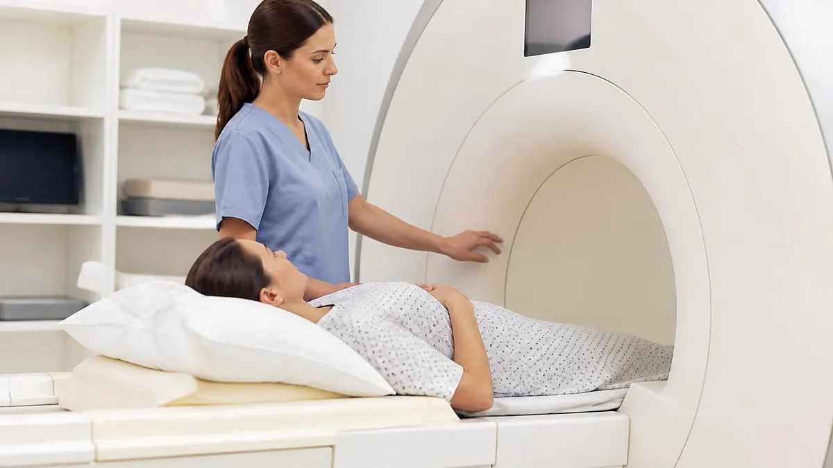
DWI MRI: Diffusion-Weighted Imaging Explained (2026 Guide)
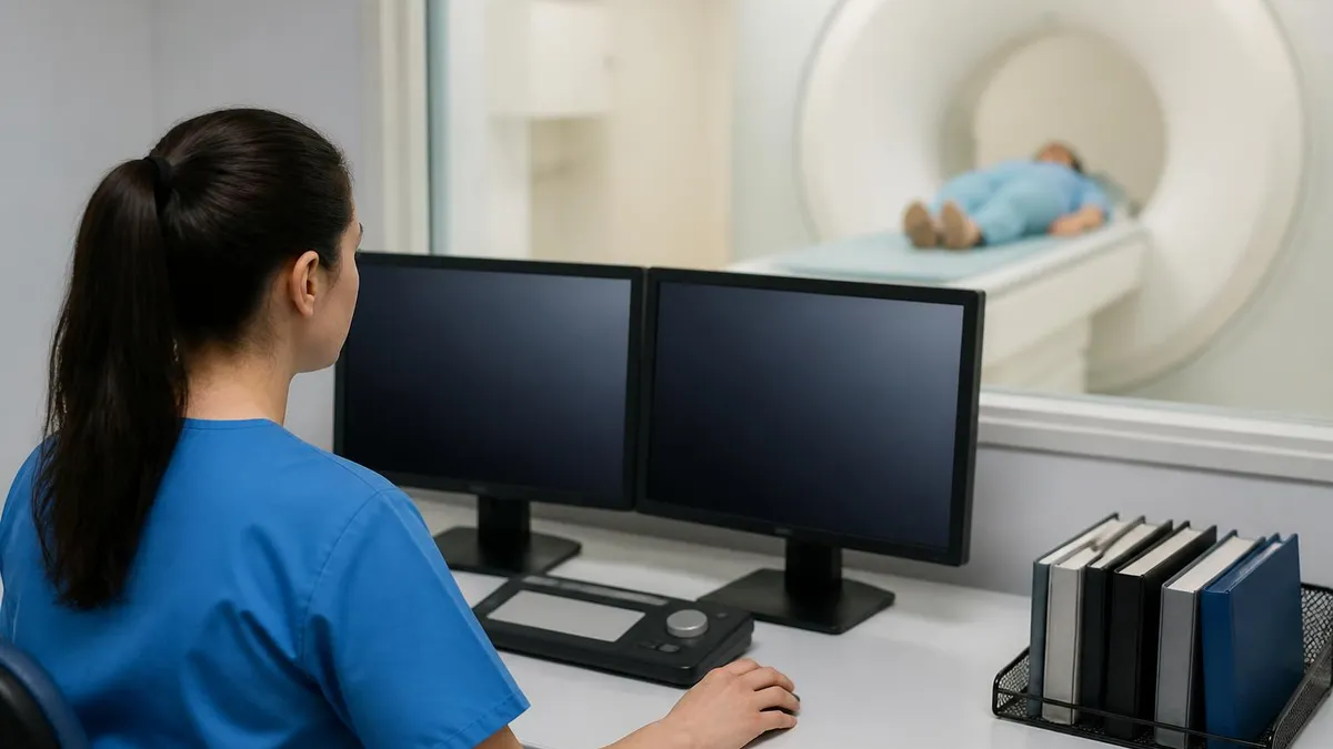
MRI Lumbar Spine Without Contrast: Complete Guide to the Scan, What It Shows, and How to Prepare
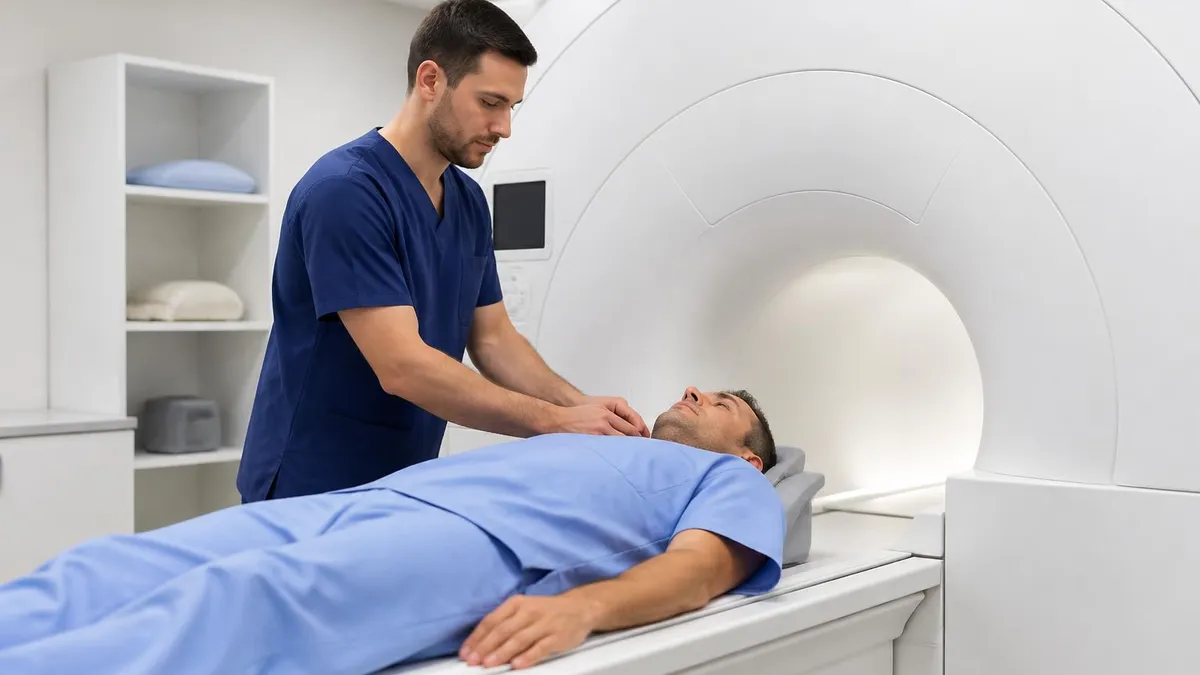
MRI History: From Nuclear Resonance to Modern Scanners

MRI of Cervical Spine: Complete Guide to Imaging, Anatomy, Pathology, and Patient Preparation
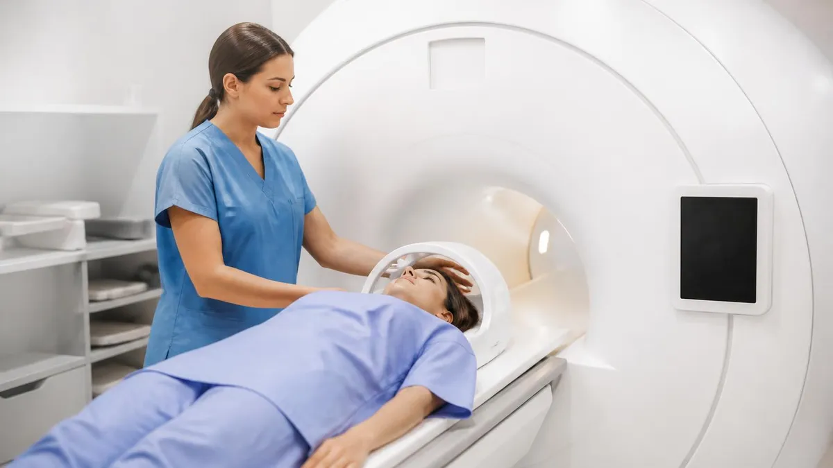
Where to Get an MRI: Facilities, Costs, and How to Choose
About the Author

Medical Laboratory Scientist & Clinical Certification Expert
Johns Hopkins UniversityDr. Sandra Kim holds a PhD in Clinical Laboratory Science from Johns Hopkins University and is certified as a Medical Technologist (MT) and Medical Laboratory Scientist (MLS) through ASCP. With 16 years of clinical laboratory experience spanning hematology, microbiology, and molecular diagnostics, she prepares candidates for ASCP board exams, MLT, MLS, and specialist certification tests.
Join the Discussion
Connect with other students preparing for this exam. Share tips, ask questions, and get advice from people who have been there.
View discussion (8 replies)