
MRI Brain Without Contrast: What It Shows, When It's Used, and What to Expect
🆕 Learn what an MRI brain without contrast shows, when doctors order it, and how to prepare. Complete guide for patients and MRI students.
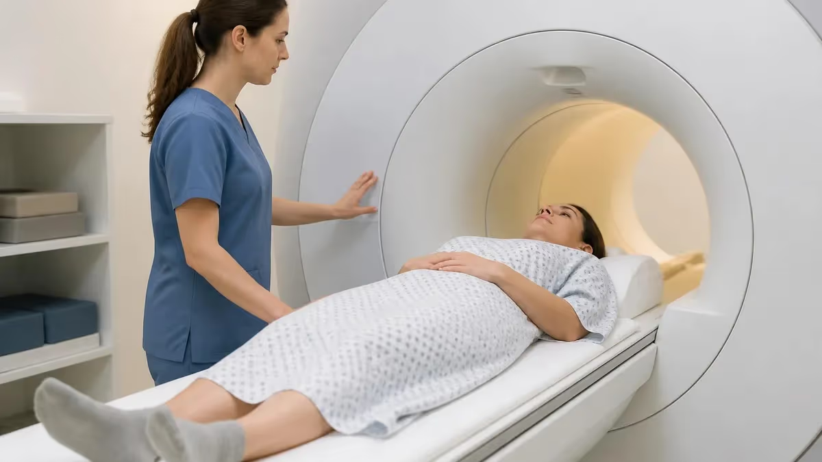
An MRI brain without contrast is one of the most commonly ordered neuroimaging studies in the United States, performed millions of times each year across hospital systems, outpatient imaging centers, and emergency departments. Unlike CT scans that use ionizing radiation, this study relies entirely on magnetic fields and radiofrequency pulses to generate detailed cross-sectional images of brain tissue, cerebrospinal fluid pathways, white matter tracts, and vascular structures — all without exposing the patient to radiation or injecting any chemical agent into the bloodstream.
Physicians order non-contrast brain MRI to evaluate a wide range of clinical concerns: new-onset headaches, suspected multiple sclerosis, seizure disorders, memory loss, dizziness, and suspected stroke among the most frequent. Because the scan requires no contrast agent, it avoids the small but real risks associated with gadolinium-based contrast agents, including nephrogenic systemic fibrosis in patients with impaired kidney function and, more recently discussed, gadolinium deposition in brain tissue after repeated administrations.
For MRI technologists preparing for registry exams or seeking to strengthen their clinical knowledge, understanding the pulse sequences, image weighting, and diagnostic limitations of non-contrast brain protocols is foundational. Sequences like T1-weighted, T2-weighted, FLAIR, diffusion-weighted imaging (DWI), and gradient echo each reveal different tissue properties, and knowing when each sequence fires and why it produces the contrast it does separates competent practitioners from truly skilled ones.
Patient preparation for a brain MRI without contrast is comparatively straightforward. Because no contrast injection occurs, patients with kidney disease, contrast allergies, or implanted devices that are MRI-conditional need not worry about the additional screening questions that accompany gadolinium administration. However, standard MRI safety screening — checking for pacemakers, aneurysm clips, cochlear implants, and metallic foreign bodies — remains absolutely mandatory before any patient enters the magnet room.
The diagnostic information yielded by a well-executed non-contrast brain MRI rivals and in some cases surpasses contrast-enhanced studies for certain pathologies. Acute ischemic stroke on DWI, for example, is detected within minutes of symptom onset without any contrast. White matter demyelinating lesions in MS appear conspicuously on FLAIR sequences. Hemorrhage and hemosiderin deposits are exquisitely sensitive on gradient echo or susceptibility-weighted imaging. Knowing this information about a brain mri without contrast helps technologists advocate for appropriate protocols and answer patient questions confidently.
This guide covers everything a patient, student, or practicing MRI technologist needs to know about non-contrast brain MRI: the sequences included in a standard protocol, what the study can and cannot diagnose, how field strength affects image quality, how to prepare your patient, and how this foundational knowledge maps to registry exam questions. Whether you are a patient scheduled for your first scan or an MRI student working toward ARRT certification, this comprehensive resource will give you a clear, accurate, and clinically relevant foundation.
The following sections walk through the imaging sequences in detail, compare non-contrast and contrast protocols, outline what conditions the study diagnoses well versus poorly, and offer practical preparation checklists. Statistical benchmarks, expert tips, and realistic practice questions are woven throughout to reinforce learning and help readers convert knowledge into confident clinical performance.
MRI: upcoming dates and official changes
- Aug 17, 2026: ARRT launched a separate entry-level Imaging Assistant, Magnetic Resonance (I.A.) credential. A reduced $75 fee applied to applications made Aug. 17–Oct. 14, 2026, for testing Oct. 15–Nov. 30, 2026. Later applicants pay $225 and can test on or after Jan. 1, 2027.[4]
- The ARRT MRI exam for postprimary-pathway candidates has 220 questions: 200 scored and 20 unscored pilot items.[1]
- The ARRT MRI exam for primary-pathway candidates has 230 questions: 200 scored and 30 unscored pilot items.[2]
- Postprimary-pathway candidates get 210 minutes for the ARRT MRI exam, inside a 230-minute (3 hours, 50 minutes) appointment.[1]
- Primary-pathway candidates get 230 minutes for the ARRT MRI exam, inside a 250-minute (4 hours, 10 minutes) appointment that also covers the tutorial, the NDA and a survey.[2]
- To pass the ARRT MRI exam you need a total scaled score of 75 on ARRT's 1–99 scale. This is not a percentage.[3]
- The ARRT MRI exam through the postprimary pathway costs $225, or $450 for candidates whose supporting credential is from NMTCB or ARDMS.[4]
- The ARRT MRI certification exam costs $225 to apply for through the primary eligibility pathway.[4]
- ARRT MRI candidates have three attempts to pass the exam, all within three years of the date their first exam window opens.[2]
- ARRT MRI certification and registration lasts 10 years, even with annual renewal. After that, holders must complete Continuing Qualifications Requirements (CQR).[5]
Numbers in brackets link to the official sources listed under Sources & last verified. Dates that have passed are removed automatically.
MRI Exam at a Glance
| Item | Detail | Source · Verified |
|---|---|---|
| Number of questions (Postprimary pathway) | 220 questions (200 scored + 20 unscored pilot)[1] | assets-us-01.kc-usercontent.com · Oct 15, 2026 |
| Number of questions (Primary pathway) | 230 questions (200 scored + 30 unscored pilot)[2] | assets-us-01.kc-usercontent.com · Oct 15, 2026 |
| Time limit (Postprimary pathway) | 210 minutes test time (230-minute appointment)[1] | assets-us-01.kc-usercontent.com · Oct 15, 2026 |
| Time limit (Primary pathway) | 230 minutes test time (250-minute appointment)[2] | assets-us-01.kc-usercontent.com · Oct 15, 2026 |
| Passing score | Scaled score of 75 (scale 1–99)[3] | The American Registry of Radiologic Technologists · Oct 15, 2026 |
| Exam fee (Postprimary pathway) | $225 application fee ($450 with an NMTCB or ARDMS supporting credential)[4] | The American Registry of Radiologic Technologists · Oct 15, 2026 |
| Exam fee (Primary pathway) | $225 application fee[4] | The American Registry of Radiologic Technologists · Oct 15, 2026 |
| Retake policy | 3 attempts within 3 years of the first exam window[2] | assets-us-01.kc-usercontent.com · Oct 15, 2026 |
| Validity | 10 years, then Continuing Qualifications Requirements (CQR)[5] | assets-us-01.kc-usercontent.com · Oct 15, 2026 |
| Renewal | Annual renewal, $65/year; 24 CE credits every 2 years[5] | assets-us-01.kc-usercontent.com · Oct 15, 2026 |
| Delivery | Computer-based at Pearson VUE test centers[2] | assets-us-01.kc-usercontent.com · Oct 15, 2026 |
| Eligibility (Postprimary pathway) | Supporting ARRT credential (or NMTCB/ARDMS) + structured education + clinical experience[1] | assets-us-01.kc-usercontent.com · Oct 15, 2026 |
| Eligibility (Primary pathway) | ARRT-recognized MRI program + associate degree or higher + ethics[2] | assets-us-01.kc-usercontent.com · Oct 15, 2026 |
| Score release | Preliminary score at the test center; official results in about 10 business days[3] | The American Registry of Radiologic Technologists · Oct 15, 2026 |
| Testing window | One-year (365-day) exam window per application[2] | assets-us-01.kc-usercontent.com · Oct 15, 2026 |
Brain MRI Without Contrast by the Numbers
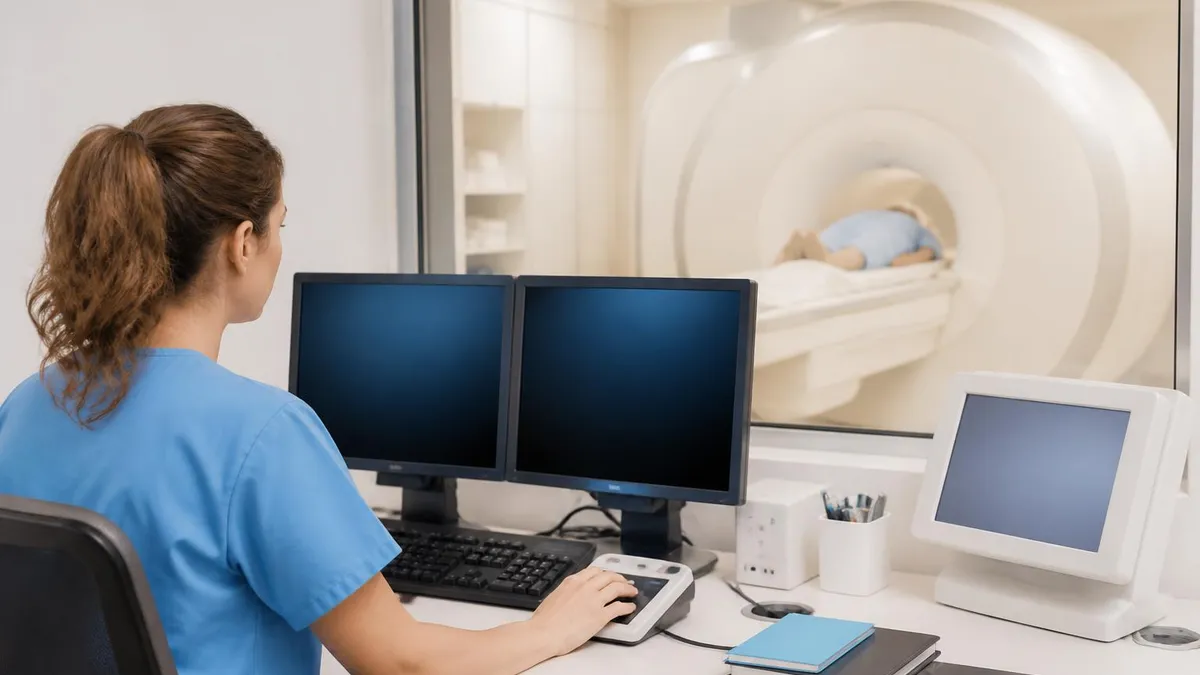
Standard Non-Contrast Brain MRI Protocol Sequences
T1 sequences show anatomy in sharp detail. Fat and subacute blood appear bright; CSF appears dark. Used to assess gray-white differentiation, detect lipomas, identify subacute hemorrhage, and provide a baseline anatomical roadmap for all other sequences.
T2 sequences make fluid bright, making pathological edema, demyelinating plaques, and cystic lesions highly conspicuous. T2 is the workhorse for detecting white matter disease, herniated disc contributions at the foramen magnum, and most inflammatory conditions.
FLAIR suppresses free CSF signal while keeping pathological fluid bright, dramatically improving visibility of periventricular MS plaques, cortical infarcts, and subarachnoid hemorrhage. It is often the single most diagnostically important sequence in a non-contrast brain protocol.
DWI detects restriction of water molecule movement, flagging acute ischemic stroke, abscess, and dense cellular tumors within minutes. The companion ADC map distinguishes true restriction from T2 shine-through, a critical distinction that separates acute from chronic infarcts.
Gradient echo and susceptibility-weighted imaging (SWI) are exquisitely sensitive to blood products, calcium, and iron. Microbleeds, hemosiderin from old trauma, cavernous malformations, and amyloid angiopathy patterns are best seen on these sequences — often invisible on T1 or T2.
Understanding what a non-contrast brain MRI can confidently diagnose is critical knowledge for both ordering clinicians and MRI technologists. Acute ischemic stroke is perhaps the highest-stakes diagnosis the study handles, and the DWI sequence revolutionized stroke imaging. Before DWI became routine in the 1990s, CT was the primary acute neuroimaging tool, but CT misses the majority of ischemic strokes in the first 24 hours, particularly small or posterior fossa events. DWI detects restricted diffusion — the earliest cellular sign of ischemia — within minutes of arterial occlusion, giving emergency physicians actionable data in the narrow thrombolysis window.
Multiple sclerosis is another condition where non-contrast MRI genuinely excels. The McDonald Criteria, the internationally accepted diagnostic framework for MS, defines lesion requirements based on MRI findings alone. A skilled radiologist reviewing T2 and FLAIR sequences can identify ovoid periventricular lesions oriented perpendicular to the corpus callosum (Dawson's fingers), juxtacortical lesions, infratentorial lesions, and spinal cord lesions — distributional patterns that, combined with clinical history, support the diagnosis of MS without ever needing contrast in many cases. For tracking known MS patients, many neurologists routinely alternate between contrast and non-contrast protocols depending on whether new activity is suspected.
Traumatic brain injury evaluation also relies heavily on non-contrast imaging. While a CT head remains the first-line study in acute trauma because of its speed and hemorrhage sensitivity, MRI — particularly gradient echo and SWI — detects diffuse axonal injury (DAI), small contusions, and microhemorrhages that CT completely misses. In sports medicine and military medicine, where repetitive subconcussive impacts accumulate, SWI has become an important tool for documenting injury burden not visible on CT or even conventional T2 sequences.
Brain tumor characterization is more nuanced. Non-contrast MRI can identify the presence, size, and location of most brain tumors, and it can characterize signal characteristics suggestive of specific tumor types. A well-defined, homogeneously T1-hypointense, T2-hyperintense round mass in the cerebellum of an adult suggests metastasis even without contrast. However, definitive assessment of blood-brain barrier breakdown — a key marker of high-grade glioma and active metastasis — requires gadolinium enhancement. For this reason, tumor imaging protocols almost always include contrast unless there is a specific contraindication such as renal failure or allergy.
Epilepsy evaluation with non-contrast MRI targets structural causes of seizures: hippocampal sclerosis, cortical dysplasia, cavernous malformations, and low-grade tumors. High-resolution coronal T2 and FLAIR sequences through the temporal lobes, combined with thin-slice T1 volumetric acquisitions, provide the most sensitive structural seizure workup. Many epilepsy centers use dedicated 3 Tesla protocols with phase-array coils to maximize cortical surface detail — an area where higher field strength dramatically improves sensitivity for subtle focal cortical dysplasias.
Neurodegenerative diseases, including Alzheimer's disease and other dementias, are evaluated with structural T1 volumetric imaging that quantifies regional cortical atrophy and hippocampal volume loss. Automated segmentation software running on these T1 datasets can compare a patient's hippocampal volume against age-matched normative data, providing objective biomarkers to complement clinical diagnosis. While advanced imaging biomarkers for amyloid and tau require PET scans or CSF analysis, structural brain MRI remains the first-line imaging tool for dementia workup in clinical practice.
Congenital brain malformations — Chiari malformations, corpus callosum dysgenesis, lissencephaly, heterotopias — are exquisitely characterized by MRI, and in these conditions contrast adds little diagnostic value. Pediatric neuroimaging heavily favors MRI over CT precisely because of the radiation-free nature of the study, making non-contrast brain MRI the dominant neuroimaging tool in children and a subject of significant technical development, including feed-and-swaddle protocols for infants and rapid acquisition sequences that reduce motion artifact in uncooperative patients.
MRI Practice Test Questions
Prepare for the MRI - Magnetic Resonance Imaging exam with our free practice test modules. Each quiz covers key topics to help you pass on your first try.
MRI Knowledge
MRI Exam Questions covering Knowledge. Master MRI Test concepts for certification prep.
MRI Physics
Free MRI Practice Test featuring Physics. Improve your MRI Exam score with mock test prep.
MRI Anatomy and Pathology
MRI Test Prep for MRI Anatomy and Pathology. Practice MRI Quiz questions and boost your score.
MRI Anatomy and Positioning
MRI Questions and Answers on MRI Anatomy and Positioning. Free MRI practice for exam readiness.
MRI Contrast Agents
Free MRI Quiz on MRI Contrast Agents. MRI Exam prep questions with detailed explanations.
MRI Patient Care and Positioning
MRI Practice Questions for MRI Patient Care and Positioning. Build confidence for your MRI certification exam.
Non-Contrast vs. Contrast Brain MRI: Key Comparisons
Many of the most common brain MRI indications are fully addressed without gadolinium. Acute stroke workup relies on DWI and ADC maps, which function independently of the blood-brain barrier. Multiple sclerosis diagnosis and follow-up, evaluation of seizure disorders, workup of dementia and neurodegenerative disease, assessment of headache without red-flag features, and screening for congenital anomalies all typically proceed without contrast injection. In these contexts, omitting contrast simplifies scheduling, reduces patient risk, lowers cost, and shortens overall examination time by approximately 15 to 20 minutes.
Additionally, patients with stage 3b or worse chronic kidney disease (eGFR below 30 mL/min/1.73m²) face elevated risk of nephrogenic systemic fibrosis (NSF) from certain gadolinium-based contrast agents. For these patients, non-contrast protocols are not merely a preference but a clinical necessity. Given the growing body of literature on gadolinium retention in brain tissue even in patients with normal renal function, many radiologists and neurologists apply a general principle of contrast parsimony — ordering it only when the clinical question cannot be answered without it.
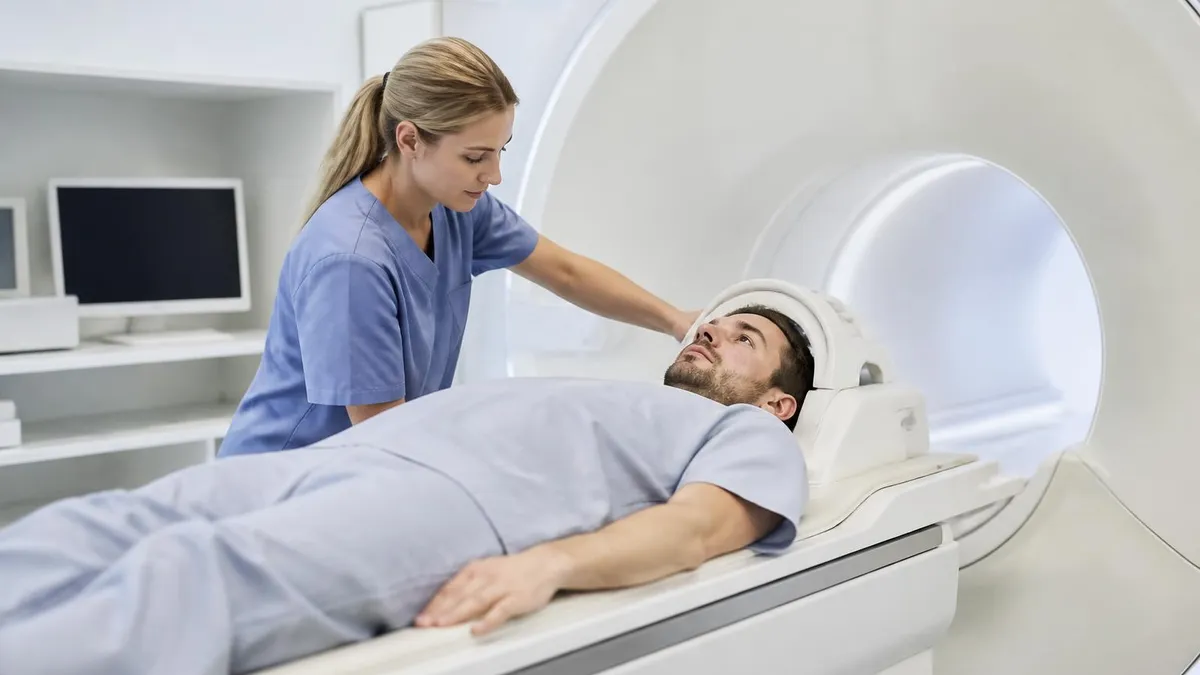
Advantages and Limitations of Non-Contrast Brain MRI
- +Zero ionizing radiation — safe for children, pregnant patients, and repeat imaging
- +No contrast injection eliminates risk of allergic reactions and nephrogenic systemic fibrosis
- +Exquisite soft tissue contrast reveals white matter, gray matter, and CSF pathways clearly
- +DWI detects acute ischemic stroke within minutes — faster than CT for most ischemic events
- +No renal function screening required before the examination
- +Lower overall cost compared to contrast-enhanced protocols
- +FLAIR and SWI sequences detect MS plaques, microbleeds, and hemosiderin with high sensitivity
- −Cannot assess blood-brain barrier breakdown — misses active tumor enhancement and acute MS lesion activity
- −Long scan times (30–45 minutes) compared to CT head (under 5 minutes)
- −Motion artifact degrades image quality — uncooperative patients may require sedation
- −Metallic implants and pacemakers may be absolute or conditional contraindications
- −Limited availability and higher cost compared to CT in emergency settings
- −Cannot distinguish viable tumor from radiation necrosis without contrast or advanced sequences
- −Susceptibility artifact from skull base and dental hardware can obscure inferior temporal and orbital structures
Patient Preparation Checklist for Non-Contrast Brain MRI
- ✓Complete the MRI safety screening questionnaire honestly, listing all implants, surgeries, and metallic foreign bodies.
- ✓Remove all metallic jewelry, hairpins, piercings, and hearing aids before entering the scan room.
- ✓Inform the technologist about any history of welding, metal grinding, or eye injury involving metal fragments.
- ✓Tell your technologist if you have a pacemaker, spinal cord stimulator, cochlear implant, or any neurostimulator device.
- ✓Wear comfortable, loose clothing without metal zippers or underwire bras, or change into a provided gown.
- ✓No fasting is required for a non-contrast brain MRI — eat and take medications as normal unless told otherwise.
- ✓Inform staff about any history of claustrophobia so they can discuss anxiolytic medication options in advance.
- ✓Arrive 15 minutes early to allow time for paperwork, safety screening, and changing if needed.
- ✓Ask whether your specific implant model is MRI-safe or conditional — bring the implant card if available.
- ✓Practice lying still and breathing quietly through your nose to minimize motion artifact during the scan.
DWI Is the Most Time-Critical Sequence in Stroke Imaging
Diffusion-weighted imaging can detect acute ischemic stroke within 30 minutes of symptom onset — long before T2 or FLAIR sequences show any abnormality. In a patient presenting with sudden focal neurological deficits, a negative DWI with positive FLAIR suggests a subacute stroke (greater than 4–6 hours old), directly influencing thrombolysis eligibility and triage decisions. MRI technologists must prioritize DWI acquisition and flag any motion degradation immediately so the study can be repeated before the patient leaves the scanner.
For MRI students and technologists preparing for the ARRT MRI registry examination, brain imaging sequences represent one of the highest-yield content areas in the entire exam. The ARRT Content Specifications for MRI list image production as the dominant exam domain at approximately 40 percent of all questions, and within image production, pulse sequence parameters — TR, TE, TI, flip angle, and their effect on tissue contrast — are tested repeatedly. Understanding how each brain sequence achieves its contrast is not rote memorization; it is applied physics.
Consider FLAIR as an example of registry-critical sequence knowledge. FLAIR is an inversion recovery technique, meaning it uses a 180-degree inversion pulse followed by an inversion time (TI) timed specifically to reach the null point of free water — approximately 2,500 ms at 1.5 Tesla. When the sequence readout fires at that TI, CSF contributes no signal, appearing dark.
Pathological fluid in lesions, however, has a shorter T1 due to protein content or cellular density, so it has already recovered past the null point and appears bright. The registry exam tests this principle both directly (what does FLAIR suppress?) and indirectly (why does a lesion appear bright on FLAIR but dark on T1?).
Gradient echo sequences and their role in hemorrhage detection form another heavily tested registry topic. Conventional spin echo sequences refocus static magnetic field inhomogeneities using the 180-degree refocusing pulse, which partially cancels susceptibility effects from iron-containing blood products. Gradient echo sequences omit this refocusing pulse, making them exquisitely sensitive to local field distortions caused by deoxyhemoglobin, methemoglobin, hemosiderin, and calcium. This explains why cavernous malformations — benign vascular hamartomas filled with blood products at various stages of evolution — appear as striking low-signal blooming artifacts on GRE while being nearly invisible on T2-weighted spin echo images.
DWI physics is another registry staple. The diffusion-weighted image is generated by applying a pair of balanced gradient pulses (the diffusion-sensitizing gradients) around a 180-degree refocusing pulse. Protons in freely diffusing water accumulate phase from the first gradient and are fully rephased by the second; protons in restricted environments (cytotoxic edema, abscess pus, dense tumor cellularity) cannot fully rephase, resulting in signal loss that paradoxically makes restricted areas appear bright on DWI due to the underlying T2 contribution.
The b-value — typically 0 and 1,000 s/mm² in routine brain protocols — controls the degree of diffusion weighting, with higher b-values increasing sensitivity to restriction at the cost of lower SNR.
MRI physics questions on the registry also address artifact recognition, which is clinically essential in brain imaging. Chemical shift artifact along the frequency-encoding direction creates a dark band at fat-water interfaces — seen, for example, at the interface between orbital fat and the optic nerve, which can mimic pathology. Gibbs ringing artifact (truncation artifact) appears as parallel lines near sharp signal transitions like the corpus callosum and can simulate syrinx or pseudolesion. Motion ghosting in the phase-encoding direction from vascular pulsation is managed by appropriate phase-encode direction selection, saturation bands, and cardiac gating in sensitive areas.
Understanding brain anatomy on MRI is equally critical for registry success. The exam tests identification of normal structures — the caudate nucleus, putamen, globus pallidus, thalamus, internal capsule, hippocampus, corpus callosum, and brainstem divisions — and their signal characteristics on each sequence type. The basal ganglia appear isointense to cortex on T1 but may show slight T2 hyperintensity in pathological iron deposition states. The posterior limb of the internal capsule, a critical structure in stroke assessment, should always appear T1-isointense in adults and shows characteristic diffusion restriction in acute lacunar infarcts.
Practicing with realistic registry-style questions remains the single most effective preparation strategy. Rather than re-reading textbook chapters, experienced MRI educators recommend spending at least 60 percent of study time in active practice mode — answering questions, reviewing explanations, and identifying knowledge gaps.
The ARRT reports a first-time pass rate of approximately 85 to 90 percent for MRI candidates, but this figure masks wide variation between candidates who practiced extensively and those who relied primarily on passive review. The candidates who consistently score in the upper percentiles on practice exams are the ones who understand why each answer is right or wrong, not just which answer choice is correct.
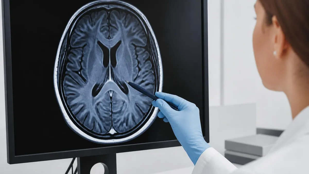
Even for a non-contrast brain MRI, a complete and thorough MRI safety screening must be performed before every patient enters Zone III or Zone IV. Patients with certain pacemakers, implantable cardioverter-defibrillators, neurostimulators, cochlear implants, or ferromagnetic aneurysm clips may be harmed or killed by the magnetic field. Never rely on verbal screening alone — confirm implant manufacturer and model through implant cards, medical records, or direct contact with the referring physician before proceeding.
The limitations of non-contrast brain MRI are as important to understand as its strengths, and recognizing when a study is insufficient — when contrast or an alternative modality is needed — is a hallmark of clinical competence. The most fundamental limitation is the inability to demonstrate blood-brain barrier breakdown.
The blood-brain barrier is formed by tight junctions between cerebral endothelial cells, normally preventing gadolinium from entering brain parenchyma. When the barrier is disrupted by tumor, inflammation, or infection, contrast leaks into tissue and creates the enhancement patterns that radiologists use to characterize pathology. A non-contrast scan simply cannot provide this information.
Active versus inactive disease in multiple sclerosis illustrates this limitation starkly. A patient with established MS may have dozens of white matter lesions on FLAIR — but are they old, stable plaques or new, actively inflamed lesions? The distinction matters enormously for treatment decisions, particularly when neurologists are considering escalation to more aggressive disease-modifying therapies. Only gadolinium enhancement reveals active demyelination, because active plaques have disrupted blood-brain barrier while older plaques do not. A non-contrast brain MRI cannot answer this question, and ordering one when active disease assessment is the clinical objective is a clinical oversight.
Leptomeningeal disease — carcinomatous meningitis from metastatic cancer spreading along the meningeal surfaces — is another condition that requires contrast for diagnosis. The leptomeninges normally receive no gadolinium signal, but tumor infiltration creates abnormal enhancement along the surface of the brain and spinal cord that is definitively absent on non-contrast sequences. By the time leptomeningeal metastasis becomes large enough to detect by structural distortion alone, the disease burden is typically very advanced. Contrast-enhanced MRI with dedicated thin-slice sequences through the entire neuraxis is the study of choice when leptomeningeal metastasis is suspected.
Small pituitary microadenomas smaller than 6 mm frequently escape detection on non-contrast sequences because their signal characteristics overlap with normal pituitary gland tissue. Dynamic contrast-enhanced sequences, which capture the differential washout kinetics of adenoma versus normal pituitary (the normal gland enhances faster than most adenomas), are standard of care for pituitary microadenoma detection in patients with hyperprolactinemia, Cushing's disease, or acromegaly. Ordering a non-contrast brain MRI when pituitary disease is the specific clinical question reflects either resource limitation or incomplete understanding of the imaging indication.
Cerebral venous sinus thrombosis (CVST) represents an interesting hybrid case. Non-contrast MRI can suggest CVST through T1 hyperintensity within the thrombosed sinus (subacute thrombus) or T2/FLAIR signal abnormality in the affected cortex from venous congestion. However, MR venography (MRV) — which can be performed as either a contrast-enhanced or time-of-flight non-contrast technique — is required to confirm the diagnosis by directly imaging venous flow. The time-of-flight MRV acquires signal from flowing blood without contrast by using flow-related enhancement; it has adequate sensitivity for large sinus thrombosis but may miss small or slow-flow thrombus better detected with contrast-enhanced MRV.
Posterior fossa imaging deserves special mention as a technical limitation of brain MRI regardless of contrast administration. The posterior fossa contains the cerebellum, brainstem, and cranial nerves — structures critical to balance, coordination, swallowing, and vital function — but it is also adjacent to the dense bone of the petrous ridges and skull base, which create susceptibility artifact on gradient echo and EPI-based sequences like DWI.
Small brainstem infarcts, the most clinically dangerous posterior fossa lesions, can be obscured by this artifact. High-resolution DWI with reduced FOV techniques and dedicated posterior fossa protocols are increasingly used at high-volume stroke centers to mitigate this limitation.
For patients and clinicians navigating the contrast-versus-no-contrast decision, the most practical framework is to let the clinical question drive protocol selection rather than defaulting to one approach. A patient with new-onset generalized tonic-clonic seizures without focal deficits generally needs a high-resolution structural non-contrast MRI.
A patient with known breast cancer presenting with headaches needs a contrast-enhanced study to rule out metastasis. When in doubt, a direct conversation between the ordering clinician and the radiologist — the radiologist consultation known as a protocoling call — typically resolves ambiguity and ensures the right imaging strategy reaches the right patient at the right time.
Practical tips for MRI technologists performing non-contrast brain examinations begin with patient positioning. The head should be centered in the head coil, with the orbitomeatal line perpendicular to the bore axis to standardize axial slice orientation. Foam padding or dedicated positioning aids should minimize residual head movement — even sub-millimeter motion during high-resolution sequences blurs cortical detail and degrades the diagnostic value of the study. Many sites use head restraint cushions or vacuum immobilization for patients with tremor or involuntary movement disorders.
Coil selection directly impacts signal-to-noise ratio and image quality. Modern brain MRI uses multi-channel phased array head coils with 20 to 64 elements that allow parallel imaging acceleration techniques like GRAPPA or SENSE. These techniques acquire partial k-space data and use coil sensitivity maps to reconstruct the full image, enabling acceleration factors of 2x to 4x that cut scan time substantially without meaningful SNR penalty. Understanding how to select appropriate acceleration factors and recognize acceleration-related artifacts (ghosting at specific locations in the phase-encoding direction) is part of the competent technologist's skill set.
Slice prescription for brain protocols follows standardized anatomical landmarks. Axial sequences are typically prescribed parallel to the anterior commissure-posterior commissure (AC-PC) line for reproducibility across serial examinations — an important consideration when tracking MS lesion load or tumor treatment response over time. Coronal sequences are prescribed perpendicular to the long axis of the hippocampus for epilepsy and memory disorder workups. Sagittal sequences, particularly T1 and FLAIR midline views, are essential for identifying corpus callosum lesions, Chiari malformations, and pineal region pathology.
Bandwidth selection affects both SNR and chemical shift artifact. Lower receiver bandwidth increases SNR but makes the image more susceptible to chemical shift artifact (misregistration of fat signal relative to water signal along the frequency-encoding direction) and geometric distortion. Higher bandwidth reduces these artifacts but decreases SNR. For standard brain imaging at 1.5T, a receiver bandwidth of approximately 16 to 20 kHz per pixel represents a practical compromise. At 3T, higher bandwidths are typically used to control the doubled chemical shift artifact that accompanies the stronger static field.
Patient communication throughout the non-contrast brain MRI directly affects image quality by reducing anxiety-driven motion. Before the scan begins, explain the sequence of sounds the patient will hear — the knocking, banging, and buzzing characteristic of gradient coil activation — and give realistic time estimates for each sequence.
Encourage the patient to breathe normally through the nose with the mouth closed, which stabilizes the jaw and reduces swallowing motion. Check in verbally or through the intercom between sequences; patients who know they are being monitored and can communicate tend to remain significantly calmer and move less than those left in silence.
Quality assurance practices for non-contrast brain MRI include checking scout images immediately after acquisition to confirm correct positioning before proceeding with the diagnostic sequences. If the orbitomeatal line is off by more than a few degrees or if motion artifact is visible in the scout, reposition or re-prescribe before investing the full scan time. Many sites use real-time motion monitoring tools — either prospective motion correction hardware or navigator echoes — to flag motion during acquisition rather than discovering it at the end of the examination when the patient has already been discharged.
Documenting scan parameters in the radiology report or in the PACS notes adds clinical value for radiologists interpreting serial studies. When a patient's brain MRI performed in 2024 used 3mm axial FLAIR slices and the 2026 follow-up uses 1mm isotropic 3D FLAIR, apparent changes in lesion conspicuity may reflect technique rather than true disease change. A brief technical note stating field strength, coil type, slice thickness, and whether parallel imaging was used provides the radiologist with the context needed to interpret apparent changes accurately and avoid clinical mismanagement based on technique-driven artifact.
MRI Questions and Answers
Sources & last verified
- assets-us-01.kc-usercontent.com — number of questions, time limit, eligibility (official source). “MRI Scored Items 200 Pilot Items 20 Total Items 220 Test Time 210 min” Verified Oct 15, 2026.
- assets-us-01.kc-usercontent.com — number of questions, time limit, retake policy, delivery (official source). “MRI Scored Items 200 Pilot Items 30 Total Items 230 Test Time 230 min” Verified Oct 15, 2026.
- The American Registry of Radiologic Technologists — passing score, score release (official source). “Our exam scoring scale ranges from 1 to 99, and you’ll need a total score of 75 to pass the exam.” Verified Oct 15, 2026.
- The American Registry of Radiologic Technologists — exam fee, other (official source). “Postprimary (M, CT, MR, BD, CI, VI, VS, BS, R.R.A.): $225 Postprimary (CT, MR, BD, or VS—using NMTCB as supporting category): $450 Postprimary (MR, VS,…” Verified Oct 15, 2026.
- assets-us-01.kc-usercontent.com — validity, renewal (official source). “ARRT certification and registration—even with annual renewal—is limited to 10 years.” Verified Oct 15, 2026.
How we verify: editorial & fact-checking policy. Report an error: contact us.

MRI of Cervical Spine: Complete Guide to Imaging, Anatomy, Pathology, and Patient Preparation

Knee MRI Images: A Complete Guide to Reading, Understanding, and Interpreting Knee Scans
Ace Your Exam with MRI Practice Test 2026 Prep

MRI Tech School 2026 — Programs, Requirements, and What to Expect
MRI Procedure: What to Expect Before, During and After Your Scan
How this page was made: written with AI tools. Exam facts are checked against the official sources listed below. Not reviewed by a subject-matter expert. Editorial policy
Sources & references
Exam facts on this page (fees, format, dates, passing scores) are checked against these official sources; the date is our last check. Editorial policy
- arrt.org — Passing score, score release, fees, exam information · verified Oct 2026
- assets-us-01.kc-usercontent.com — Renewal, score validity, test delivery, eligibility, exam format, test dates, time limit · verified Oct 2026