
EEG Database: What It Is, How It Works, and Why It Matters for Brain Science
Learn what an EEG database is, how EEG test data is stored and used in research, and what this means for diagnosis. 🧠 Complete 2026 September guide.

An EEG database is a structured collection of electroencephalography recordings, metadata, and clinical annotations gathered from patients and research participants over time. When clinicians or scientists perform an EEG test, the raw brainwave signals captured by scalp electrodes do not simply disappear after the report is written — they are preserved, indexed, and stored so that future researchers, neurologists, and artificial intelligence systems can analyze them again. Understanding what these databases contain, how they are built, and who uses them helps demystify an important but often overlooked layer of modern neuroscience and clinical neurology.
The EEG medical test has been performed for nearly a century, meaning that large institutions have accumulated thousands — and in some cases millions — of individual recordings. Each recording may span 20 minutes for a routine outpatient study or extend across several days for continuous inpatient monitoring. When organized into a searchable eeg database, these recordings become powerful resources for training diagnostic algorithms, validating new electrode technologies, comparing drug effects on brain activity, and establishing normative values for different age groups.
For anyone preparing for a career as an EEG technologist or pursuing board certification, familiarity with database concepts is increasingly important. Modern EEG departments rely on electronic health record integrations, proprietary waveform storage systems, and shared research repositories that follow international data-sharing standards. Knowing how test data flows from the bedside to long-term storage — and how it can be retrieved — reflects the kind of practical knowledge examiners now expect candidates to demonstrate.
This article explores the full landscape of EEG databases: their architecture, their role in clinical and research settings, the major publicly available collections used in neuroscience, and the privacy and regulatory considerations that govern how patient recordings are handled. We also touch on cost considerations for the underlying EEG test itself, including EEG test price ranges and typical EEG test cost in the United States, since many patients who agree to data donation want to understand the value of the procedure they have undergone.
Beyond clinical use, EEG databases have become central to the field of computational neuroscience and machine learning. Engineers building seizure-detection algorithms need thousands of labeled ictal and interictal recordings to train reliable classifiers. Sleep researchers require nights of polysomnographic EEG data from diverse populations to model normal and abnormal sleep architecture. Brain-computer interface developers depend on carefully annotated motor-imagery datasets to teach devices to recognize intentional thought patterns. In every case, the database is the foundation upon which discovery rests.
Whether you are a student just learning what is an EEG test, a technologist seeking board exam preparation materials, or a researcher evaluating which public repository best fits your study design, this guide will give you the context and vocabulary you need. We cover the technical standards that make data sharable, the ethical frameworks that protect patient identities, and the practical steps professionals follow when contributing to or drawing from an EEG database. By the end, you will have a clear picture of why these collections matter and how to navigate them confidently.
EEG Database & Testing by the Numbers

How EEG Databases Are Structured
The core of any EEG database is the time-series voltage data sampled from each electrode. Files are typically stored in open formats such as EDF (European Data Format) or BDF, which preserve channel labels, sampling rates, and recording timestamps in a standardized, software-independent way.
Neurologists and technologists add event markers — seizure onsets, artifact timestamps, activation procedure intervals — directly to the recording. These labels transform raw signal into analyzable data, allowing algorithms to learn what a spike-wave discharge or a burst-suppression pattern looks like without manual review of every file.
Age, sex, diagnosis, medication history, and recording date are stored alongside the waveform. When de-identified according to HIPAA Safe Harbor standards, this metadata allows researchers to stratify analyses by demographic group or clinical condition without exposing personal health information.
Good databases include quality scores indicating how much muscle artifact, electrode noise, or movement contamination is present. Researchers use these flags to filter out unusable segments automatically, improving the reliability of any downstream analysis and reducing the manual workload on technologists and scientists.
As annotations are corrected or preprocessing pipelines change, database managers maintain version histories so published studies can cite the exact dataset state used. This reproducibility layer is essential for peer-reviewed research and regulatory submissions involving AI-based diagnostic tools.
Clinical EEG databases and research EEG databases serve overlapping but distinct purposes, and understanding the difference helps clarify why certain recordings are collected, how they are stored, and who ultimately has access to them. A clinical database is maintained by a hospital or neurology practice primarily to support patient care — neurologists retrieve prior studies to track changes in epilepsy burden over time, compare post-treatment brain activity against baseline, and document the evolution of an encephalopathy. These systems are tightly integrated with electronic health records and picture archiving systems, and access is restricted to treating clinicians and authorized administrative staff.
Research databases, by contrast, are curated to answer scientific questions that extend beyond any individual patient. The Temple University Hospital EEG Corpus, for example, contains more than 25,000 recordings from clinical patients who consented to secondary research use. This corpus has become one of the most important training grounds for seizure-detection algorithms in the world. Similarly, the PhysioNet platform hosts several EEG collections spanning epilepsy monitoring, sleep staging, and cognitive neuroscience experiments, all freely downloadable by registered researchers.
Hybrid databases occupy a middle ground: they are built within healthcare systems but are explicitly designed to feed research pipelines. The Mayo Clinic and Cleveland Clinic have both developed internal corpora that link EEG data to genetic profiles, imaging results, and long-term clinical outcomes. These longitudinal collections are extraordinarily valuable because they allow scientists to ask whether a particular brainwave pattern in childhood predicts epilepsy severity in adulthood — a question that a single-timepoint dataset simply cannot answer.
The technical architecture of modern EEG databases has evolved considerably since the early days of paper tape and analog storage. Contemporary systems use cloud-based object storage for waveform files, relational databases for metadata and annotations, and application programming interfaces that allow authorized software to query and stream data without downloading entire archives. This shift has made it practical for a research team in Boston to analyze recordings collected in San Francisco without physically transferring terabytes of data, dramatically accelerating the pace of collaborative science.
Standardization remains one of the biggest ongoing challenges. Different EEG amplifier manufacturers use proprietary file formats — Nihon Kohden's JE-111A format, Natus's Neurofax format, and Compumedics Profusion format are just a few examples — and converting these reliably without losing channel information or timing precision requires careful engineering. The Brain Imaging Data Structure (BIDS) extension for EEG, published in 2019, has made significant headway by defining a folder hierarchy and JSON sidecar specification that most major analysis toolboxes now support, including MNE-Python, EEGLAB, and FieldTrip.
For EEG technologists entering the workforce today, practical familiarity with at least one data management platform is increasingly expected. Many epilepsy monitoring units use systems such as Persyst or Natus NeuroWorks that include built-in trend analysis and automatic seizure detection drawing on internally trained classifiers. Understanding how these classifiers were built — and what kinds of database limitations may affect their sensitivity in specific patient populations — makes a technologist a more effective clinical partner and a stronger candidate for advanced roles in neuromonitoring.
It is worth noting that the growing importance of EEG databases does not diminish the significance of individual test quality. A database is only as good as the recordings it contains, and recordings are only as good as the technologists who obtain them. Proper electrode application, accurate artifact documentation, and thorough activation procedure protocols — including hyperventilation and photic stimulation — all determine whether a recording contributes meaningful signal or just adds noise to the corpus. This is why mastery of EEG fundamentals remains the bedrock of both clinical excellence and meaningful data science in this field.
What Is an EEG Medical Test — And How Does Data Get Into a Database?
An EEG test is a non-invasive neurological procedure in which between 19 and 256 small metal electrodes are attached to the scalp using a conductive gel or cap. These electrodes detect the tiny voltage fluctuations generated by synchronized neuronal activity beneath the skull. The resulting signal is amplified, digitized at rates typically between 256 and 2,000 samples per second per channel, and displayed as a series of waves that the neurologist interprets. A routine outpatient study lasts 20 to 40 minutes and may include activation procedures such as hyperventilation and photic stimulation to provoke latent abnormalities.
Once the study is complete, the digitized waveform file is saved to the institution's EEG management system. A technologist completes a technical description documenting electrode impedances, patient behavior, and any artifacts observed during recording. The neurologist then reviews the study and generates an interpretation report. Both the waveform file and the report are retained in the electronic health record, and if the facility participates in a research database, a de-identified copy is also deposited into the research archive after patient consent is confirmed.

Advantages and Limitations of EEG Databases
- +Enable large-scale validation of diagnostic algorithms across diverse patient populations
- +Accelerate rare-disease research by aggregating recordings that no single center could collect alone
- +Support reproducibility in neuroscience by providing shared benchmarks for algorithm comparison
- +Allow longitudinal tracking of how brainwave patterns change with age, treatment, or disease progression
- +Reduce patient burden by enabling secondary analysis without additional testing
- +Drive development of AI seizure-detection tools that can flag critical events in real time
- −Historical databases over-represent certain demographics, introducing bias into trained algorithms
- −Inconsistent electrode placement and amplifier settings across sites complicate cross-database comparisons
- −De-identification protocols may remove clinically meaningful metadata, reducing research value
- −Large file sizes and proprietary formats create significant storage and interoperability challenges
- −Patient consent frameworks vary by jurisdiction, limiting international data sharing
- −Annotation quality depends heavily on individual technologist and neurologist expertise, introducing label noise
Pre-Test Checklist: Contributing Quality Data to an EEG Database
- ✓Verify all electrode impedances are below 5 kΩ before beginning the recording to minimize noise.
- ✓Document the exact electrode cap or paste brand used so downstream users can account for signal differences.
- ✓Record patient age, sex, and primary clinical indication in the metadata form before the study starts.
- ✓Note all current anti-seizure medications and dosages, including timing of last dose relative to recording.
- ✓Perform all activation procedures (hyperventilation 3 minutes, photic stimulation full frequency ramp) and mark their exact start and end times.
- ✓Flag any technical artifacts — electrode pop, chewing, 60 Hz noise — with event markers at the moment they occur.
- ✓Confirm patient consent for research data use is documented in the electronic health record before uploading.
- ✓Export the waveform file in EDF or EDF+ format with channel labels conforming to the 10-20 naming convention.
- ✓Complete the technical description within 24 hours of the study, noting any deviations from standard protocol.
- ✓Verify the de-identification script has removed all 18 HIPAA identifiers before depositing the file into the shared repository.
Database Quality Starts at the Bedside
No amount of sophisticated post-processing can recover signal quality lost during a poorly conducted EEG test. Electrode impedance above 10 kΩ, incomplete hyperventilation, or unlabeled artifacts permanently degrade every downstream analysis. The single highest-leverage action a technologist can take to improve the scientific value of an EEG database is to conduct each recording as if it will be reviewed by both a neurologist today and an AI algorithm ten years from now.
Privacy and ethical governance are the most complex dimensions of EEG database management, and they present challenges that are simultaneously legal, technical, and philosophical. In the United States, EEG recordings qualify as protected health information under HIPAA, which means that any database intended for research use must either obtain explicit informed consent from each participant or apply a rigorous de-identification process that satisfies the Safe Harbor or Expert Determination standards defined in the Privacy Rule.
Safe Harbor de-identification requires removing all 18 categories of direct identifiers, including names, dates of service (shifted to relative days in many implementations), geographic information below the state level, and any unique identifiers embedded in waveform file headers.
However, even after removing direct identifiers, EEG data carries subtle risks of re-identification that researchers are only beginning to quantify. A 2021 study demonstrated that machine learning models trained on EEG could identify individual participants with accuracy exceeding 95 percent across sessions recorded months apart, raising profound questions about whether brainwave patterns should be considered biometric identifiers akin to fingerprints. This finding has prompted some institutional review boards to require more restrictive access controls — such as controlled-access tiers where researchers must submit data use agreements — rather than making recordings fully open.
The European Union's General Data Protection Regulation (GDPR) takes an even stricter position: EEG data may qualify as a special category of health data, requiring explicit consent for each specific research purpose and giving participants the right to request deletion of their records from any database at any time. For multi-national collaborations, harmonizing HIPAA and GDPR requirements has become a significant logistical challenge, often requiring data enclaves where analysis is performed on servers located within the jurisdiction of the data's origin rather than transferred internationally.
Despite these complexities, the research community has made substantial progress in developing ethical frameworks that protect participants while enabling scientific discovery. The Global Alliance for Genomics and Health (GA4GH) has published a Framework for Responsible Sharing of Genomic and Health-Related Data that several major EEG repositories have adopted as a governance model. This framework emphasizes proportionality — restricting access only as much as necessary to manage identifiability risk — and transparency, requiring repositories to publish clear policies about who can access data, for what purposes, and under what oversight conditions.
Patients who participate in EEG testing and consent to research use often have nuanced preferences that standard blanket consent forms fail to capture. Some participants are comfortable with their data being used to train seizure-detection algorithms but not with their recordings being shared with commercial entities developing brain-computer interface products. Tiered consent models, in which participants indicate specific acceptable uses at the time of enrollment, are gaining traction as a more respectful approach, though they significantly complicate database architecture because different subsets of recordings carry different permission profiles that must be enforced at query time.
For EEG technologists working in clinical settings, understanding these governance frameworks has practical implications. When a patient asks whether their brainwave data will be kept private, a knowledgeable technologist can explain not just that HIPAA applies, but how de-identification works, what the institution's specific research consent process involves, and what rights the patient retains after the study is complete. This level of transparency builds trust, improves consent rates, and ultimately contributes to larger, more representative databases that benefit future patients who share similar neurological conditions.
Regulatory oversight of EEG databases is also evolving as AI diagnostic tools derived from these collections approach clinical deployment. The FDA's Software as a Medical Device framework requires developers to document the training datasets used, the patient populations represented, and the known performance gaps in underrepresented groups. This means that the ethical and technical quality of the database directly determines whether an AI tool can receive market authorization — creating a clear economic incentive, in addition to a moral one, for institutions to invest in rigorous data governance from the moment a recording is captured.

Recent research shows that machine learning models can identify individual patients from de-identified EEG recordings with greater than 95% accuracy. Before depositing EEG data into any publicly accessible repository, confirm that your institution's IRB has reviewed the re-identification risk and that the access tier (open vs. controlled) is appropriate. When in doubt, use controlled-access distribution with signed data use agreements rather than unrestricted public release.
The application of artificial intelligence and machine learning to EEG databases has transformed what is possible in both clinical care and fundamental neuroscience research. Convolutional neural networks, recurrent architectures, and transformer-based models trained on tens of thousands of labeled recordings can now detect seizures with sensitivity and specificity that rival or exceed experienced neurologists in specific, well-defined tasks. Understanding how these systems are built — and the role the database plays in their performance — is increasingly relevant for technologists and clinicians who interact with AI-assisted interpretation tools on a daily basis.
The quality and composition of the training database determines almost everything about an AI model's real-world behavior. A classifier trained exclusively on recordings from a single center using one amplifier brand may perform poorly when deployed in a different hospital using different equipment, even if the underlying patient population is similar. This domain shift problem is one of the most active areas of research in clinical EEG informatics, and addressing it requires databases that deliberately include recordings from multiple sites, multiple equipment vendors, and multiple demographic groups.
Transfer learning has emerged as a powerful technique for addressing the scarcity of labeled EEG data. Rather than training a model from scratch on a small annotated dataset, researchers first pre-train a large neural network on a massive collection of unlabeled or weakly labeled recordings, allowing it to learn general representations of brainwave structure, and then fine-tune it on a smaller set of carefully labeled examples.
The PhysioNet Sleep-EDF database, the TUH EEG Corpus, and the TUAB (Temple University Hospital Abnormal EEG) corpus have all been used extensively as pre-training datasets in this paradigm, with the pre-trained models then fine-tuned for tasks as varied as anesthesia monitoring and neonatal encephalopathy detection.
Beyond seizure detection, EEG databases are enabling breakthroughs in sleep medicine, psychiatric diagnosis, and cognitive neuroscience. Researchers at the University of California San Diego have used publicly available resting-state EEG databases to identify electrophysiological signatures that distinguish subtypes of major depressive disorder, potentially enabling more targeted treatment selection. Sleep researchers have developed automated staging algorithms trained on thousands of overnight polysomnography recordings that can classify sleep stages with accuracy comparable to inter-rater reliability among human scorers, dramatically reducing the time required to analyze large epidemiological sleep studies.
Brain-computer interface (BCI) research depends entirely on carefully curated EEG databases. The BCI Competition, which has been held multiple times since 2000, publishes standardized datasets of motor-imagery EEG recordings that allow competing teams worldwide to benchmark their decoding algorithms on identical data.
This competition model has driven rapid algorithmic progress and established performance baselines that define the state of the art. The datasets themselves — covering tasks such as imagined left-hand versus right-hand movement, cursor control, and P300 speller paradigms — have been downloaded hundreds of thousands of times and remain among the most cited EEG resources in the engineering literature.
Prospective database design is now recognized as essential for generating AI-ready data. Rather than collecting recordings in whatever format a clinical system produces and attempting to harmonize them later, forward-looking institutions design data collection protocols that specify electrode placement standards, amplifier settings, artifact rejection criteria, and annotation ontologies before any data is gathered. The HBN (Healthy Brain Network) initiative, which targets 10,000 participants aged 5 to 21 years with detailed cognitive, psychiatric, and EEG assessments, is an example of this prospective approach that will eventually support developmental neuroscience research for decades.
For technologists and clinicians navigating this landscape, the practical takeaway is clear: the standards you apply today when conducting and documenting an EEG test directly shape the science that will be possible tomorrow. Whether you are preparing patients for a routine outpatient study or conducting continuous monitoring in an epilepsy monitoring unit, your technical precision and annotation thoroughness are contributions to a collective knowledge base that extends far beyond any individual patient encounter or clinical question.
For EEG technologists preparing for board certification exams, understanding EEG database concepts can give you a meaningful edge. Questions about data formats, quality standards, and the clinical context for various recording types appear with increasing frequency on credentialing exams as the field modernizes. Examiners are not simply testing whether you can apply electrodes correctly — they expect candidates to understand the broader informational ecosystem in which EEG data lives, moves, and is used.
One practical study strategy is to access publicly available EEG databases yourself and explore real recordings in open-source software such as MNE-Python or EEGLAB. Seeing a genuine epileptiform discharge in raw EDF data, scrolling through a night of sleep EEG, or examining the montage reference choices made by different research teams builds intuition that textbook descriptions alone cannot provide. The PhysioNet platform offers free access to dozens of datasets without institutional affiliation, making it an accessible starting point for independent learners at any career stage.
When reviewing for exam sections that cover activation procedures, it is particularly useful to find database recordings that include well-labeled hyperventilation and photic stimulation segments. Comparing how different patients respond — some showing pronounced build-up of slow activity during hyperventilation, others remaining relatively unchanged — reinforces the range of normal and abnormal responses that you need to recognize confidently in clinical practice. Seeing these patterns across hundreds of examples in a database is more efficient than waiting to encounter them organically during clinical rotations.
For technologists already working in clinical settings, advocating for better data practices within your department is both professionally valuable and scientifically important. Simple improvements — adopting consistent channel naming conventions, using event markers more systematically, ensuring consent forms clearly explain research data use — can substantially increase the scientific value of your institution's recordings without requiring additional resources or technology investments. Many departments are receptive to these improvements once staff understand that their own clinical observations may eventually help train the algorithms that assist future colleagues.
If you are considering specialization in neuromonitoring or epilepsy monitoring, developing familiarity with at least one major EEG database platform will strengthen your professional profile considerably. Experience contributing to or analyzing data from a repository like the TUH EEG Corpus or PhysioNet demonstrates initiative and positions you as a technologist who understands the intersection of clinical skill and research utility — a combination that academic medical centers and research-intensive private practices increasingly seek.
Staying current with developments in EEG informatics does not require a computer science background. Professional organizations including the American Society of Electroneurodiagnostic Technologists (ASET) and the American Clinical Neurophysiology Society (ACNS) publish guidelines and educational materials that address data standards and emerging technologies in accessible language designed for clinicians rather than engineers. Subscribing to relevant journals and attending annual conferences where database-related research is presented keeps your knowledge current without requiring deep technical expertise in machine learning or database architecture.
Ultimately, the most effective way to prepare for a career in EEG — whether in clinical care, research, or the growing field of neurotechnology — is to master the fundamentals while remaining curious about how those fundamentals connect to larger systems and future possibilities. Every EEG test you perform, every artifact you document, and every activation procedure you conduct with precision is a small but real contribution to the collective scientific enterprise that EEG databases represent. That perspective transforms routine clinical work into something with enduring significance.
EEG Questions and Answers
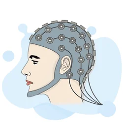
EEG Electroencephalography Practice Test PDF (Free Printable 2026)

Seizure EEG: What the EEG Test Shows, How It Works, and What to Expect
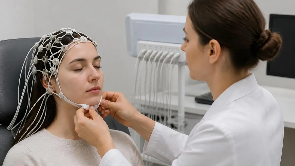
EEG Test Cost in 2026: Prices, Insurance Coverage, and How to Save

EEG Coherence: What It Is, How It Works, and What It Reveals About Brain Connectivity
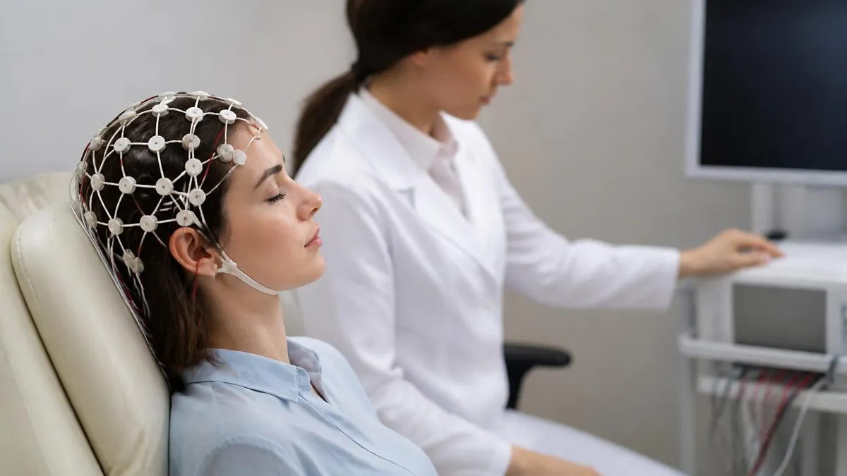
EEG vs ECG vs EKG: What Each Test Measures, How They Differ, and What to Expect
About the Author

Educational Psychologist & Academic Test Preparation Expert
Columbia University Teachers CollegeDr. Lisa Patel holds a Doctorate in Education from Columbia University Teachers College and has spent 17 years researching standardized test design and academic assessment. She has developed preparation programs for SAT, ACT, GRE, LSAT, UCAT, and numerous professional licensing exams, helping students of all backgrounds achieve their target scores.